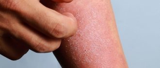
Itchy skin
Itchy skin:
Occurs in 2% of pregnant women in the third trimester of pregnancy.
Pathogenesis:
Presumably, itching is caused by increased excretion of bile acids under the influence of endogenous hormones (estrogen and progesterone). Its development is also associated with recurrent cholestasis.
Clinical picture:
Usually the itching is localized in the abdominal area, but it can also be generalized. Some patients have pronounced scleral icterus and an increase in the content of alkyl phosphatase in liver tests. After childbirth, the itching disappears, but may reappear during the next pregnancy or use of oral contraceptives.
Differential diagnosis:
- Scabies.
- Toxicoderma.
- Hepatitis.
Prurigo of pregnancy occurs at an earlier stage (from the 25th to the 30th week) and is not urticarial in nature, unlike polymorphic dermatosis. The clinical picture is characterized by disseminated itchy pruriginous rashes in the form of excoriated nodules with a hemorrhagic crust at the top. The rash persists throughout pregnancy and is difficult to treat (see Pruritus); After childbirth it regresses on its own.
Make an appointment for a consultation by phone. +7 (495) 589-85-42, +7 (495) 589-85-43.
Introduction
There are itchy rashes that are characteristic of pregnancy and the postpartum period.
In 1983, Holmes and Black proposed a classification of skin diseases of pregnancy, which included pemphigoid gravidarum, multiform dermatosis of gravidarum, prurigo of gestation, and pruritic folliculitis of gestation. In 1998, Shornick proposed adding cholestasis of pregnancy to this list. The latest classification was proposed by Ambros-Rudolph et al in 2006, based on a large retrospective study of 505 patients, and includes four dermatoses: pemphigoid gravidarum, multiform dermatosis gravidarum, atopic dermatitis of pregnancy (including prurigo gravidarum and pruritic folliculitis of gestation), and intrahepatic cholestasis of gestation. .
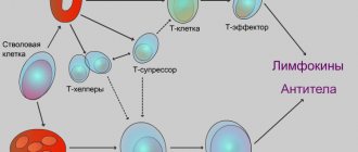
The main reason for skin changes during pregnancy is considered to be changes in the pregnant woman's immune system. To prevent fetal rejection, an imbalance develops between the cellular and humoral immunity. The production of cytokines by T helper type 2 (Th2) cells predominates over Th1, an increase in humoral immunity and a delay in the growth of cellular immunity are observed. The influence of changes in the level of maternal hormones is assumed, because many skin conditions develop during the third trimester. This article is devoted to skin diseases characteristic of pregnancy, with an emphasis on clinical manifestations, possible complications for the fetus, pathogenesis, diagnosis and treatment.
Discussion
Pemphigoid gravidarum (PG)
Pemphigoid gravidarum, formerly known as herpes gravidarum, is the rarest of the skin disorders of pregnancy, with an incidence of 1:2000 to 1:60,000. PG initially appears as papules and plaques that transform into vesiculobullous elements. PG is characterized by the appearance of rashes in the navel area, spreading to the chest, back and limbs. The palms and soles may be involved, but usually the mucous membranes rather than the face. The rash most often develops in the third trimester. Dermatosis exists throughout pregnancy and in 75% of patients, exacerbation occurs during childbirth. PG usually resolves spontaneously within a few months after delivery. Typically, there is a recurrence of dermatosis during subsequent pregnancies, with earlier onset of dermatosis and greater severity compared to the previous pregnancy. There are also reports of exacerbations during menstruation or when using oral contraceptives. There is an increase in the incidence of prematurity, especially with more severe cases, blistering and the onset of dermatosis before the third trimester.
Approximately 10% of children develop a temporary, bullous rash due to the transfer of antibodies across the placenta. PG is an autoimmune disease in which there are antibodies against the NC16A region of collagen XVII (BPAG2, BP180), which is present in amniotic, placental, and umbilical tissue, in addition to the basement membrane of the skin. Antibodies activate the complement cascade with inflammation and blistering.
IgG4 antibodies are detected. Women with this disorder are at high risk of autoimmune diseases, especially Graves' disease. An association with HLA-DR3 and HLA-DR4 is observed. Histologically, bullous PG is characterized by cutaneous edema and perivascular inflammation with lymphocytes, histiocytes, and eosinophils. Subepidermal blisters are observed in vesiculobullous lesions with a predominance of eosinophilic infiltrates.
Direct immunofluorescence shows linear deposition of complement 3 (C3) along the basement membrane zone in all patients. Some patients also have IgG deposits along the basement membrane. Enzyme-linked immunosorbent assay (ELISA) can detect specific antibodies against collagen XVII, which correlates with disease activity and can be used to monitor the effectiveness of treatment. Treatment of PG should be aimed at reducing itching and blistering. In mild cases, topical corticosteroids and antihistamines are effective. In severe cases of bullous PG, it is advisable to use systemic corticosteroids. The dose can be reduced once adequate control is achieved, however, it is often not reduced due to the high risk of exacerbation. The use of systemic corticosteroids does not increase the risk to the fetus.
Polymorphic dermatosis of pregnancy (PEP)
PEP is a benign, pruritic, inflammatory condition with an incidence of 1 in 160 pregnancies. It usually occurs in the late third trimester or immediately after delivery in the first pregnancy, with an increased risk in multiple pregnancies and rapid weight gain. Urticarial papules and plaques appear first on the abdomen and, unlike PG, do not affect the umbilicus area. The rash usually extends to the thighs and buttocks and is rarely generalized. Blisters with a diameter of 1-2 mm may develop, but unlike PG, bubbles are not observed. Rashes with clear boundaries regress spontaneously within 4-6 weeks without connection with treatment.
PEP does not pose a risk to the fetus. The histological picture is similar to PG. At the onset of PEP, superficial to mid-skin perivascular infiltrates of lymphocytes, histiocytes, and eosinophils are observed with dermal edema. Late stages of PEP are characterized by epidermal spongiosis. In contrast to PG, the immunofluorescence assay is negative. Treatment of PEP is based on symptomatic relief using topical corticosteroids and antihistamines. If the rash becomes generalized, a short course of systemic corticosteroids may be used.
Atopic dermatitis of pregnancy (AEP)
AEP is the most common skin disease in pregnant women, accounting for almost 50% of all dermatoses. It has also been called by other names, including prurigo gravidarum, prurigo gravidarum, Spangler's papular dermatitis of gestation, pruritic folliculitis of gestation, and eczema of gestation. AEP is a benign condition characterized by a pruritic, eczematous or papular rash. Develops until the third trimester, unlike other dermatoses of pregnancy. Two thirds of AEP cases are characterized by eczematous skin changes localized to atopic areas of the body such as the neck and flexor surfaces of the extremities. The remaining cases are characterized by a papular rash in the abdomen and limbs. The lesions usually respond well to treatment and resolve spontaneously after delivery. However, AEP is likely to recur in subsequent pregnancies. Dermatosis does not significantly affect the fetus, but there is an increased risk of developing atopic dermatitis in the infant.
It is believed that the development of AEP is initiated by pregnancy-related immune changes in the body. In this case, there is a shift towards humoral immunity with increased activation of Th2. Pregnant women with AEP are predisposed to atopic dermatitis, but 80% of these pregnant women develop these skin changes for the first time during pregnancy. There is often a family history of atopic dermatitis in relatives.
AEP is usually a diagnosis of exclusion because Diagnostic testing is nonspecific. Serum IgE levels are elevated in 20-70% of patients. AEP is differentiated from ICP, scabies and drug allergies. The mainstay of treatment is topical corticosteroids. In severe cases, a short course of systemic corticosteroids and antihistamines may be used. Phototherapy may also be used.
Intrahepatic cholestasis of pregnancy (ICP)
ICP, formerly known as obstetric cholestasis, cholestasis of pregnancy, and jaundice of pregnancy, is a reversible cholestasis that is likely due to hormonal changes late in pregnancy in predisposed women. ICP is characterized by acute-onset itching that often begins on the palms and soles and then generalizes. The skin contains mainly secondary lesions, such as excoriations, but there may also be papules. In 10%, jaundice develops due to concomitant extrahepatic cholestasis. After childbirth, the itching goes away within a few weeks. There is a risk of recurrence in subsequent pregnancies and when using oral contraceptives.
Diagnosis of ICP is important because... may be associated with serious consequences. Possible fetal complications include preterm birth, intrauterine fetal distress, and intrauterine fetal death. The incidence of complications in the fetus correlates with the total level of bile acids in the maternal blood serum. In cases of severe ICP complicated by jaundice, there is a risk of bleeding in the mother or fetus due to impaired absorption of vitamin K. Severe itching in ICP is associated with increased levels of bile acids in the blood caused by impaired secretion, a multifactorial process resulting from genetic disorders, the influence of environmental factors environment and endogenous hormones. There is a high incidence of ICP in multiple pregnancies. ICP is diagnosed based on the detection of elevated bile acid levels. Hyperbilirubinemia is seen only in the most severe cases, about 10-20%, and liver tests may be normal in 30%. Histology is nonspecific and immunofluorescence is negative.
Treatment is aimed at normalizing serum bile acid levels to reduce risk to the fetus and to control maternal symptoms. Treatment with ursodeoxycholic acid (UDCA) is recommended. Other drugs that reduce itching, such as antihistamines, S-adenosyl-L-methionine, and dexamethasone, may be used. Anion exchange resins such as cholestyramine may cause vitamin K deficiency regardless of the presence of ICP and should therefore be avoided.
Conclusion
The four skin diseases characteristic of pregnancy, pemphigoid gravidarum, polymorphic dermatosis of pregnancy, atopic dermatitis of pregnancy, and cholestasis of pregnancy can be distinguished by clinical presentation, histopathology, pathogenesis, and risk of fetal complications. Only pemphigus of pregnancy and intrahepatic cholestasis of pregnancy are associated with a significant risk to the fetus. Because all of these dermatoses are pruritic, careful evaluation of any pruritic pregnancy is necessary. Adequate treatment of the pregnant woman and prevention of any potential risk to the fetus is imperative.
Polymorphic dermatosis
Polymorphic dermatosis of pregnant women:
Developing in the third trimester of pregnancy, on average at the 36th week of pregnancy, usually within 1-2 weeks. before giving birth; Regresses on its own within a few days after birth.
Frequency:
One case in 120-240 pregnancies.
Pathogenesis:
The disease is associated with an abnormal increase in body weight of the pregnant woman and fetus, and overstretching of the abdominal skin.
Clinical picture:
Red papules, 1-3 mm in size, appear on the skin of the abdomen, buttocks, thighs, occasionally the face, palms and soles, which quickly merge into plaques resembling large blisters, in places forming confluent polycyclic lesions. The rash is accompanied by severe itching. The mucous membranes are not affected.
Diagnostics:
Usually based on the clinical picture.
Differential diagnosis
- Herpes in pregnant women.
- Drug toxicoderma.
- Diffuse neurodermatitis.
Course and prognosis:
All symptoms usually disappear 7-10 days after birth. Most women do not experience relapses in subsequent pregnancies. No adverse effects on the pregnant woman or fetus were noted.
Synonyms:
- Pruritic urticarial-papular and plaque dermatosis of pregnant women.
- Toxic erythema of pregnancy.
Make an appointment for a consultation by phone. +7 (495) 589-85-42, +7 (495) 589-85-43.
Herpes in pregnant women
Herpes in pregnant women:
Dermatosis, manifested by a polymorphic rash and severe itching, occurring during pregnancy and the postpartum period.
Frequency:
One case in 4,000–50,000 pregnancies.
Risk factors:
- Episodes of illness during a previous pregnancy.
- Use of oral contraceptives.
- Tumors from trophoblast.
Etiology and pathogenesis:
An autoimmune disease involving complement-fixing IgG antibodies. In patients, alleles HLA-B8, HLA-DR3 and HLA-DR4 are detected with high frequency.
Clinical picture:
The disease usually begins in the 4th to 7th month of pregnancy, but can occur in the first trimester and in the postpartum period. Manifests itself in the form of papules and vesicles. There are also small and large red blisters, large tense blisters, erosions and crusts. The elements are arranged in groups, which gives rise to the name “herpes of pregnant women.” The process most often begins from the navel area, and then spreads to the skin of the abdomen, thighs, palms and soles, and rarely to the mucous membrane of the mouth.
Differential diagnosis:
- Polymorphic dermatosis of pregnant women.
- Polymorphous exudative erythema.
- Dühring's dermatitis herpetiphora.
- Drug toxicoderma.
Treatment:
For mild cases, corticosteroid ointments are used topically in combination with oral antihistamines. For bullous rashes, prednisolone is prescribed orally 40 mg/day. followed by a reduction to a maintenance dose. During childbirth, a significant increase in the dose is sometimes necessary due to a sharp increase in rashes and itching.
Make an appointment for a consultation by phone. +7 (495) 589-85-42, +7 (495) 589-85-43.
Atopic dermatitis in pregnant women
Fundamentally important for pregnant women. Those suffering from Atopic Dermatitis (hereinafter referred to as “AD”) are required to strictly adhere to sleep, rest, and food intake. Contact with dishwashing detergents must be completely avoided. household chemicals. The furnishings in the room where the pregnant woman spends most of her time should be “Spartan”, that is, a minimum of furniture, wet cleaning and ventilation should be carried out daily.
It is recommended to refrain from using antihistamines, especially in the early stages. In especially severe cases, it is possible to use loratodine, but under the supervision of a doctor.
A pregnant woman's nutrition should be complete, so prescribing a very strict diet is not required, but complete dietary freedom cannot be given either. Usually, an elimination diet is sufficient (i.e., eliminating from the diet foods to which the patient notes allergic reactions) and limiting foods that are aggressive to the gastrointestinal tract.
To correct the condition of the gastrointestinal tract, choleretic and hepatoprotective drugs (Hofitol, Hepabene Essentiale), probiotics are usually used. Long-term use of probiotics by a pregnant woman reduces the risk of developing AD in the child. Normalizing the activity of the biliary tract has a beneficial effect on the skin, so it is advisable to use preparations from artichokes (Hofitol). But only after consulting a doctor.
Pregnant women with AD need to adhere to a protective regimen for the skin and regularly use emollients (in case of exacerbation of AD, it is desirable that the drug contains antipruritic and healing components). Preference should be given to pharmaceutical cosmetics lines due to the absence of dyes and fragrances and a balanced composition. Basic care preparations: “Emolium”, “Sensaderm”, “La Roche Posay”, “Uriage Topi” cream, “Bioderma”. To cleanse the skin, cleansers with an acidic pH and a creamy additive / “soapless soap” are used.
In AD, there are several mechanisms of skin barrier disruption, so a basic care product should contain a natural moisturizing factor, lipids, and have an antipruritic effect. All these requirements are met by the above mentioned care lines.
Creams are designed for application to small areas of the skin and have a richer structure, while emulsions are convenient for application to common lesions.
In addition to moisturizing-obesifying preparations, all cosmetic lines include hygiene products: creamy gels for washing, emulsion for baths, cleansing oils for baths. They are used both during exacerbation and during remission of the disease.
In cases of severe exacerbation, it is permissible, with caution, to take short courses of topical steroids: Elokom, Advantan, Lokoid, only after consulting a doctor! For mild exacerbations, Locobase Repea cream is used, preferably at night, once a day.
In all cases of AD, both during exacerbation and remission, regular use of basic care products is necessary. This will not only improve the physiological condition of the skin, but also reduce the risk of developing both bacterial and fungal complications of the disease.
Barzeeva N.D.
Atrophic stripes
Atrophic stripes:
They appear in most pregnant women on the skin of the abdomen, buttocks, and mammary glands.
Pathogenesis:
Apparently, in addition to the mechanical factor (stretching), degeneration of the dermis caused by endocrinopathy, autonomic disorders, etc. is of certain importance.
Clinical picture. Atrophic stripes of skin up to 4-6 mm wide, located symmetrically. Areas of band-like atrophy are sharply defined, with a wrinkled surface of a whitish-lilac hue, slightly sunken.
Treatment:
Use of Mederma or Contratubes creams after childbirth.
Synonym:
Striae of pregnant women.
In addition, during pregnancy, more pronounced general skin pigmentation is observed, especially in brunettes, developing in the area of the nipples of the mammary glands, genitals, and midline of the abdomen. In many women, the number and size of melanocytic nevi increase. The hair thins somewhat, even diffuse alopecia is possible, but after childbirth, over time, the properties of the hair are restored. Sometimes there is some hypertrichosis in combination with acne, usually in the second half of pregnancy. This may be the result of an androgen-secreting tumor, luteoma, luteal cysts, or polycystic ovarian disease and requires careful evaluation. Vascular changes are associated with the action of estrogens; they manifest themselves in 70% of pregnant women with erythema of the palms, which in some cases can be expressed only in the thenar and hypothenar areas. Small hemangiomas may occur, most often on the scalp and neck. Varicose veins of the extremities, hemorrhoids, and, less commonly, deep vein thrombophlebitis are often observed. Swelling of the face, eyelids, feet, and hands is also possible, which occurs in the morning and disappears in the evening. 80% of pregnant women develop gingivitis (swelling and redness of the gums), sometimes accompanied by pain and ulceration. In 2% of cases, changes in the gums are accompanied by the appearance of small vascular formations such as pyogenic granuloma, which bleed when touched (granuloma of pregnancy). These symptoms are somewhat relieved by taking vitamin C.
During pregnancy, there may be an increase in the frequency of infectious lesions, especially candidiasis, genital warts, herpes simplex, bowenoid papulosis. During pregnancy, autoimmune diseases (especially lupus erythematosus), lymphomas, and atopic dermatitis worsen. The effect of pregnancy on psoriasis can be different, and some patients may develop an acute pustular form.
II. Skin diseases relatively often reported during pregnancy:
This group includes a variety of skin changes and diseases that often accompany pregnancy, as well as some common dermatoses, the course of which usually worsens during gestation. The main mechanism is a change in hormonal ratios in the pregnant woman’s body. This, in turn, affects the functionality of the immune and nervous systems, the state of the gastrointestinal tract, kidneys, cardiovascular activity, water-salt metabolism, etc.
This group includes: hyperhidrosis
- hypertrichosis
- Palmoplantar telangiectasias
- erythema of the palms
- alopecia – hair loss
- onychodystrophy - changes in the nail plates
Most of these and other conditions usually disappear after childbirth. If necessary, their treatment is symptomatic.
The course and severity of symptoms of other skin diseases also change during pregnancy. These primarily include eczema, atopic dermatitis, psoriasis, lichen planus, acne, and Dühring's dermatitis herpetiformis. During pregnancy, they often worsen, and exacerbations are usually more pronounced and more severe than before gestation. Sometimes, on the contrary, during pregnancy a remission occurs, which continues until childbirth, after which another exacerbation occurs. Correcting exacerbations of these dermatoses during pregnancy is very difficult. Many medications used under normal conditions affect fetal development. Therefore, external symptomatic therapy becomes the leading method.