Epstein-Barr virus belongs to the herpes virus family. As a rule, infection with this virus occurs in childhood, after which the infection remains in a latent form in the human body throughout life.
Transmission of the virus occurs through airborne droplets, that is, through kisses, saliva, and blood transfusions. A distinctive feature of the disease is that it does not cause the death of the infected cell, but changes its structure and function.
The danger of the Epstein-Barr virus is that the infection is hidden. The virus remains hidden in the human body until a decrease in immunity occurs, after which the infection again becomes active.
The Epstein-Barr virus can cause mononucleosis, adenoiditis, Hodgkin's disease, nasopharyngeal cancer, hepatitis, tonsillitis, sclerosis, chronic fatigue syndrome and many other diseases.
Introduction
Infertility is a pressing medical and social problem.
In Russia, every fifth married couple is infertile [1]. Moreover, the male factor is responsible for half of these cases. This is associated with a progressive decline in reproductive function. There is numerous data on the negative impact of viruses on the state of the male reproductive system. Human herpes viruses (HHV) are widespread in the human population [2]. Herpes viruses cause lifelong infection and are the cause of many diseases, half of which have a relapsing course. From 95% to 100% of the population are infected with HHV, and at different periods of life a person is infected with at least one, and more often, several types of viruses belonging to the herpesvirus family. In the second half of life, almost all people have antibodies to most HHVs [3]. 8 types of herpes viruses are pathogenic for humans.
Transmission routes
Herpes type 4 is found in the body of almost every person. It is one of the most common viruses. About 90% of all people on the globe over the age of 20 are its carriers.
How is it transmitted:
- during sexual contact with an infected person;
- in contact with personal items;
- during blood transfusion;
- when breastfeeding;
- through contaminated air.
The entry of the virus into the body does not mean the immediate development of the disease. It can be inside and not cause problems. However, with a decrease in immunity, it enters an active state and leads to the appearance of various diseases.
Types of human herpes viruses
The herpes simplex virus (HSV) was first isolated by W. Goiter in 1912; HSV is presented in two types: 1 and 2. Carriage of the HSV-1, -2 virus occurs in more than 90% of the population. These are DNA-containing viruses that infect the skin, mucous membranes, vascular endothelium, blood cells, and the central and peripheral nervous system.
HHV type 3 - Varicella
-zoster
virus
, varicella zoster virus (VZV) - discovered in 1911 by N. Aragao and is a DNA virus. It is the causative agent of chickenpox and herpes zoster and can affect the skin, mucous membranes, vascular endothelium, blood cells, liver, central and peripheral nervous system.
HHV type 4, Epstein-Barr virus (EBV), was isolated in 1964 by Epstein and Barr. EBV has tropism mainly for B lymphocytes, the epithelium of the upper respiratory tract and the epithelium of the gastrointestinal tract. It can act as an etiological agent in infectious mononucleosis and Burkitt's lymphoma.
HHV type 5 - cytomegalovirus (CMV) was isolated in 1956 by M.G. Smith, contains double-stranded DNA surrounded by a glycoprotein shell. It can affect almost any cell of the human body, causing a wide range of diseases.
HHV type 6 (roseolovirus) was identified in 1986 and is associated with sudden exanthema and febrile seizures in children.
HHV type 7 (roseolovirus) was isolated in 1990 from peripheral blood CD4+ T lymphocytes. The virus is associated with various diseases of the central nervous system and skin of an autoimmune nature.
HHV type 8 is associated with Kaposi's sarcoma and was discovered in 1994, is a new transforming human virus that promotes malignant transformation of cells.
HHVs are common sexually transmitted viruses [4], with possible effects on male factor infertility. The pathogenic potential can be realized either directly by viral toxic effects on cells of the genital tract, or indirectly by local or systemic infectious or immunological reactions. The herpesvirus family (HSV-1, -2, VZV, EBV, CMV, and HHV-6) is commonly found in semen, but the effects of these viral agents on male fertility have not been widely studied.
Herpes simplex viruses types 1 and 2
and the varicella-zoster virus
(HSV) affect the skin and mucous membranes, being the main cause of genital ulcers. HSV types 1 and 2 are often detected in the spermoplasm, on the surface and inside sperm [5].
Transmission of HSV-1, -2 has been recorded during insemination with donor sperm. Despite the fact that the frequency of detection of HSV-1 and -2 in semen is relatively low (less than 5%) [6], HSV is the pathogen that has the strongest effect on sperm parameters: sperm concentration, their total number, motility, concentration of citrate and neutral α-glucosidase. These pathogenic properties were also identified in later studies. It is known that antiviral treatment of infertile men with HSV in the ejaculate led to successful pregnancies [7].
In 2000 E.E. Bragina et al. For the first time, evidence of sperm infection with HSV-1, -2 was presented. These viruses were detected in urethral smears only in persons with manifest genital herpes; in the ejaculate they were detected in 33–54% of those examined. Electron microscopy of the ejaculate made it possible to detect spermatozoa containing HSV-1, -2 nucleocapsids of varying degrees of maturity. HSV can infect sperm and disrupt spermatogenesis by sharply reducing the concentration of sperm and their motility, increasing the number of pathological forms, including pathology of the head. Among married couples with genital herpes, there is a higher incidence of miscarriage compared to healthy spouses [8].
A study was also conducted to study blood antibodies to CMV, EBV and HSV-1, -2 in men with and without leukospermia. It turned out that anti-HSV-1, -2 IgM were associated with leukospermia, although polymerase chain reaction (PCR) could not detect HSV-1, -2 in the ejaculate [9].
In a prospective randomized study by E. Neofytou et al. (2009) included 172 men, of whom 143 (83.1%) had HHV detected in their ejaculate. In the group of men with normal sperm parameters versus the group with pathospermia, the virus content was: HSV-1, -2 - 2.5% / 2.1%, VVZ - 1.2% / 3.2%, EBV - 45% / 39 .1%, CMV - 62.5% / 56.5%, HHV-6 - 70% / 66.3%, HHV-7-0% / 0%, respectively. Thus, no association was found between viral agents and pathospermia, but VZV was detected almost three times more often [10].
A decrease in the concentration of neutral α-glucosidase and citrate indicates that infertility occurs due to dysfunction of the epididymis and prostate under the influence of HSV.
Cytomegalovirus
A number of studies have assessed the prevalence of CMV. Thus, in many countries in human sperm samples this figure ranged from 0% to 62.5% [11].
CMV is a member of the HHV family and can cause a variety of teratogenic lesions in newborns and clinical cases of the disease in adults such as infectious mononucleosis. Its presence and persistence in sperm have been described in various studies [9, 12–14]. CMV causes asymptomatic infection in healthy individuals, including infection of the urogenital tract. Like all herpes viruses, CMV has the ability to reactivate and cause chronic inflammation [15, 16].
The content of CMV in the ejaculate of healthy donors usually ranges from 3–5%, in the ejaculate of patients with infertility - from 1.4% to 56.5% [11].
In a study conducted by M. Habibi et al. Screening of ejaculates from patients with infectious inflammation of the prostate gland using the PCR method revealed that CMV is more common in infertile men with chronic inflammatory diseases of the genitourinary system, and CMV DNA is associated with the cellular fraction of the ejaculate. The prevalence of viral DNA was 3.4% for EBV, 5.2% for CMV, 6.5% for HHV-6, 0.43% for EBV + CMV, 0.87% for EBV + HHV-6 , 1.3% - for CMV + HHV-6. Detection of CMV was significantly more frequent in the group of patients with infertility and was associated with a decrease in sperm count. The results obtained indicate that both CMV and HHV-6 can contribute to the development of infectious inflammation of the prostate gland and male infertility [9].
The concentration of CMV in sperm can vary from 110 to 12 million. In this case, there is a possible tendency to reduce sperm motility and the concentration of neutral α-glucosidase, a marker of damage to the epididymis. Thus, CMV may have an impact on sperm quality due to damage to the epididymis [17, 18].
More convincing results were obtained in the work of M. Mohseni et al. This study determined the detection rate of CMV using real-time PCR in 100 infertile men who presented to an in vitro fertilization clinic with low motility (less than 40%) or sperm concentration (less than 15 million/ml), a history of infertility of more than 5 years, at the same time, their wives were healthy and without apparent causes of infertility. The control group included 100 healthy sperm donors. The detection rate of CMV in the study group was 23%, in the control group - 7%, the difference was significantly significant (p <0.01). Thus, the significant role of CMV in infertility in men has been demonstrated. The authors propose to include the detection of CMV in the diagnostic search for infertility, and to exclude sperm donors with detected CMV from the list of donors [19].
Studying the ways in which viruses penetrate male gametes still arouses the interest of researchers, and until recently it was not possible to infect mature sperm with CMV and HSV. However, E.A. Malolina et al. (2016) developed a model of sperm infection in vitro
and for the first time the fundamental possibility of infection of mature gametes with CMV was proven [16].
A study by Muhsin et al. (2019) was conducted on 90 men, among whom there were 35 (38.9%) fertile men with normal sperm quality, 55 (61.1%) infertile men. Detected IgG antibodies in spermoplasm to EBV (38%), CMV (43%) and HHV-6 (53%) were significantly associated with idiopathic infertility. The identification of immunological markers of viral infection was accompanied by an increase in the levels of pro-inflammatory cytokines: interleukin 10, γ-interferon, which affected the state of antioxidant protection: an increase in the content of 8-OH-deoxyguanosine in sperm plasma and a significant decrease in the total antioxidant capacity of sperm were observed. The results of the study demonstrated that 45 (50.0%) infertile men had a reduced sperm count, 52 (57.8%) had a decreased number of motile sperm, and 33 (24.4%) had abnormal sperm morphology. The data confirm a negative correlation between the level of inflammatory biomarkers and standard spermogram indicators: sperm concentration, motility, and the proportion of cells with normal morphology. The authors of the study suggested that chronic viral infection occurs against the background of increased concentrations of cytokines, disrupts the oxidative status of sperm, the antioxidant defense system, which leads to damage to sperm DNA [20].
The relationship between herpesvirus infection and male infertility was demonstrated using PCR and DNA sequencing in infertile men aged 21–49 years in China (n=151). Herpesviruses were identified in 59 patients (39%). The number of carriers was 39 (25%) for HSV-1, 6 (4%) for EBV, 33 (22%) for CMV, and 3 (2%) for HHV-6. Dual infection was detected in 22 individuals (59 samples, 37%), the majority of whom (n=15) were infected with HSV-1 and CMV [21].
Epstein-Barr virus
EBV causes infectious mononucleosis and Burkitt's lymphoma. EBV has been detected in semen and there is strong evidence that it is sexually transmitted. The frequency of its detection in ejaculate is 1–2 orders of magnitude higher than HSV-1, -2 [11]. In a study by E. Moretti et al. (2017) the detection of anti-EBV antibodies in the blood of 47% of individuals was associated with a decrease in sperm concentration and motility, as well as an increase in the frequency of necrozoospermia, which led to a decrease in the fertility index [14]. It is known that EBV persists in B lymphocytes, which are found only in small numbers among all leukocytes in sperm. The median concentration of EBV in semen was 500 copies/ml [17]. However, there is also directly opposite evidence that in the presence of EBV in the ejaculate, a paradoxical twofold increase in type A motility was observed in a study by V. Naumenko et al. (2016) [17]. Apparently, the phenomenon of increased sperm motility provides evolutionary advantages to some viral agents, which use it to spread more widely in the population.
EBV is the etiological agent of B-cell lymphoma, and the primary testicular variant of this disease leads to infertility [22]. Studies have shown that the presence of EBV in semen is associated with a 4.8-fold increase in the likelihood of testicular cancer [23]. Systematic review and meta-analysis by A. Garolla et al. confirmed a significant connection between EBV infection of sperm and testicular germ cell tumors, while CMV did not have oncogenic potential [24].
Human herpesviruses type 6
An important property of HHV-6 is the chromosomal integration of its genetic material, which can be observed in 0.8–1.5% of carriers. It is possible to suspect chromosomal integration if the concentration of HHV-6 in the blood is more than 105 copies/ml. A direct method for diagnosing chromosomal integration is fluorescent in situ
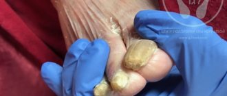
- inaccessible and labor-intensive. Indirect confirmation of this condition is possible by detecting HHV-6 in hair follicles and nails, i.e., in samples in which HHV-6 is usually not detected [25]. Genetic integration of the virus occurs in the terminal regions of chromosomes - telomeres, which is extremely alarming regarding the effect on cell division and apoptosis. The integrated virus is transmitted to descendants according to Mendelian laws, i.e., half of the children inherit the chromosomal integration of HHV-6 [26].
The effects of HHV-6 on reproductive function have been studied in several studies. Thus, in the work of V. Evdokimov et al. (2016) HHV-6 was significantly more often associated with infections of the accessory gonads in men (19% versus 6.3% in the control group) [27]. In this study, the median concentration of HHV-6 was only 250 copies/ml, thus, with chromosomal integration of HHV-6, where the viral load is measured in millions of copies per milliliter, significant abnormalities in sperm parameters can be expected [28].
As we see, in the literature there is no clear opinion about the effect of HHV on sperm, and some researchers cannot establish a connection between the presence of viruses in the ejaculate and the deterioration of spermogram parameters, others note a decrease in the concentration and motility of sperm in ejaculates positive for HHV [11, 29] .
It is possible that there are different variants of sperm infection by the virus. In the first scenario, already mature gametes are infected with HHV as they pass through the genital tract. In the second, infection is possible at the stage of sperm maturation, during which the damaging effects of viruses will be more significant [11].
Thus, there is important evidence of the influence of various HHVs on sperm quality and reliable associations of these viruses as an etiological factor in the development of infections of the accessory gonads, leading to infertility in men. However, further research in this direction is required to determine the exact mechanisms of the pathogenesis of viral infections as etiological agents of infections of the accessory gonads and infertility in men.
Symptoms of the disease
The symptoms of herpes type 4 depend on the disease caused by the virus.
Infectious mononucleosis
Initially, the disease manifests itself as decreased appetite, weakness and mild fever. At further stages, the disease is characterized by signs of ulcerative necrotic or catarrhal tonsillitis. In this case, fibrous films appear in the patient’s oral cavity.
Why can our articles be trusted?
We make health information clear, accessible and relevant.
- All articles are checked by practicing doctors.
- We take scientific literature and the latest research as a basis.
- We publish detailed articles that answer all questions.
Type 4 herpes is accompanied by the following symptoms:
- muscle pain;
- dizziness;
- gagging ;
- loss of appetite;
- drowsiness;
- swelling of the pharynx;
- nasal congestion
- headache ;
- sweating;
- hyperemia of the oropharyngeal mucosa;
- general weakness;
- high temperature (up to 40 degrees);
- decreased performance.
These symptoms can easily be confused with a sore throat or pharyngitis. Such signs are observed in the first stages of the disease (on the second to fourth days after infection).
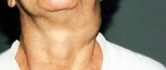
Enlarged lymph nodes are also considered an important symptom when diagnosing pathology. Their compaction, mobility, and pain on palpation are observed. Lymphadenopathy spreads to the submandibular, posterior cervical, and sometimes inguinal nodes.
In some cases, hemorrhagic (subcutaneous hemorrhages in the form of dots) or urticarial rashes (itchy pink blisters) appear on the skin and mucous membranes.
Internal organs also suffer - the spleen and liver become significantly enlarged. In this case, patients experience a change in the shade of urine and yellowness of the sclera of the eyes. These signs are observed for a long time - almost up to one month.
Burkitt's lymphoma
According to statistics, the disease is a common complication of infection with the Epstein-Barr virus. This pathology is accompanied by hyperthermia and increased sweating.
The disease is characterized by the formation of multiple or single tumors on the jaw. Burkitt's lymphoma causes difficulty swallowing, shortness of breath, weight loss, and tooth loss.
There are two forms of this disease:
- Generalized. The oncological process spreads to the spinal cord and spine. It manifests itself as a violation of motor coordination and decreased sensitivity of the legs and arms.
- The abdominal form, which affects the digestive organs (liver, intestines, pancreas). With pathology, dyspeptic disorders and pain in the abdominal area occur. Intestinal obstruction and ascites may develop.
The disease occurs in four stages. In the final stages, bone tissue and the nervous system are affected.
Nasopharyngeal carcinoma
With this pathological condition the following symptoms occur:
- difficulty breathing through the nose;
- noise and ringing in the ears;
- loss ;
- purulent nasal discharge with bloody clots;
- headache .
If symptoms of mononullosis and malignant processes appear, it is important to consult a specialist in a timely manner. After diagnosis, appropriate treatment is prescribed that will help prevent unwanted consequences and alleviate the patient’s condition.
Own results
We conducted a study that included 287 men with chronic prostatitis/chronic pelvic pain syndrome IIIA, 23 of whom had infertility. Diagnoses were verified according to the criteria of the European Urological Association [30].
In addition to standard diagnostic procedures, HHV types 4–6 were detected in samples from the urethra, prostate secretion, and ejaculate.
When HHV was detected, patients with infertility (group 1) were prescribed valacyclovir 1000 mg/day for 3 months. and the drug VIFERON® (interferon α-2b in combination with antioxidants (vitamins E and C)) in the form of rectal suppositories. Based on the authors’ personal experience, patients were prescribed the following regimen of drug use: 3 million IU 2 times a day for 10 days, then 3 times a week (also 2 times a day) for the next 3 weeks. Viral load was monitored at the end of 3 months of treatment. The remaining patients in whom HHV types 4–6 were not detected (group II) were prescribed levofloxacin 500 mg for 30 days, tamsulosin 0.4 mg for 3 months, diclofenac suppositories 50 mg for 10 days.
In addition to the acyclic nucleoside, which has a direct antiherpetic effect and is prescribed for the treatment of abacterial prostatitis associated with herpes viruses, VIFERON® was chosen as a drug that can provide a long-term antiviral effect. The drug contains interferon α-2b, which is a human recombinant analogue of a substance produced in any cells of the body. It has antiviral, immunomodulatory, antiproliferative properties, suppresses the replication of RNA and DNA viruses. Interferon α-2b in cells triggers a sequence of reactions leading to suppression of the synthesis of viral proteins, assembly and release of virions into the intercellular matrix. In addition to influencing protein synthesis, interferons have epigenetic activity, activating hundreds of genes that play an important role in the antiviral defense of the cell [31]. In addition, interferon α-2b limits the spread of viral particles by activating the so-called “genome guard” protein p53, which leads to apoptotic death of the infected cell [32]. Ascorbic acid and α-tocopherol acetate, which are part of the drug, are highly active antioxidants, have anti-inflammatory, membrane stabilizing, and regenerating properties.
In 11 patients with identified herpes viruses, electron microscopic examination of sperm was performed (JEM 1400, JEOL, Japan).
We noted that out of 103 patients with detected HHV types 4–6, 8 had infertility (group I), while among 184 patients without herpes viruses, infertility was verified in 15 (group II).
Of the 24 urogenital samples from 8 patients in group I, 12 virus-positive samples were obtained: 7 - ejaculate, 1 - urethral smears, 4 - prostate secretions. CMV and EBV were detected in 4 and 3 samples, respectively, including samples with combinations of 2 HHVs. HHV-6 was much more common - in a total of 7 samples out of all 12. The median viral load of herpes viruses types 4–6 was 1770 copies per 100,000 cells.
As a result of antiviral treatment, by the end of the 3rd month, control studies detected the virus in only 5 of 8 patients in group I, in whom herpes viruses were detected in 5 bioassays. At the same time, the median viral load in them decreased to 48 copies per 100,000 cells.
As a result of therapy for 6 months. after the end of treatment, pregnancy was observed in the spouses of 10 out of 23 men, who were distributed into groups as follows: 6 out of 8 from group I and 4 out of 10 from group II. Statistical analysis showed that differences in pregnancy rates reached significant significance between groups I and II (χ2 criterion value was 4.960, critical χ2 value at the significance level p=0.05 was 3.841, significance level p=0.026).
The effect of the course of antiviral therapy was also assessed by electron microscopy. A study of spermatozoa of 11 patients with infertility showed that if before treatment, HHV capsids of types 4–6 were found in 8 patients, then after treatment in 4 of these patients, viral capsids were no longer detected in spermatozoa, and in 3 other patients the number of spermatozoa with capsids was on average decreased by more than 2 times: from 12% to 5% of infected gametes in the ejaculate (Fig. 1).