A choroidal nevus is a benign tumor of the choroid. Such tumors are detected in almost 10% of people with white skin color and are practically not found among dark-skinned people.
Choroidal nevus is a pigmented formation of a cluster of melanocytes (pigment cells). As a rule, nevi are formed from birth, however, their pigmentation appears later, in the prepubertal period. Therefore, they are found, in most cases, in adults. In women and men, nevi occur with the same frequency.
The usual location for choroidal nevus is the posterior fundus region, behind the equator. However, sometimes it is found in the preequatorial zone and even directly in the equator zone.
At the early stages of their development, such nevi are localized on the superficial layers of the choroid, with age, spreading into its deeper tissues.
Pathogenesis.
The development of a birthmark is associated with the period of intrauterine development (formed from the neural crest). The neural crest is a collection of nerve plexus cells called new cells. These cells remain immature and move to the epidermis layer (mostly the deep layer).
Some reach only the dermis. The role of new cells is that they produce a certain pigment. This pigment colors a specific area of the skin. Subsequently, a pigmented nevus is formed.
The classification of nevi (ICD 10 code) is as follows:
- D22.0 Melanoma of the lip;
- D22.1 M. century;
- D22.2 M. ear;
- D22.3 M. other parts of the face;
- D22.4 M. head;
- D22.5 M. trunk.
- D22.6 M. upper limbs;
- D22.7 M. lower extremities;
- D22.9 M. unspecified nature.
Nevi on the head
Such formations are most often removed precisely for cosmetic reasons. If a mole is located on the surface of the body, no matter what size it is, it can almost always be hidden under clothing. Nevi on the head cannot be hidden. Some patients believe that a mole emphasizes their individuality, others are sure that such a formation only spoils their beauty. As a result, patients often seek removal of even those nevi that will never become malignant.
Often, not only moles on the head are removed, but also tumors on the neck. The main danger of such moles is that they often rub against the surface of clothing and are eventually damaged, which increases the risk of degeneration. On the head, nevi also appear in the hair area. They are practically invisible to the naked eye and are often felt by the patient during hair washing and other procedures. The main danger of neoplasms is that they can be easily damaged during banal combing of hair. Nevi, which include cells of the sebaceous glands, also often appear on the scalp in the hairline area. They are characterized by the shape of a wart with no hair on the surface, as well as an irregular shape. Human papilloma viruses, as well as hereditary factors, can cause the appearance.
Most moles on the head appear at birth. The formation is examined and research is done to make a final diagnosis. In childhood, neoplasms are rarely removed, since they are not subject to degeneration during this period. It occurs most often during puberty. The removal method is selected depending on the size of the formation. If the nevus is small, you can get rid of it with laser therapy; to combat large formations, surgery will be required.
It is important to remember that at the moment there is no magic pill that could remove all dangerous moles from human skin that can become malignant. It is important to monitor the condition of nevi and pay attention to any changes.
Moles are successfully removed. It all depends on the size, qualifications of the medical specialist, as well as the location of the nevus.
Reasons for appearance
The presence of a nevus on the skin is normal. It is diagnosed in 85% of the country's population. An adult has an average of twenty-five nevi.
Their appearance depends on hormonal changes in the body (for example, adolescence or pregnancy). Before it appears on the human body, a nevus goes through certain stages:
- located inside the epithelium;
- then goes into a borderline state;
- and already in adulthood it is located inside the dermis.
80% of nevi are acquired.
The reasons for the development of a birthmark are as follows:
- A defect was obtained in the prenatal period;
- Heredity;
- UV radiation;
- Hormonal changes in the body;
- Various infections;
- Traumatic factor.
The resulting defect.
The pathogenesis of nevus development is as follows: cell division is disrupted at the very last stages of the child’s development. A nevus forms by the age of four years of a child’s life (about 70% of all moles in a child).
Heredity
All information in a person is embedded in DNA. After the genetic material is transferred, the development of the organism occurs, which is embedded in this chain. This formation will be benign. It is worth noting that those moles that a person acquires throughout his life will not be inherited.
UV radiation.
Leads to the fact that melanocytes begin to multiply rapidly in the skin. Melanotropic hormone is produced. This nevus will be acquired. People who frequently visit solariums are among those at risk.
Traumatic factor.
Insect bites can mechanically damage the skin at various levels. The formation of biologically active substances begins, they, in turn, begin to increase cellular growth. This situation occurs extremely rarely.
Hormonal changes.
This should include the period of adolescence, pregnancy, as well as patients with thyroid diseases. The nature of the course is benign.
Infectious factor.
The impact of various pathogenic microorganisms on the body can act as a provoking factor so that the inflammatory process in the body is started. As a protective response - the appearance of pigment spots and papillomas.
Who gets these spots more often?
- Visitors to solariums, as well as workers who are often exposed to UV radiation;
- People working with chemicals and carcinogens;
- People with hormonal imbalances who have been using certain hormonal drugs for a long time;
- Having problems with the thyroid gland, as well as weakened immunity;
- Those who have skin cancer or are prone to this disease.
What is the risk that your nevus will develop into skin cancer?
The presence of at least one nevus on the skin creates the preconditions that you may develop skin cancer. In 40% of cases, the presence of a nevus on the skin can be considered a precancerous disease.
The risk increases if your nevus is one of the following two types in nature: congenital or dysplastic. If the size of the nevus is more than three cm (up to 25%). If most nevi are located on the face, chest, shoulders. If you have more than 40 nevi, your risk increases sixfold.
What to do if a child scratches or tears off a mole?
In most cases, nevi are not a cause for concern. The appearance of moles in a child of any age is absolutely normal. They will gradually increase along with the growth of the child himself, and the location of the moles can be anywhere, including on the head, soles and even the genitals. Moles in children can easily be irregular in shape, have uneven borders, varied color patterns and large size, especially for congenital nevi.
It is also normal that moles will arouse your child's curiosity. If a child scratches or even picks off a mole, do not panic. This is only a reason to contact a dermatologist for dermatoscopy and further observation. So the answer to the question of what to do if your baby picks a mole is very simple: don’t worry and make an appointment with a specialist. ⠀
Parents need to be wary only in the following cases:
- rapid, sudden growth of formation in volume or diameter;
- the appearance of bleeding or a crust on the surface of the mole without previous injury;
- a rare type of mole;
- a large number of moles (>50) or cases of melanoma in the family;
- positive “ugly duckling” symptom (one formation is very different from other moles).
If you or your child just touched a mole, there is no need to worry.
What types of nevi exist.
Pigmentary.
This is an embryonic defect that has caused cells to begin to divide, multiply and distribute incorrectly in the skin. And this pigment is a consequence of this defect.
Pigmented nevi include the following:
- border,
- complex,
- leather inside. These species have the ability to move from one to another.
Borderline.
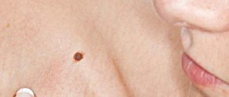
A higher percentage of occurrence occurs in childhood. They look like flat spots, brown in color, round in shape, the edges are smooth and regular. Acceptable dimensions are up to 1 cm; if the stain exceeds these dimensions, immediate removal is indicated.
Difficult.
- Located in the deep layer of the epidermis;
- Different colors of the two halves;
- Not flat, there may be indentations;
- It is necessary to conduct a study such as a biopsy of this area of skin.
Congenital.
- It is detected while the child is in the womb.
- They look like small tubercles or nodules.
- Covered with black hairs.
- Available in sizes from 1 cm to 30 cm.
It is worth paying attention to the size of the nevus because the larger it is, the higher the risk of developing cancer. When diagnosing, it is necessary to perform a biopsy, since it is not possible to remove the nevus in all cases.
Inside the skin.
It looks like a lump that is dark brown or black in color. She is covered with hair.
Atypical.
- The shape has irregular edges;
- No hair;
- Incorrect coloring;
- Throughout life it does not undergo any changes.
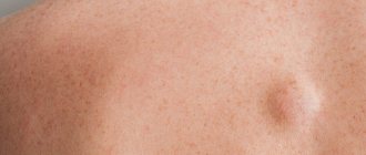
A mole with a rim around it.
- The rim is located at the edges of the nevus;
- The skin around the rim becomes white;
- It fades gradually from several weeks to a year.
Papillomatosis.
- Soft;
- Elastic;
- The presence of scales on the formation itself is noted.
Nevus in the form of a spot.
- Brown color;
- In the center there are foci resembling grain;
- Sizes up to eight cm;
- It is also possible to see a blue tint in the center.
Nevus Spitz.
- Cone;
- Color varies from red to brown;
- Most often occurs in childhood (and subsequently remains for life);
- Has blood vessels.
Nevus located in the dermis.
- Blue or light blue tint;
- In the form of a tubercle, bump;
- On the head or buttock area;
- It quickly increases in size and reaches several cm.
Seborrheic nevus.
- It is detected already in infancy;
- Sizes up to ten cm;
- Smooth or lumpy;
- Hue from yellow to orange;
- High risk of developing skin cancer.
What are the types of congenital moles in a child?
VMN come in various sizes, and depending on the diameter they are divided into:
- small - up to 1.5 cm;
- medium - from 1.5 to 10 cm;
- large - from 10 to 20 cm;
- giant - more than 20 cm.
One of the most typical symptoms of VN is hypertrichosis, that is, hair is present on the surface of the formation, most often dense and black. It is worth noting that the appearance of some VN is formed gradually, the nature of the coloring may become uneven, tubercles may appear on the surface, and over time, hair begins to grow.
For VN, such changes are a variant of the norm, but they require monitoring by a doctor.
Photos of some types of nevus
Inside the epidermis.
Small size. Takes up space in the skin itself. Smooth.
Blue.
Small in size. Always located singly on the body.
Borderline.
The peculiarity of this nevus is that its color intensifies (becomes darker) from the outskirts to the center.
Dysplastic.
Uneven edges, surface, coloring.
Nevus of Otta.
In the area of the eyes, cheeks. Possible location on mucous membranes.
Stages of changes in nevi
Melanocytic nevi , both acquired and congenital, are characterized by staged changes throughout life. The classic path of development includes 3 stages, which differ in the depth of the formation:
- a simple nevus or borderline nevus is a superficial flat brown spot;
- a complex or combined nevus is located deeper and looks like a brown spot or raised formation on the skin;
- An intradermal nevus is a light-colored, raised formation located deep in the skin.
Diagnostics
To diagnose this disease, a doctor must:
- fully interview the patient (heredity characteristics in the family, whether there have been cases of skin cancer in the family, we ask about the reactivity of the body: state of health, immune system, etc., we ask if there are any chronic diseases);
- fully examine the patient (study of the nevus itself: localization on the skin, mucous membranes, scalp, etc.);
- examine the skin and mucous membranes (the doctor should note the following: how many moles are on the body, their size, where they are located, what their color is, what their size is, whether new ones have appeared, the dynamics of change);
- examine the condition of the soft tissues around the affected area (thickness, size, blood supply);
- perform an x-ray, measure body temperature (we study in detail the structure of the affected tissue. If the course is malignant, a heterogeneous band with compaction can be seen on the x-ray; with a malignant course, body temperature rises by two to three degrees);
- take a biopsy for histological analysis (the most accurate and accurate way to determine the type of birthmark).
Types of biopsy:
- Needle biopsy.
- Total excisional biopsy.
An alternative to a biopsy may be to examine the imprint of the spot itself. It will be especially useful if there are discharges on the surface of the stain, etc. It is also necessary to perform OAC, blood biopsy, and OAM.
How they appear
Moles are divided into congenital and acquired.
Congenital nevi
This species represents a defect of embryonic development. Because of it, the transition of melanoblast cells into the skin is disrupted. Nevi appear due to the accumulation of these cells.
Moles appear in the first years of a person’s life.
Small-diameter nevi almost never degenerate. In larger ones, this process is most likely, so if there are such nevi, you need to be constantly monitored by specialists. It is forbidden to irradiate these nevi under ultraviolet light, and, if possible, it is better to remove them in a timely manner.
Acquired nevi
Nevi form in new places on the skin, changing contours, relief, and color. It is necessary to monitor these processes and have follow-up examinations with a doctor.
The appearance of moles can be genetic and can be passed on from parents to children.
Often moles begin to appear due to changes in the endocrine system associated with pregnancy or teenage changes. Also, the number of moles increases due to excessive exposure of the skin to sunlight, so excessive exposure to solariums and sunbathing negatively affects health.
By adolescence, the number of nevi increases, but by old age it decreases.
Treatment and prevention
There are three main treatment options:
- deletion;
- traditional therapy;
- prevention if removal was not performed.
Delete.
According to indications:
- possibility of malignancy;
- malignancy;
- cosmetic defect.
Here are some removal methods:
- we excise the changed tissues (we excise with a scalpel, the doctor cuts out all the cells that have changed their color and a small area of healthy skin (from one to three centimeters). Under anesthesia (it depends on the location and size of the nevus). After the nevus is removed, over time in its place it is possible to observe the formation of a scar).
- we freeze the tissue (indicated for those spots that do not affect the deep layers of the skin. Since the method is as follows: we freeze the birthmark, thanks to this the cells can no longer divide and increase in size, and we remove it without affecting the skin. Huge advantages of this technique amount to: no pain and no scars in the end).
- laser treatment (the advantage of this technique is that there is no need to anesthetize or excise tissue. All the liquid evaporates from the nevus. And in its place a small mole remains. Disadvantages: the operation takes a very long time, the mole may degenerate back into a nevus. B Due to this, it is advised to remove moles after evaporation).
- the use of an electrocoagulator (if the nevus is small in size. The cells die under the influence of electric current. It is somewhat reminiscent of laser treatment).
Traditional therapy.
They use products that have disinfectant, anti-inflammatory properties, and also have the most important effect: they cauterize the birthmark.
Medicines are applied to cleansed skin up to five times a day. The procedure is painful. It is advisable to lubricate the affected area with fatty oils after the procedure to eliminate the pain effect. What means of folk therapy are considered as a treatment for this problem:
- lemon juice;
- celandine juice;
- lapis pencil;
- various pharmaceutical preparations containing herbal ingredients.
Important! Practice has shown that traditional therapy copes with this problem in only 20% of all cases. In addition to burning the skin, you can get a burn; by injuring the skin, there is a risk of early introduction of one or another pathogenic flora, which can cause inflammation. Nowadays, it is advisable to consult a doctor of this specialty (a dermatologist), it is much safer and more reliable.
If the mole was not removed for any reason, in order to avoid cancer, it is necessary to take a number of preventive measures, for example:
- limit contact with the sun's rays (outside, stop visiting solariums);
- avoid dry skin (baby creams, lotions, etc.);
- if there is any skin disease, treat it immediately;
- protect the skin from mechanical injuries;
- protect your skin from contact with chemicals;
- lead a healthy lifestyle (give up all bad habits,
- do sports and walks in the evenings);
- increasing immunity (taking vitamins).
Remember, your health is in your hands. If it has already happened that you notice a nevus on your body, do not delay, but consult a doctor as soon as possible for a full examination and diagnosis.
Non-pigmented moles
Most species are characterized by a color ranging from light pink to black. There are a few exceptions to the rule. So, sometimes a nevus may not have pigment. Such moles are easy to identify; they appear as a whitish or lightish spot with unclear boundaries and an irregular shape. Moles without pigment can only appear on the body of Europeans.
Moles without pigment are characterized by a small size of up to 2 centimeters. Basically, only the cosmetic side of the issue becomes an indication for removal. If a person often sunbathes, the nevus remains light in color and, as a result, stands out on the skin and attracts attention.
Clinical manifestations
A nevus on the eye can be located on the outer or inner part of the tunica albuginea, the lacrimal caruncle, the limbus, and sometimes on the retina. True, in the latter case it is not visible to the naked eye and is detected only during ophthalmoscopy (examination of the fundus). An ocular nevus can have different colors (pinkish, black, brown, yellow), shape and size.
As statistics show, the more melanin a person has in his skin, the lower the likelihood of developing a nevus on his eye. That is why pathology is more often observed in fair-skinned and blue-eyed people, in particular residents of Scandinavian countries.
What to do with an ocular nevus?
If you suddenly notice a nevus appearing on your eye or your loved ones, then under no circumstances try to get rid of it using home methods. This can cause irreparable damage to your visual function!
You should contact an ophthalmologist and be registered with him at the dispensary. If the doctor has doubts about the benign quality of the formation or if it suddenly begins to change its color and size, the doctor will most likely recommend removing it, as this will prevent the development of melanoma.
Causes of retinitis pigmentosa
Retinitis pigmentosa, also known as pigmentary abiotrophy, is one of the degenerative diseases of the retina of the eye that is hereditary in nature. This pathology is accompanied by the occurrence of severe visual impairment and most often ends in complete blindness.
This disease has been known since ancient times as one of the causes of complete loss of vision at different ages. However, its name, “retinitis pigmentosa,” was proposed by the Dutch ophthalmologist F. Donders in 1857. The development of scientific thought and the emergence of genetics made it possible to prove that this disease includes a whole set of retinal pathologies with similar pathogenesis and completely different etiology.
To date, several dozen genes have been identified that, in hundreds of mutations, can lead to retinitis pigmentosa. The mechanism of inheritance of this disease is also different. Currently, autosomal dominant forms of the pathology, autosomal recessive, and X-linked forms have been described. The latter include both recessive varieties (only men are carriers) and dominant varieties (when both sexes are affected).
The incidence of retinitis pigmentosa in the population is on average approximately 1 case per 5 thousand people. Moreover, some forms of the disease are more common, others are very rare. There are medical statistics according to which almost 100-120 million people on the planet are carriers of genetic abnormalities (including asymptomatic carriers).
Due to the genetic heterogeneity of retinitis pigmentosa, the etiology of the disease is very diverse. Today, a huge number of its forms are known, which is caused by mutations of various genes. However, it has been reliably proven that the cause of its occurrence is metabolic disorders in the pigment epithelium and photoreceptors of the retina, which lead to the accumulation of decay products and harmful toxins.