Correct histological diagnosis has a decisive role in the treatment of melanoma and the prognosis of the disease.
Histology helps determine:
Type of melanoma
Melanoma stage
Classification of melanoma
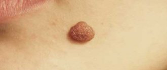
Moles are small, flesh-colored to dark brown spots. In shape, they can be in the form of papules or nodules, consisting of a cluster of pigment cells. Their main medical significance (other than cosmetic) is their resemblance to melanoma. Pigmented lesions are assessed by a set of characteristics (appearance, borders, color, pruritus, bleeding, etc.) that help exclude atypical nevi or melanoma.
Removing a mole is a relatively simple procedure. It is performed for aesthetic purposes or to eliminate pathological changes in the skin that may turn out to be malignant. The only way to know for sure whether a suspicious lesion will require further treatment depends on pathological examination of the tissue. Analysis of moles and other suspicious areas of skin, sending tissue material for histology makes it possible to answer these questions.
What does the histology of a mole show?
This is a more accurate tissue diagnostic method than cytology. It is carried out in a special laboratory, after pre-treatment, staining of a tissue sample, using a powerful microscope. Intradermal nevi, atypical moles, and non-pigmented skin formations can be differentiated by many distinctive features during histology.
Stages of histological examination: timing and reasons for delays
There are several stages of histological examination. I won’t go into detail about their details, I’ll just list them and briefly explain the essence:
1. Biopsy, i.e. removal of a mole. The main thing I want to note here is that there is never “too little” material for research. They often write to me: “The doctor said that the formation was too small, there would be nothing to send for examination, so I did not have a histology.” In my practice, there have been cases when a normal conclusion was received for a mole up to 1 mm in size. Therefore, no matter how huge a “soldering iron” a mole is removed, it is important to send a fragment of the largest possible size for histology. This is certainly worse than examining the entire nevus, but much better than nothing.
2. Fixation of the material. To prevent the mole from decomposing while it is being transported to the laboratory, it is necessary to destroy microorganisms and deactivate the enzymes of the mole cells. This is done using special solutions. Immediately after removal, the mole is placed in a formaldehyde solution. You can replace it with alcohol, but this option is less common
3. Washing the material. To remove traces of the fixing liquid, the material is washed, as a rule, for several hours in running water.
4. Dehydration . Preliminary preparation for paraffin filling.
5. Compaction (filling). The washed and dehydrated mole is filled with liquid paraffin. This is necessary so that the thinnest sections can be made, suitable for examination under a microscope. The result is paraffin blocks like these.
6. Preparation of sections. Using a special device - a microtome - sections are made with a thickness of about 0.05 mm.
7. Coloring the material. This is necessary to distinguish different cells and tissues under a microscope. In an unpainted material, all structures refract light equally and nothing can be seen.
8. Conclusion of sections. This is the name for the arrangement of colored sections between two glasses.
9. The final stage - the pathologist places the glass under the microscope lens and makes his conclusion on the material being studied.
All stages of histological examination take no more than 10 days. The fastest stage, in my opinion, is the examination of the preparations by a pathologist. In some advanced laboratories, due to the high automation of all of the above processes, histological examination is completed within 48 hours.
And sometimes I am surprised by patients who tell me that the histological examination took two months or more. If this happens to you, you should call the laboratory and find out what the difficulties are.
I monitor one mole, remove another: how not to miss dangerous neoplasms on your skin
Where do they come from
On the Internet you can find a version that our moles initially grow... due to tanning. They say that a person is born with clear skin, and over time it is because of the sun that moles appear on it.
“In fact, the formation of moles and their number is determined primarily by heredity,” says dermato-oncologist at the Clinic of Skin Diseases, assistant at the Department of Skin Diseases at Sechenov University, candidate of medical sciences Ekaterina Vertieva.
- That is, those who have a lot of new growths on the skin in their family - mom, dad, grandmother or grandfather - will have more moles.
The second factor is the sun. Under the influence of its rays, the number of moles increases.
At the same time, there are several peaks in a person’s life when most moles appear. Such surges are associated with hormonal changes: at a younger age (3 - 6 years), adolescence and from 25 to 35 years. Women may experience additional peaks during pregnancy.
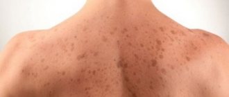
— Is it true that there is a “safe limit” of moles, and if there are more than normal, then a person has an increased risk of skin cancer?
— If there are more than a hundred moles on the skin, then the risk of malignant neoplasms is really higher. But not so much that those with a rich scattering of moles should immediately sound the alarm. The logic is simple: the more tumors there are, in principle, the higher the likelihood that among them there will be nevi with malignant potential (how to protect yourself from danger - see below).
STAY IN TOUCH
What is what
Among the names of growths on the skin you can find the classic, familiar word “mole,” as well as nevus and keratoma. What is the difference?
“ Nevus is the medical name for the same mole,” explains Ekaterina Vertieva. - Essentially, this is a benign skin tumor. A mole consists of pigment cells of melanocytes, which produce the coloring pigment melanin (because of it, our skin darkens when tanning. - Ed.) A mole is a cluster of such melanocyte cells. That is why the appearance of nevi is associated, among other things, with exposure to the sun.
— Keratoma is a benign superficial formation on the skin that can never degenerate into something malignant. Keratomas occur more often in old age. They consist of keratinocyte cells (these are the surface cells of the upper layer of the skin of the epidermis). Externally, a keratome usually looks like a brown formation rising above the surface of the skin. Keratomas often become crusty and flaky. Pieces can fall off, which scares people. In fact, this is a natural desquamation process. There is no need to be scared, the doctor reassures.
On a stem and with hairs - what can you expect from them?
- There are moles that seem to move away from the skin and rise above it - is this dangerous?
- Look: we have superficial layers of skin, the epidermis, and deep layers, the dermis. Common, classic dark moles are superficial, so-called border nevi. They are found in the epidermis. Over time, some superficial nevi absolutely physiologically, that is, safely, naturally grow into the deep layers of the skin, the dermis. And then they begin to rise above the surface of the skin. Nothing wrong with that.
Moreover, over time, by about 50-60 years, dermal nevi completely lose their pigment. They cease to be brown and acquire the color of healthy skin. This is the so-called mole involution. All this is absolutely safe.
- When do hairs grow from a mole? They say this is a good sign, such nevi are definitely safe and will not degenerate into melanoma.
- This is a misconception. Melanoma can easily grow hair. Therefore, if a nevus has suspicious signs (see below), then the hairs are not a reason to calm down and not check the mole.
By the way, doctors categorically do not advise pulling hair out of a nevus; this is unnecessary trauma. You can only cut it carefully.
Meyerson phenomenon
This is the name of the condition when redness appears around the mole, like an inflamed pink corolla, our expert explains. And it reassures: as a rule, it is not dangerous. Most often, the Meyerson phenomenon occurs in patients with a tendency to skin diseases: eczema, atopic dermatitis. If you experience any discomfort or itching, you should consult a doctor. Usually, dermatologists prescribe atopic corticosteroids to relieve such symptoms; with their help, the symptoms are easily removed.
— If you damage a mole and it bleeds, do you need to see a doctor urgently?
— It is necessary, but not necessarily right on the same day or the next day, up to 7 days is quite normal. There is no need to be afraid that after injury, even with bleeding, your mole will definitely degenerate into something bad. Do not believe the stories about a grandmother who picked off a mole, it immediately metastasized into the blood, and death quickly occurred. It is a myth. If a person injures a benign mole, then nothing bad will happen to it. Adverse consequences occur if it was initially melanoma.
— What to do immediately after a mole is damaged, before going to the doctor?
- Treat with a clear antiseptic. Hydrogen peroxide, for example. Just don’t smear it with fucorcin, iodine or brilliant green. Because it will then be difficult for the doctor to examine and assess the condition of your mole due to staining with these substances.
How often to check with a doctor
There are tips on the Internet to show your moles to a doctor every 6 to 12 months.
“If a person has 100 or more nevi and is advised to visit a doctor every six months or a year, then this is still too much,” says Ekaterina Vertieva. - In most cases, moles will not turn into anything bad. But, to be sure of this, it is important to undergo a full examination once by a dermatologist, or even better, an oncodermatologist. Have a specialist carefully check all moles and confirm that none of them are cause for concern. Or he indicated those that could potentially be dangerous, and then told how often they need to be monitored.
If the doctor, after examination, says that all moles are calm, then, as a rule, it is recommended to see the next time in 3 - 4 years. At the same time, no one canceled the self-examination.
THIS WILL BE USEFUL
The golden rule for self-examination
This is the international standard for assessing moles. It can be used as a test at home. The mole is tested according to the ABCDE criteria.
A (asymmetry, asymmetry) - has the mole become asymmetrical?
B (border) - have the borders of the mole become uneven?
C (colour) - has the color of the mole changed: several shades of brown, black, blue, pink have appeared?
D (diameter, diameter) - has the mole grown more than 6 mm in diameter?
E (evolution, evolution) - does the mole change intensively over 4 - 6 months (in shape, color, size)?
Important : if the answers to all questions are “yes,” this is reason to suspect the mole is malignant and immediately consult a doctor. If you have several of the symptoms, then the risk is lower, but you also need to see a doctor.
Delete cannot be saved
“There are three reasons for removing moles,” says Dr. Vertieva. — Cosmetic removal — if a person simply doesn’t like the way a mole looks. We also recommend removing benign moles if they are located in places where they are often injured. For example, for men in the beard area, for women in the bra clasp area. The third option is if we see suspicious symptoms. In these cases, the doctor conducts an examination using a dermatoscope (a device similar to a microscope. - Ed.). If a mole is causing concern, removal is done for medical reasons.
— What are the current methods for removing moles?
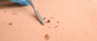
- Laser, radio wave method, electrocoagulation (cauterization of the nevus with high-frequency current) are used - these methods destroy the mole. As well as surgical excision.
If the patient has a suspicious nevus, then surgical removal is necessary. Only in this case, the tumor tissue is fully preserved, and histologists will be able to give a reliable conclusion as to whether there are malignant cells in the mole.
In other cases, when the mole is definitely calm, the least traumatic method is the radio wave method or laser (coagulation is a rougher method). After removing the nevus, a neat wound remains; it must be treated with a solution of potassium permanganate or fucorcin. A crust forms, which falls off after an average of two weeks. You can wet the wound site with water from 3 to 4 days.
I strongly do not recommend self-medicating at home and using celandine to remove tumors. At best, this technique leads to a chemical burn, and sometimes to more serious consequences in the case of malignant potential of the formation.
QUESTION ON THE TOPIC
Is it possible to sunbathe if you have a lot of moles?
“Yes, this is not a contraindication for tanning,” our expert rejoices. — The main thing is to follow safety rules: stay in the sun before 11 a.m. and after 4 p.m. And use sunscreen.
BREAKTHROUGH AND PROSPECTS
“Now there is a real revolution in the treatment of melanoma, the most aggressive skin cancer,” says Ekaterina Vertieva. — Previously, chemotherapy was used, which was very toxic and at the same time extended the life of patients by only a couple of months. Since the beginning of the 2010s, targeted drugs have been used. They do not hit all cells of the body, like chemotherapy, but selectively hit specific targets. These drugs are available in Russia under compulsory medical insurance and prolong the life of our patients very significantly. The main trend now is the study of new targets for the fight against melanoma. And also immunotherapy (the method for which the Nobel Prize in Medicine was awarded this year. - Ed.).
CONGRATULATIONS!
The oldest department of Sechenov University is 150 years old
On May 27, the Department of Skin and Venereal Diseases named after. V.A. Rakhmanov Sechenov University - 150 years. Already in the first years of its work, the department was recognized as the best in Europe. Over a century and a half history, vast experience in teaching the science of dermatovenereology has been accumulated here. Today, about 2,500 skin and venereal diseases are known, say department employees. The most common are psoriasis, atopic dermatitis, eczema and fungal infections; In recent decades, there has been a steady increase in the incidence of allergic dermatoses.
The head of the department, Professor Olga Olisova, lists modern promising scientific developments of the department's employees: improving the early diagnosis of lymphoproliferative skin diseases (T-cell lymphomas) using genetic markers; development of hardware methods for diagnosing melanocytic skin neoplasia; studying the pathogenetic connections of HPV with the development of skin tumors using molecular genetic detection methods; development of diagnostic markers for severe bullous dermatoses and orphan skin diseases (using the example of mastocytosis).
BY THE WAY
At the Department of Skin and Venereal Diseases of Sechenovka, a unique plaster museum has been created, containing more than 3,000 wax plaster casts with images of various skin and venereal diseases. In terms of the quality and quantity of exhibits, the dummy museum is a national treasure that has no equal not only in Russia, but throughout the world.
One mole – one container with full name!
If histological examination of the mole reveals melanoma, the removal site should be re-excised more widely. The capture of healthy tissue is determined depending on the Breslow thickness.
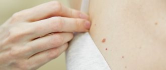
I would like to draw your attention to the fact that if all removed moles are placed in one container, they will be able to undergo histological examination. But in this situation it will no longer be possible to establish where exactly the melanoma was and it will be necessary to do a wide excision at the sites of all removed moles. To have a correct idea of the volume of tissue removed, you can look at the photo and see what an average scar looks like.
Important! If you have several moles removed, when sending them for histology, the location of each nevus must be clearly indicated.
But it’s not worth removing so many moles at once, as in the photo. Why? I'll tell you in the following publications.
“Can I not take away the answer? Will they call me if something is wrong?”
Of course, you don’t have to take the answer, and then write messages like this on the forums:
Please don't think I'm laughing at tragedy. This is just a way to get your attention.
As far as I know, there is no law according to which the morphology laboratory must notify the patient that cancer has been detected. At the end of the conclusion after the examination, moles may be written: “Re-examination with the results of histological examination.” But no one reads these conclusions.
And then everything happens according to the classic scenario, even if in an institution calls of this kind are in the order of things, the human factor comes into play. For example, the person responsible for this quit, or someone replaced him, or he got sick, didn’t make an appointment, it was a birthday, etc. - in the end, no one called, and the patient didn’t even know that they could have done this. Remember that your health is yours and no one owes you anything.
Please always take the result of the histological examination and always show it to the oncologist who performed the removal.
“I took the histological answer, what should I do with it? Will I figure it out myself?”
No. If you received the result of the study in paper or electronic form, you must show it to the oncologist who performed the removal. Let me give you an example.
Here are two diagnoses:
- Dysplastic nevus with severe dysplasia. There are tumor cells at the resection margin.
- Squamous cell papilloma with inflammatory component. Removed within unaffected tissues.
In the first case, repeated excision of the mole removal site is required, but in the second - not. Therefore, do not be lazy, show the conclusion to the doctor.
Signs of malignancy
The clinical signs of a malignant mole are very diverse. This is manifested in size, shape, outline, surface character, consistency, color, and dynamics of change.
Common to all forms is a set of characteristics, which is expressed by the initial letters by the abbreviation “AKORD”, developed by dermatologists and cosmetologists:
- Asymmetry (A) - lack of symmetry in the shape and contours of a spot, with the exception of birthmarks present on the child’s body at birth.
- Edges (K) are most often uneven and indistinct (blurry).
- Coloring (O) - uneven; The presence of dots and stripes of various tones of dark brown and black is noted.
- Size (P) - in diameter from 7 mm or more.
- Dynamics (D) of development - an increase in the previous birthmark or a rapid increase in the size of a new pigmented formation.
Are there errors in the histological examination of nevi?
Yes, unfortunately, they are possible. I wrote about this. Histological examination is carried out by people, and they can make mistakes. If you have the slightest doubt about the results of the conclusion, take your histological preparations and send them for review to another laboratory. The preparations include both paraffin blocks with mole tissue and glasses with sections.
You ask: “How can I go and pick something up? They’ll send me…” If they don’t give you histological preparations, then you need to go to the head doctor and demand it. In my experience, after that everyone gives away.
Immunohistochemistry of melanoma
An effective diagnostic method that allows one to determine the properties of a tumor, as well as its sensitivity to certain types of antitumor drugs, is immunohistochemistry. More than half of cutaneous melanomas have mutations in the BRAF and NRAS genes. Thanks to immunohistochemical analysis, it is possible to identify specific mutations in the nucleus of tumor cells, which makes it possible to assess the degree of effectiveness of certain target drugs and prescribe the most accurate therapy.
The method is also effective for the morphological diagnosis of anonymous metastatic neoplasms.
Specialists at the Israeli Oncology Center have extensive experience in histological diagnosis. The diagnosis of melanoma in Russia and the CIS countries differs from the diagnostic protocols used in Israel. The lack of clinical experience and the lack of high-tech equipment in most medical institutions in the CIS leads to cases of discrepancies in diagnoses and stages in the expert opinions of specialists.
In Israel, if doubts arise about the morphology and stage of the tumor, other specialists are brought in for examination (second opinion). This approach allows us to reduce the likelihood of subjective error in diagnosis to zero.
Is a histological examination of a wart necessary?
To be honest, I’m already tired of answering this question. I will simply give 2 histological conclusions, which are united by a clinical (on examination) diagnosis - a wart.
First case:
I was unable to trace the fate of the patient and the result of the immunohistochemical study. However, in a conversation with a morphologist, I realized that the outcome here can be very different and not necessarily successful.
And the second case:
Intraepithelial carcinoma is not the worst thing that can happen. Bowen's disease (and that's exactly what it is) is just stage zero squamous cell carcinoma, which can be successfully cured by removal within healthy tissue. However, another year or two, and the consequences for the man could be dire.
Conclusion: warts should be sent for histology. Always. No matter how close they may seem to you.
How long does it take to perform histology?
The duration of processing of tissue samples should be checked with your doctor. There are 2 types of analysis:
- Emergency - the cut is examined during the operation. This allows, if necessary, to expand the scope of surgical measures and remove more tissue. And immediately after removal of the tumor, begin drug treatment. Used in controversial cases, when new circumstances are identified during surgery.
- Planned - used when the diagnosis is beyond doubt, and an emergency study confirmed the doctor’s assumption. The processing time for results is up to 7 days.
The timing of the study is influenced by the following factors:
- legal status of the laboratory or clinic - private or public;
- analysis methods and equipment used in a particular medical center;
- laboratory workload;
- force majeure circumstances.
A long sample processing time is not synonymous with bad news!