Fungal infections (mycoses) are diseases that are caused by different types of microscopic fungi.
Mycoses of the skin, mucous membranes, and nails are the most common among all human fungal diseases. Systemic mycoses (affecting several body systems) are predominantly found in people with immunodeficiency. Most often these are HIV-infected patients, patients after organ transplantation, cancer patients, etc.
The main causative agents of skin mycoses: trichophyton fungi Trichophyton rubrum, Trichophyton mentagrophytes, var. interdigitale, Epidermophyton floccosum and candida Candida.
Infection with fungi occurs through direct contact with a patient, through shoes, clothing, when using general hygiene products (washcloths, manicure tools), when visiting gyms, baths, saunas, and swimming pools. The main factor of infection is the presence of abrasions and cracks in the skin, which occur with increased sweating, dryness, and poor circulation in the extremities. People with diabetes, immune disorders, blood diseases; Those taking antibiotics, glucocorticosteroids, and cytostatics for a long time are especially susceptible to the development of mycoses.
Most often, microscopic fungi cause damage to the skin of the feet and large skin folds (axillary, groin and others).
Main symptoms and complaints
When infected with fungi, ring-shaped inflammatory foci appear on the skin and in the folds, with clear boundaries and a raised ridge along the edges. There may be small scales and bubbles on the surface. If large folds are affected, the surface may become weeping and macerated. Often observed in the groin areas, scrotum, armpits, and under the breasts.
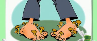
Fungal disease of the feet is manifested by peeling of the skin in the spaces between the toes, soles, thickening of the stratum corneum of the soles and lateral surfaces of the feet, cracks.
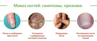
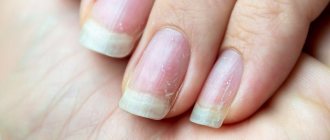
With onychomycosis (“nail fungus”), yellowish and whitish stripes form in the thickness of the nail, the nail plate thickens, crumbles, and the edges of the nail become jagged. Sometimes thinning of the nails is observed, and detachment of the nail from the bed may occur.
Itching is the main complaint with fungal skin diseases. When the surface macerates (especially in folds), a burning sensation occurs. Fungal infections develop slowly and have a long course. Exacerbation usually occurs in the summer. It is important to remember: the use of ointments and creams containing hormones causes worsening of the condition and is strictly contraindicated for mycoses.
Among fungal diseases of the scalp, the most common is microsporia , which is caused by fungi of the genus Microsporum. Microsporia is a highly contagious disease. In Russia, Microsporum canis fungi are widespread and often infect animals (cats, dogs, rabbits, hamsters, guinea pigs). Human infection occurs through contact with a sick animal. Peaks of incidence are May-June and September-November. Children most often get sick, not only because of their ardent love for animals, but also because of the structural features of their skin - more delicate than that of adults. A symptom of microsporia is the appearance of round or oval lesions, with raised clear boundaries, covered with scales or crusts. When the scalp is affected, the hair in the lesion is broken off.
Yeast-like fungi are the cause of candidiasis. Urogenital candidiasis is an inflammatory disease of the genitourinary tract caused by fungi of the genus Candida. It is believed that 70-75% of women develop vulvovaginal candidiasis (“thrush”) at least once during their lives. The main symptoms in women are profuse white, cheesy or creamy discharge from the genital tract, itching and burning in the external genital area. Relapses of the disease often occur due to a decrease in immune defense, the use of antibiotics, or a change of sexual partner. In men, manifestations of urogenital candidiasis may include redness and swelling of the genitals, with a white coating on the surface of the rash.
Candida can affect the mucous membranes of the oral cavity, most often in young children. Erosed elements with a white coating appear on the mucous membrane, accompanied by a burning sensation and discomfort when swallowing.
Dermatomycosis
Mycoses of the scalp
They appear as bright red infiltrated plaques, which are covered with gray scales on top. The elements are mainly formed around the hair in the form of a muff. Occasionally, deep inflammatory foci are possible, which are large in size and covered with massive grayish-yellow crusts. At the site of a fungal infection, hair breakage is observed at a height of 5-8 mm or at the very root. A person complains of severe itching and rapid contamination of hair after washing.
Mycoses of skin folds
The main representative of this group is dermatophytosis inguinalis. The disease affects the inguinal folds and adjacent areas of the skin: the inner surface of the thighs, the perianal area, and the perineum. Due to self-infection, the process may spread to the armpits, elbows, popliteal fossae, and in severe cases to any area of smooth skin. The frequency of inguinal dermatophytosis in the structure of all dermatomycosis is up to 10%.
At the initial stage, the pathology is represented by pink swollen spots that have a round shape, clear contours and a smooth surface. If treatment is not carried out, the lesions merge to form large polygonal spots, the marginal zone of which is covered with polymorphic elements: bubbles, erosions, crusts. Most of all, patients are worried about the painful itching, which interferes with sleep and daily activities, forcing them to scratch the affected area until it bleeds.
Mycoses of the feet
The clinical picture of epidermophytosis of the distal extremities depends on the form of the lesion. As a rule, the pathology begins with an erased form, in which there is slight peeling in the interdigital spaces, which does not cause any concern to the patient. Sometimes, against the background of peeling, superficial skin cracks form, which do not become inflamed or bleed.
In the squamous-hyperkeratotic form, abundant peeling is accompanied by reddish-bluish plaques and yellowish-gray calluses, which arise due to excessive keratinization of the skin. With the dyshidrotic form, multiple blisters with a thick tire appear, when opened, bright pink weeping erosions are formed. For mycoses of the feet, secondary allergic rashes, called dermatophytids, are typical.
Onychomycosis
Fungal infection of nails has different symptoms, which depend on the shape and depth of the lesion. The pathology is mainly manifested by grayish-yellow stripes on the nail plate, its increased fragility, tendency to deformation, transverse striations and cracks. At an advanced stage of the disease without treatment, significant destruction of the nail is observed, in addition to a cosmetic defect, itching and painful sensations occur.
The normotrophic form is characterized by decreased transparency of the nail and thickening of its edges due to subungual hyperkeratosis. In the hypertrophic variant, the thickening of the nail plate is more pronounced; in severe cases, the nail acquires a curved beak-like shape (onychogryphosis) and a dirty gray tint. For atrophic onychomycosis, total destruction and detachment of the nail plates is typical, and onycholysis is considered the extreme degree of this process.
Mycoses of smooth skin
Classic dermatomycosis is characterized by the appearance of flat, scaly spots of pink or red color with a raised border. There may be inflammatory papules or vesicles along the edges of the lesions. Over time, the central part of the elements becomes brown due to hyperpigmentation, and the edges continue to grow, so that the lesions merge into large polygonal spots. Subjectively, patients experience itching, burning, and pain in the affected area.
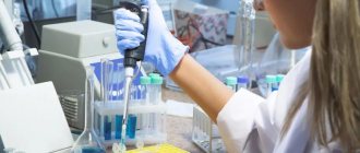
Laboratory research
- Nail plate screening is a study during which the content of micro- and macroelements is determined. The goal is to see the level of vital substances: iron, potassium, calcium, magnesium, zinc, copper, sodium, selenium and others. And also determine the presence of toxic elements: barium, beryllium and aluminum, cadmium, arsenic, mercury, lead.
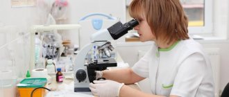
- Test for the presence of fungus. A piece of the plate itself and the subungual contents are examined under a microscope.
- Sowing. The material affected by the fungus is placed in a nutrient medium to grow colonies of fungi. In this way, the type of fungi and their resistance to drugs are determined. The process takes three weeks to a month.
Development of the disease
Nail mycoses, or onychomycosis, affect every tenth representative of the globe. The main pathogens are dermatophytes, such as T. rubrum. But there are forms caused by mold or yeast-like fungi. Factors that provoke onychomycosis: wearing uncomfortable, tight shoes and neglecting hygienic care of hands and feet.
Onychomycosis occurs in several stages:
- Spots of a yellowish or gray-white hue appear on the platinum.
- The plate becomes brittle, flakes, crumbles, and some areas thicken.
- The bed takes on a brownish tint, and the nail plate comes off.
Pathological changes can be observed both in a local area and on the entire surface. The fungus gradually spreads to neighboring fingers.
Diagnosis and choice of doctor
Nails are a derivative of the skin, so the examination should begin with a visit to a dermatologist. At the first stage, the doctor will conduct a visual examination. Next, you will need to undergo general tests and conduct laboratory tests directly on the nails.
At the medical center, they will do a microscopic examination of the plates for the presence of pathogenic fungi, take a scraping and conduct a screening. All types of diagnostics are carried out in our own laboratory in the shortest possible time - from two working days. The clinic is attended by an experienced dermatologist who will find the cause of the problem. If necessary, refer you for consultation to other specialists.
Tests for skin fungus
Fungal skin diseases (dermatomycosis) are the most common among skin diseases. Fungi have been known since ancient times; they are a special type of microorganisms that combine features of both plants and animals, and fungi feed on organic substances. They are tenacious, capable of forming spores and persisting in the environment for a long time, and grow very slowly. Therefore, fungal diseases can occur without manifestations for a long time, and their detection is not an easy task for doctors.
Mostly dermatophyte fungi are causative agents of superficial fungal diseases, in particular such as microsporia, trichophytosis (ringworm), epidermophytosis. They constantly live on the skin and hair of humans and animals, some in the soil. Human contact with fungi occurs. However, the skin is normally a powerful protective barrier and does not allow fungi to exhibit pathogenic properties.
The following factors contribute to the development of fungal skin diseases:
- mechanical damage to the protective barrier of the skin: cracks, microtraumas, including those resulting from cosmetic procedures.
- failure to comply with the rules of personal hygiene at home and when visiting public saunas and baths, fitness rooms, if individual towels, slippers, and hats are not used.
These factors are not leading; the skin will not allow the enemy to penetrate if the immune system works well. A decrease in local and general immunity of the body is a decisive trigger. This is facilitated by:
- Genetic causes (congenital immunodeficiencies)
- Lack of vitamins and microelements
- Chronic skin diseases
- Endocrine diseases: diabetes mellitus primarily, thyroid dysfunction, obesity, other hormonal problems
- Long-standing chronic diseases (gastrointestinal tract, lungs, heart; tuberculosis, AIDS)
- Long-term use of antibiotics, glucocorticoids, antitumor drugs.
If the fungus has overcome all the obstacles in its path, a disease develops. The peculiarity of skin fungi is damage to the stratum corneum (surface) layer. Fungi feed on the keratin that is part of it. Skin, nails and hair are affected.
How do fungal skin diseases manifest?
Since only the superficial layers of the skin are involved, manifestations may include:
- Itching and peeling skin without spots
- The formation of red spots with clear edges in the form of one or multiple foci, usually accompanied by peeling and itching
- Nails become thicker, deformed, and split
- Hair loss due to trichophytosis
- Inflammation of hair follicles with crusting and flaking
Manifestations are typical for many skin diseases. The examination data does not allow us to determine whether there is a fungal infection of the skin or not.
To establish a diagnosis, the following laboratory methods are used:
Microscopic examination of skin, nail plates and hair for the presence of pathogenic fungi (performing time 2 days)
An important point for the correctness of the study is to take material at the border of healthy and affected tissues, where there is a higher probability of detecting active growth of fungi. The microscopic method allows you to see a pathological fungus under a microscope or exclude its presence, but does not allow you to determine the type of pathogen. Identification of fungal mycelium is a criterion for confirming active mycosis. The presence of only spores requires detailed study and confirmation by other methods.
Culture for fungi (causative agents of mycoses) (up to 30 days) is a more accurate study. Skin scrapings, nails and hair are placed in special media enriched with nutrients specifically for skin fungi. The doctor observes the growth of fungi for up to a month, since they grow very slowly and it is this period that allows an adequate examination and not to miss the disease. Thanks to culture, the specific type of dermatophyte fungus is determined, which makes it possible to prescribe the correct antifungal drug.
Please note that the malasezia furfur fungus, the causative agent of dandruff in animals and humans, does not grow in crops. This is his peculiarity!
In case of fungal diseases, determination of sensitivity to antifungal drugs is carried out only for scientific purposes in order to introduce a new medicine, since this is a long, labor-intensive process and it is impractical in medical practice. The fact is that known antifungal drugs act on certain types of fungi, and when a fungal disease is confirmed, the doctor will not wait for further progression of the process for another month, but will prescribe a drug depending on the type of fungus determined as a result of the analysis.
How to properly prepare for research?
Skin scrapings are performed only by a doctor. Hair and nails can be collected independently and placed in a special container, if possible at the border of healthy and affected tissue. The study is carried out before starting to take antifungal drugs or 2 weeks after the end.
Get examined competently and in a timely manner with the KDL laboratory!
Fungal skin infections
Fungal infections of the skin and its appendages have been known since ancient times. Doctors gave skin diseases with different symptoms from others separate designations (for example, favus), not yet knowing that they were caused by fungi. The history of the science of dermatophytes began with the discovery of the favus pathogen Achorion schoenleinii in 1839 by Schonlein JL. Schonlein found fungal mycelium in the skin rashes of a patient whom he had mistakenly diagnosed as having impetigo. In 1841, Grubi D. established the relationship between skin diseases and fungi. He described the clinical picture of microsporia and the morphological features of the fungus that causes this disease. As a result of the use of the special nutrient medium proposed by Sabouraud R. for the cultivation of mushrooms, favorable conditions were created for mycological researchers. Relevant clinical and laboratory studies began to be carried out, and the era of discovery of new types of pathogens began. In the field of medical mycology, significant changes have occurred in various directions: dermatophytes, molds, yeast-like fungi, the pathogenesis of mycotic infections were studied and described in detail, the nature of actinomycosis was determined, the treatment of fungal diseases was improved, etc. Russian scientists also made a significant contribution to the development of medical mycology , among whom the most famous are P. N. Kashkin, A. M. Arievich, N. D. Sheklakov, O. K. Khmelnitsky, A. N. Arabian, Z. G. Stepanishcheva, N. A. Krasilnikov, G. O Suteev, V. M. Leshchenko and many others.
Fungal diseases have long gone beyond the specialty of a dermatologist, but dermatologists-mycologists are still doing a lot of work to combat dermatomycosis, which ranks first in prevalence in all countries.
Superficial mycoses (dermatomycosis) include infections that affect the skin, nails and hair. The main pathogens are dermatophytes, fungi that can absorb keratin. These include fungi of the genera Microsporum, Trichophyton and Epidermophyton, which, depending on the source of infection, are divided into anthropophilic, zoophilic and geophilic. The habitat of zoophilic dermatophytes (Trichophyton mentagrophytes v. gypseum, T. verrucosum, Microsporum canis, etc.) is animals, anthropophilic (Trichophyton rubrum, T. mentagrophytes v. interdigitale, Microsporum ferrugineum, Epidermophyton floccosum, etc.) is human, geophilic species ( Microsporum gypseum) live in the soil. Dermatophytes are highly contagious and can be transmitted to humans from humans, animals or soil, causing disease. The main “spreaders” of infection are anthropophilic species.
Superficial mycoses also include keratomycosis: pityriasis versicolor and piedra, which affect the most superficial areas of the stratum corneum and hair cuticle. The causative agent of pityriasis versicolor and seborrheic dermatitis, the yeast-like fungus Malassezia furfur, lives on the skin of humans and animals and, under favorable conditions, can affect the stratum corneum of the epidermis and the mouth of the follicles. The disease in most cases is not contagious. White and black piedra are usually found in countries with hot and humid climates. The diseases are less contagious. The causative agent of black piedra, Piedraia hortae, is found only on hair. Trichosporon beigelii is widespread in the environment and, in addition to white piedra, can also cause skin and nail lesions.
In addition to true dermatophytes, which infect only the skin and its appendages, superficial mycoses can also be caused by other fungi, isolated from a variety of localizations during systemic mycoses. Candida spp. - the second most common discharge in dermatomycosis and onychomycosis after dermatophytes. Up to 40% of cases of onychomycosis of the hands are caused by Candida. Lesions of yeast-like fungi on the scalp with a clinical picture of seborrheic eczema were noted. Superficial forms of candidiasis also include lesions of the mucous membranes of the oral cavity and genital organs.
In cases of onychomycosis caused by molds, there is still doubt about the ability of these opportunistic pathogens to independently infect nails due to their weak proteinase and keratinase activity. It is known that mold fungi can manifest themselves as a secondary infection, penetrating into tissues already affected by dermatophytes. However, as a result of many years of research, it has been proven that some molds can penetrate into the intercellular nail space using perforating organs. The most common causative agents of mold lesions of the skin and nails are Scopulariopsis brevicaulis, Pyrenochaeta unguis-hominis, Aspergillus spp., Fusarium spp., Alternaria spp., Cladosporium spp. and etc.
There are numerous options for the classification of fungal infections, which to a greater or lesser extent take into account the etiology, pathogenesis, clinical picture and features of the epidemiology of diseases. In domestic dermatology, the classification of N. D. Sheklakov is most often used:
- Keratomycosis (lichen versicolor, piedra, imbricated mycosis).
- Dermatophytosis (athlete's foot, trichophytosis, microsporia, rubromycosis, favus, etc.).
- Candidiasis (superficial candidiasis of the skin and mucous membranes, visceral, etc.).
- Deep mycoses (chromomycosis, sporotrichosis, etc.).
- Pseudomycosis (erythrasma, actinomycosis, nocardiosis, etc.).
However, many countries around the world have adopted a classification of fungal diseases according to the localization of the pathological process:
- Tinea pedis - mycosis of the feet.
- Tinea corporis is a mycosis of the smooth skin of the trunk.
- Tinea cruris - inguinal mycosis.
- Tinea capitis is mycosis of the scalp.
- Tinea unguim - onychomycosis.
- Tinea manum - mycosis of the hands.
- Tinea barbae is mycosis of the face.
This classification is convenient from a practical point of view, but does not take into account the etiological features of dermatophytosis, which can determine the nature of epidemiological measures and treatment features.
Main clinical features of fungal infections
Mycoses of the feet (Fig. 1). The interdigital folds and soles are mainly affected. In the interdigital folds, slight peeling with minor inflammatory phenomena, moderate maceration, cracks, and blisters are observed. On the sole there is thickening of the stratum corneum, flour-like peeling in the skin grooves, small cracks on a slightly hyperemic background. In the dyshidrotic form, numerous blisters form on the skin of the arch and inferolateral surface of the foot, which then merge to form large blisters. In place of the opened blisters, erosions with an uneven edge remain.
Mycosis of smooth skin of the body (Fig. 2). With tinea versicolor, brownish and white spots are usually localized on the skin of the chest, back, neck and shoulders. Scaly lesions have clear boundaries and are not accompanied by inflammatory phenomena.
When the skin is damaged by other pathogenic fungi, clearly demarcated, round, swollen lesions with a raised ridge are formed. The center of the lesion is flattened, with slight peeling. The lesions increase due to peripheral growth.
Mycosis inguinal. Typical localization is the inner thigh, lower abdomen, buttocks. The lesions are clearly defined, scaly, erythematous, with an inflammatory ridge. Over time, the general moderately erythematous background gives way to a brownish one.
Mycosis of the scalp. Most often observed in children. The disease manifests itself as large, round, clearly demarcated, scaly patches of baldness. Inflammatory phenomena are mild. Discolored hair within the lesion is broken off a few millimeters above the skin level (with microsporia) or, breaking off at the skin level, leaves a stump in the form of a black dot (with trichophytosis). Zoophilic pathogens can cause the development of an infiltrative-suppurative form of dermatophytosis: the lesion protrudes above the surrounding skin, is covered with purulent-bloody crusts, and hair falls out.
Onychomycosis. Various types of fungal infection of the nail plates are characterized by loss of transparency, discoloration (whitish, yellowish), thickening, subungual hyperkeratosis, crumbling or destruction down to the nail fold.
Mycosis of the hands. In the squamous form of palm lesions, the disease manifests itself in fine-lamellar mealy peeling in the skin grooves. Cracks may form, accompanied by pain and itching. In the dyshidrotic form, vesicles form, which often cluster and can coalesce to form blisters.
Mycosis of the face. Most often, lesions are localized in the neck, chin and lower lip. The infiltrative-suppurative form of the lesion manifests itself in the formation of large bluish-red nodes with a bumpy surface. Numerous pustules merge to form abscesses. Changed dull hair in the lesion is removed easily and without pain. The superficial version resembles mycosis of smooth skin.
Laboratory diagnostics
Microscopy of clinical material is a quick and simple method for preliminary diagnosis of the disease. In cases where there is no growth of the pathogen in culture, a positive result of direct microscopy can be an undoubted confirmation of a mycotic infection. In scrapings from skin and nails, dermatophytes are usually represented by thin, 2–4 µm in diameter, straight and rarely branched mycelium. Often in the preparation one can find atypical forms of dermatophyte mycelium - chains of round arthrospores (Fig. 3).
M. furfur, when microscopy of skin scales, is revealed in the form of round cells with a diameter of 3–8 μm, collected in clusters, and short curved mycelial hyphae of a characteristic “banana-shaped” shape. In most cases, the diagnosis of lichen versicolor can be established precisely by microscopy of the native specimen, since culture of the material usually does not give results.
Scopulariopsis brevicaulis in nail scales is represented by characteristic cup-shaped spores with a rough shell.
Fungi of the genus Candida form typical budding yeast cells and pseudomycelia (branching chains of long cells).
Identification of other pathogens in native preparations is difficult.
When hair is damaged by dermatophytes, several tissue forms of the fungus are observed: 1) endoectothrix - spores with a diameter of 2–3 microns are “mosaic” located inside and mostly outside the hair, forming a muff on the root (“Adamson’s sheath”). Inside the hair you can also see mycelium, which, with light pressure on the preparation, comes out of the hair in the form of a fringe - “Adamson's brush”. The causative agent is Microsporum spp.; 2) endothrix - spores with a diameter of 4–6 microns are located inside the hair in longitudinal chains, completely filling it. The causative agent is Trichophyton spp. The final identification of fungi is carried out by cultural studies.
For cultural studies of pathological material, Sabouraud's medium with chloramphenicol and gentamicin is used, as well as for the selective isolation of dermatophytes - with cycloheximide (actidione), to inhibit the growth of fast-growing saprophytic fungi.
Dermatophyte genera are distinguished by the presence and morphology of multicellular macroconidia and unicellular microconidia.
Characteristics of the most important pathogenic fungi
Epidermophyton floccosum. Anthropophil. Affects the skin of the groin folds and legs. Colonies grow slowly, grayish-brown, lemon-olive, later white, folded-lumpy in the center. The surface of the colony is leathery or velvety-mealy. By microscopy: in mature cultures there are chains of intercalary chlamydospores. Macroconidia are 4–5-celled, club-shaped, smooth, with rounded ends. Arranged in bunches of 3–5 pieces. Microconidia are absent.
Microsporum canis (Fig. 4). Zooanthropophil. The most common pathogen of microsporia in Russia. Colonies are fast-growing, flat, radiant-hairy. The mycelium is grayish-white, against the background of a brownish-red or orange reverse side, the general hue of the colony is salmon. Under microscopy: it forms a characteristic bamboo-like mycelium, there are combs, short spirals, intercalary chlamydospores. Macroconidia are fusiform, pointed, spinous, multi-chambered (4–12 cells) with a distinct double-circuited shell. Microconidia are pear-shaped and occur inconsistently.
M. gypseum. Geophilic pathogen. It affects the skin and hair primarily of people who cultivate the soil. The colonies are fast-growing, flat, mealy (powdery), later with a small velvety elevation in the center. The color is yellowish-pink. The reverse side is yellow. Under microscopy: numerous macroconidia (4–6 cells) spindle-shaped, wide, blunt-ended, smooth. Microconidia, if present, are numerous and pear-shaped or oval.
Trichophyton rubrum (Fig. 5). Anthropophil. The most common causative agent of dermatomycosis. Affects toenails, hands and skin anywhere on the body.
The colonies are velvety, white, sometimes waxy at the beginning of growth; later they can acquire a pinkish or purple-red color. The reverse side is yellow, red or cherry red. Under microscopy: abundant microconidia of elongated, drop-shaped or pear-shaped are located on the sides of the mycelium. Macroconidia are 5–6-celled, blunt-pointed.
T. mentagrophytes var. interdigitale. Anthropophil. Affects the nail plates and skin of the feet.
Colonies are fast-growing, velvety, white, sometimes pinkish. With age, the colonies of different strains become fluffy or densely powdery. The reverse side is colorless or brownish. Under microscopy: microconidia are round, located on the sides of the mycelium, singly and in clusters. Cigar-shaped, 3–5-celled macroconidia with a rounded end are rare. In mature cultures there are many whorls and spirals, nodular organs and intercalary chlamydospores are formed.
T. mentagrophytes var. gypseum. Zooanthropophil. Affects skin and hair. In terms of frequency of isolation, it ranks second after T. rubrum.
Colonies are fast-growing, flat, granular-powdery, white, cream, yellowish. The reverse side is brownish-red. Under microscopy: microconidia are abundant, round, located on the sides of the mycelium singly and in the form of clusters. Macroconidia are cigar-shaped, 3–8 cells with rounded ends.
Treatment
Therapy for various mycotic lesions of the skin and nails is carried out with antifungal drugs, which can be systemic or applied externally. In practice, both monotherapy and various combinations of antifungal drugs are used. In most cases, treatment should be comprehensive using external antifungal agents, systemic drugs, and symptomatic treatment. Methods and means of topical therapy are mandatory components of the treatment of various mycotic skin lesions. If the fungal process is in the initial stage and there are minor skin lesions, then it can be cured by prescribing only external antifungal drugs. With a widespread or deep mycotic process, as well as in patients with damage to hair and nails, treatment may be difficult.
Antifungal drugs or antimycotics are specific agents used to treat fungal infections of the skin, nails, hair, etc. They may have fungicidal and fungistatic properties. The fungicidal effect of the antifungal agent leads to the death of fungal cells, while the fungistatic effect stops the formation of new fungal cells. Antimycotics are conventionally divided into 5 groups: polyene antibiotics, azole compounds, allylamine drugs, morpholine derivatives and medications without a clear relationship to any specific group.
When prescribing topical therapy, it is important to take into account the nature of the specific effect of the antifungal agent. It is known that griseofulvin preparations are active only against dermatophytes. External products containing polyene antibiotics - against yeast and mold fungi, preparations of selenium, zinc, benzyl benzoate - fungi of the genus Malassezia and the causative agent of erythrasma Corynebacterium minnutissium. Modern antifungal agents of the azole, allylamine, morpholine series, thiocarbamycins and pyridine compounds have a significantly wider spectrum of antifungal activity, as well as anti-inflammatory and antipruritic properties and are well tolerated.
Onychomycosis is the most treatment-resistant disease. The basis for success is an individually selected treatment regimen. When treating, it is necessary to take into account the patient’s age, concomitant diseases, the number of affected nails, and the degree of involvement of the nail plates in the pathological process.
Currently, mycologists have a large arsenal of agents with a wide spectrum of fungicidal action, accumulation in therapeutic concentrations in the nail plate and nail bed. The greatest preference is given to drugs that meet the requirements for therapeutic effectiveness, as well as aesthetics and ease of use.
Treatment of onychomycosis is divided into local, systemic, and combined.
Local treatment allows you to create high concentrations of the drug on the surface of the nail plate. However, the active ingredients of the antimycotic do not always penetrate into the nail bed, where the most viable fungi are located, in effective concentrations.
Local antimycotics: varnishes - Loceryl, Batrafen; creams - Lamisil, Nizoral, Mikospor, Mifungar, Travogen, Ecozax, Exoderil, Pimafucin, sprays - Daktarin, Lamisil, etc. It is necessary to take into account that they are not intended specifically for the treatment of onychomycosis, but they can be used in the treatment of fungal infections of the skin of the feet, interdigital gaps, often combined with onychomycosis.
To treat nails, local antiseptics are also used - alcohol solutions of iodine and dyes.
Multicomponent preparations contain an antimycotic or antiseptic in combination with an anti-inflammatory agent. In the treatment of skin infections accompanying onychomycosis, the following are used: Triderm, Travocort, Mikozolon, Pimafucort, Lorinden S, etc.
Azole, allylamine, morpholine compounds, as well as mixed group drugs are active against a large number of pathogens. Considering that quite often mycoses of the feet are caused by mixed fungal flora, it is preferable to prescribe these drugs, which are broad-spectrum antimycotics. Most of them damage the cytoplasmic membranes of fungal cell walls, suppressing the synthesis of their main components, in particular ergosterol.
Currently, the mycologist has highly effective systemic antimycotics: itraconazole (Sporonox, Orungal), fluconazole (Diflucan, Forkan), terbinafine (Lamisil), the antifungal effect of which is reflected in the table.
One of the important principles of topical therapy for mycotic skin lesions is the alternation of external antifungal drugs, which avoids resistance of dermatomycetes to them.
For squamous-keratotic forms of skin lesions, keratolytic agents are used as part of collodion peels or ointments: Arabian, Arievich, Andriasyan ointment or 5-10% Salicylic ointment. For candidal lesions, use Nystatin, Levorin, Amphotericin ointments, Pimafucin 1-2 times a day for 10-15 days. As a result of treatment, foci of candidal intertrigo, interdigital candidal erosions, and paronychia are resolved.
For acute skin mycoses with a pronounced inflammatory component, treatment begins with the elimination of swelling, hyperemia, exudation, eczematization, and allergic rashes. Lotions and wet-drying dressings with disinfectant and astringent compositions are prescribed: tannin, ethacridine, boric acid, etc. Then 2–5% boron-naphthalan paste, 5% ASD paste, as well as combinations of antifungal and corticosteroid agents in creams are used: Mycozolon, Travocort, Triderm. At the same time, acute inflammatory phenomena are quickly eliminated, which makes it possible to subsequently switch to treatment with fungicidal agents. It is recommended to use external agents with corticosteroids for 7–8 days to avoid activation of the mycotic process.
After acute inflammatory phenomena have subsided or after detachment of the stratum corneum in hyperkeratosis, azole antimycotic drugs can be used: Canesten, Clotrimazole, which are used 1 to 3 times a day, applying a thin layer to the lesions.
External dosage forms of terbinafine have high therapeutic activity: Lamisil (1% spray, cream), Lamisil Dermgel (gel). All forms have pronounced antimycotic and antibacterial properties. The presence of three dosage forms allows the mycologist to use the drug with the greatest benefit. Thus, Lamisil spray is indicated for acute mycoses with hyperemia, swelling, and rashes. The areas irrigated with the spray are covered with a thin film and isolated from the environment. Lamisil spray does not cause irritation and leads to a rapid resolution of mycosis areas: pain, itching, burning disappears, the lesions turn pale and dry out. With the help of Lamisil spray, foci of erythrasma are cured within 5 days. Within 7–10 days, recovery occurs in patients with various forms of lichen versicolor. Lamisil Dermgel, like the spray, is more indicated for acute mycoses, as it has a pronounced cooling effect and is easily applied to the affected areas. For erythematous-squamous and infiltrative manifestations of skin mycoses, the use of Lamisil in the form of a cream is indicated. The gel and cream of this drug are also effective for microsporia, lichen versicolor, candidiasis of large skin folds and periungual ridges. When using Lamisil cream for one week, a concentration of the drug is created in the skin that retains its fungicidal properties for another week after its discontinuation. This circumstance justifies short courses of treatment with Lamisil compared to other local antifungal agents.
Therapy for mycoses of the scalp, as well as in the treatment of smooth skin, is carried out with systemic and external antimycotics. Apply 2–5% tincture of iodine to the foci of mycosis and apply antifungal ointment in the evening. In cases of significant inflammation, combination drugs are used that contain, in addition to antimycotics, corticosteroid hormones. During an infiltrative-suppurative process, remove crusts with 2-3% Salicylic ointment and use disinfectant solutions (Furacilin, potassium permanganate). To increase the effectiveness of treatment, it is recommended to shave the hair on the head every 10 days.
For questions regarding literature, please contact the editor.
I. V. Kurbatova , Candidate of Biological Sciences G. A. Plakhotnaya , Candidate of Medical Sciences
IMPiTM im. E. I. Martsinovsky, MMA named after. I. M. Sechenova, Moscow
. Spectrum of activity of systemic antimycotics
Other problems
Nails change color not only due to fungal infection. The cause may be injury and lack of vitamins. This also happens when internal organs malfunction and due to diseases:
- Brittle nails are a sign of zinc and iron deficiency. Fragility can also indicate thyroid problems and a predisposition to diabetes. But it is possible that excessive fragility is your physiological feature.
- White spots and stripes are the consequences of microtraumas or a signal that you need to adjust your diet, check for heart failure or nervous exhaustion.
- Small depressions that strew the plate indicate psoriasis or arthritis.
- Deformations of the nail plate and grooves indicate that you are not eating well enough. These are common consequences of strict diets.
- Spoon-shaped, curved nails indicate problems with the endocrine system and a lack of iron.
- Nails are white at the base and almost brown at the tips - a symptom of kidney failure.
Mycosis: causes and treatment
Mycosis is a fungal infectious disease. Its causative agents are pathogenic and opportunistic microorganisms. Fungal diseases are very common these days; Potentially dangerous places include public beaches, swimming pools, bathhouses, and gyms . The fungus enters the human body through the respiratory system , microcracks in the skin and through mucous membranes.
Mycosis is a highly contagious disease - it can spread quite quickly not only among people, but also among animals.
Here are the reasons
that may contribute to the development of
fungal diseases
: • Weak immunity (promotes the engraftment of the fungus and the development of symptoms). • Frequent use of antibiotics (reduces immunity). • Varicose veins. • Diabetes mellitus (poorly healing wounds and cracks are the gateway for fungal penetration). • Abrasions, ulcers. • Narrow interdigital space (increased sweating in this area is a favorable environment for the development of fungus). • Increased body humidity, sweating. • Contact with an infected person or animal. • Using someone else's shoes, toiletries, towels. • Non-sterile manicure/pedicure instruments.
The signs of a fungal infection are well known:
with mycosis of the feet (dermatomycosis) - itching, redness, peeling of the skin.
With nail fungus (onychomycosis) - a change in the color of the nail (yellow, brown, gray or black) and its structure, which, under the influence of the disease, becomes loose and layered, thins or thickens. However, onychomycosis
is easily confused with other disorders. For example, with neurotic lesions of the nails or psoriasis, in which the nail becomes like a thimble (into a small hole) or turns yellow. Orthopedic problems, including lumbosacral osteochondrosis, disorders of peripheral innervation and blood supply, as well as vascular diseases, thyroid gland and other endocrine pathologies, greatly change the appearance of the nail. In addition, today a blurred picture of the disease is often encountered, which leads to late referral to specialists and ultimately to the development of advanced forms of fungal disease. Typically, there are two stages of toenail fungus, and it is easy to miss at the initial stage.
The first signs of fungus:
• small white dots on the nails; • changing the shape of the nail; • appearance of white stripes; • nail fragility; • change in surface, it can become hard and ridged; • The skin around the affected nail often turns red and peels.
In the advanced stage, the color of the nail plate changes, it becomes rougher and thicker, and the nail begins to crumble. Gradually, the fungus can spread to neighboring nails.
Tips on hygiene, prevention and treatment
Since a combination of mycosis of the skin and nails often develops, treating these processes separately is ineffective.
required :
a cream for the affected skin of the feet, a deeply penetrating solution and/or varnish for the treatment of nails.
If you request varnishes/solutions for the treatment of mycoses or if you have complaints about fungal infections of the nail plate, we recommend Mikoderil solution
or
Onihelp varnish
Exol
kit for removing fungal infections from the nail plate and its further restoration.
an antifungal cream (Terbized) or gel (Lamifungin)
is required . And don’t forget that you need to prepare for long-term treatment.
NB! External remedies can be effective only if no more than ⅓ of the nail plate and no more than 3 nails are affected by the fungus. To combat an advanced infection, long-term oral use of systemic antimycotics, which are prescribed by a doctor based on test results, will be required.
To maintain foot hygiene, broad-spectrum antiseptics ( MestaMidin-sens solution)
/
MykoStop
spray ) are effective preventative agents. Such preparations are also necessary when using varnishes and solutions, because when filing the nail plate, fungal spores can get onto the surrounding skin.
One of the reasons for the occurrence of mycoses may be reduced immunity. To maintain the body's defense system in the spring-summer period, it is recommended to take antioxidant vitamins C, D, Zn, Se ( VerrumVit
*,
Selenium WTF
*) or immunostimulants (
Ingaron
) and immunoadaptogens (
Trekrezan
). This will have a beneficial effect on susceptibility to any kind of infections and diseases.
Consequences of the disease
- not only severe itching, but also an unsatisfactory appearance of the nail plate.
Therapeutic and prophylactic varnishes will help mask these manifestations, and sedatives will help to cope with the nervousness and irritability that accompany the disease ( Passnovel*/Passiflora plus*
).
Among other things, fungi are allergens and produce mycotoxins that harm almost all body systems. They contribute to the development and complicate the course of bronchial asthma, urticaria, atopic dermatitis, allergic rhinitis, nasal polyps, and conjunctivitis. Therefore, it would be useful to offer sorbents in the form of tablets ( Normasorb
*,
Nor Masorb-Lacto
*) or powders (
Sorboxan
*,
Liquid Charcoal
*) for oral administration.
If hygiene rules are not followed, relapses of the disease may develop. To ensure dry feet, if we are talking about skin and toenail fungus
(after all, a humid and warm environment is an excellent environment for fungal growth),
DryDry
,
Salton
, and
Scholl deodorants are suitable.
And we definitely recommend treating shoes where fungal spores can remain for a long time.
For this we offer antiseptics (MestaMidin-sens)
or special sprays (
MikoStop
). To avoid contracting fungal infections, you should adhere to generally accepted rules of hygiene and avoid contact of spores with mucous membranes.
THERE ARE CONTRAINDICATIONS, YOU MUST READ THE INSTRUCTIONS OR CONSULT WITH A SPECIALIST. Dietary supplement NOT A MEDICINE.
How to collect material for analysis
- A week before the start of the study, you should not use creams or other care products. During this period, you should not cover your nails with varnish, including medicinal ones.
- Before collecting biomaterial, hands should be washed thoroughly, rinsed off with detergent and completely dried. You cannot use a nail file! The scissors or blade must be disinfected.
- If a test is needed to identify a fungus, a sample is taken from the most affected areas. If ME analysis is required, samples from all fingers will be needed. More details will be provided in the laboratory.
- The cut parts of the nail plate are placed in a sterile test tube or a special envelope. The containers are signed.