Sometimes after removing a mole, an unpleasant aftertaste may remain in your soul. Either the doctor was fussy and unsure, or the administrator was not very polite, or maybe they just noticed dust in the corners.
It often happens, after removal, people find on the Internet an opinion according to which histology is mandatory for all moles. Otherwise, cancer or melanoma may develop. And the doctor before the operation simply looked at the mole and said: “Histology is not needed - it is 100% benign.” A state close to neurosis sets in...
Should moles be removed at all?
A mole needs to be removed only in two cases - if it causes physical or psychological discomfort. When a neoplasm is regularly injured during changing clothes or performing hygiene procedures, it is better to resolve the issue of removal for the sake of your safety. Moles on the face and other exposed parts of the body spoil the appearance and cause complexes and self-doubt. This problem especially worries young girls who always want to remain attractive and not think about how to disguise external defects. You should also remove moles that, without injury, have changed their shape and color, began to hurt and increase in size. It is advisable to remove moles that are located on the palms, soles of the feet, and scalp, where there is a high risk of injury. If you often go on seaside holidays, sunbathe a lot and have large moles on exposed areas of your body, you may want to consider removing them for your own safety. But only after consultation with a tumor removal specialist.
Indications for removal of moles and papillomas
- Suspicion of a malignant disease
During the examination, the doctor can identify pathological structures in the formation, and therefore urgent surgical removal is necessary.
- Education is subject to frequent trauma
The inconvenient location of the mole, which in turn is often subject to friction and trauma, causes discomfort and bleeding of the formation in the patient.
In this case, there is a need for removal.
- Aesthetics
Some formations carry a cosmetic defect for the patient, and therefore require an removal procedure. After examination, the doctor selects an individual tactic for removing the formation, with maximum cosmetic effect.
Examination of a mole before removal?
Before removing a nevus, it must be examined to exclude the malignant nature of the neoplasm (consultation with an oncologist, histology). Each mole that you want to remove on your own initiative should be at least visually examined. If the mole is on a hidden part of the body, small in size and completely small in size, you can simply observe it for a while. In any case, the decision to remove any skin growths should be made together with your doctor.
Make an appointment with a dermatologist
CLINIC21 has everything you need for an effective examination and painless removal of tumors. Removal is carried out with preliminary dermatoscopy, the excision method using the Fotek electrosurgical device with mandatory examination of the removed tumor for its cellular composition.
During the procedure, the doctor completely removes the mole and coagulates the wound to prevent bleeding and relapse. The destruction zone is the diameter of the mole plus 1-2 mm.
Make an appointment
Where can you check moles in St. Petersburg for malignancy?
If you have a problematic mole and would like to have it checked for malignancy and removed, please contact our clinics. Removal of tumors is carried out by experienced specialists who treat each patient carefully and responsibly - dermatocosmetologist, dermatologist, laser removal specialist, dermatologist-oncologist. Moles are tested for malignancy in the laboratory immediately after collecting the material. If you decide to remove a mole at the nearest beauty salon, they will not provide you with this service, which means there will be no opportunity to assess the nature of the neoplasm, which is very important when diagnosing cancer. Laser mole removal can be done in our clinics.
Removal methods
- Surgical method
Used for cancer-suspicious formations. Such formations require the intervention of a scalpel, as there is a risk that malignant cells may remain in the skin. The formation is excised with a slight indentation from the skin, after which the doctor applies a cosmetic suture. The minor operation is performed under local anesthesia, is painless and takes no more than 30 minutes. There is no need to stay in the clinic after the operation.
Sutures are removed 10 days after surgery. Complete healing of the wound takes 3-4 weeks.
- Radio wave method (electrocoagulation)
It is used to eliminate cosmetic defects caused by moles, papillomas and other benign skin formations.
Not suitable for removing large skin lesions.
Using a high-frequency electrical discharge, the doctor removes the formation on the skin.
Advantages of the method:
- Virtually no bleeding
- Faster healing compared to surgical method
- More pronounced cosmetic effect
- Painless method
Flaws:
- Applicable only when confirming the good quality of the formation
- A small scar may still be present
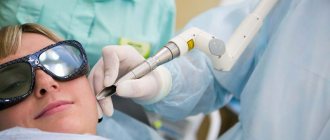
- Laser method
The doctor uses special equipment equipped with a powerful light beam that destroys tumor cells without affecting the surrounding skin tissue.
This method is used only for confirmed benign formations. Advantages of the method:
- Painless and bloodless procedure
- Pronounced cosmetic effect, does not leave scars on the skin
- Fast healing
- Removal in one visit
Flaws:
- Applicable only when confirming the good quality of the formation
How is mole histology performed?
Thanks to histological examination, cancer cells can be detected even at the stage of their inception.
Indications for mole histology:
- redness of the skin around the tumor;
- small ulcers on the surface of the nevus;
- burning or pain in the mole area;
- change in color, size, shape of the tumor.
Histology is performed with the procedure of surgical excision of the mole. A small tissue sample is removed and sent for further examination. In the laboratory, biological material is placed in a special suspension and examined under a microscope. Diagnosis is based on the analysis of pigment-forming cells - melanocytes. Laboratory testing may take up to 14 days.
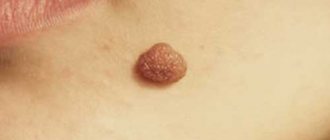
Pathological moles
There are other neoplasms that rightly cause concern in their owner. They can provoke the development of skin cancer. These moles also require removal, but this should be done by an oncologist or surgeon, with the selection of subsequent therapy. Removing pathological moles in aesthetic clinics and centers is extremely dangerous, which is why competent and conscientious plastic surgeons prescribe a series of tests at the slightest suspicion of a malignant growth.
The main signs of a “suspicious” mole:
• Abrupt appearance in adulthood;
• Intensive growth up to 1 cm in diameter;
• Rapid modification of form and structure;
• “Glossy” or rough surface, disappearance of the skin pattern from the elevation;
• Thinning texture;
• The appearance of pronounced asymmetry;
• Peeling of the surface followed by the formation of a crust;
• Presence of itching and burning in the area of the tumor;
• Partial or complete loss of hair from the surface;
• The appearance of depigmented areas, complete or partial change in color;
• Ulcerations on the surface;
• Formation of daughter nodules;
• The presence of hyperemia and swelling around the nevus;
• Formation of new nodules directly on the surface;
• Bleeding (sudden or with minor trauma);
• Separation of moisture and exudate.
Are there any restrictions after mole removal?
A small crust forms at the site of the mole, which, after complete drying, comes off on its own, leaving clean, healthy skin in its place. Under no circumstances should this process be accelerated, because an unsightly scar may form in place of the crust. You should not sunbathe for a month after removing a mole. Before going outside, if it is an open area of the body, use a cream with SPF 50+, which provides a good level of protection against ultraviolet rays.
Restrictions that must be followed for 2-3 weeks after mole removal:
- Avoid contact with water
- Refrain from visiting baths, saunas, swimming pools, and open water bodies.
- The place where the nevus used to be must be protected from any thermal effects.
- Both natural and artificial tanning in a solarium are prohibited.
Melanoma and dysplasia
If, according to histology, we receive a result - severe dysplasia or early melanoma, then this can also be regarded as a positive result, since the disease was detected in the initial stage. In this case, we refer the patient to the oncology clinic, where he undergoes further treatment.
Early (thin) melanoma is very treatable, with a good survival prognosis. In contrast to melanomas, which are left untouched, which grow and patients treat them at stages 3-4, when effective treatment is no longer possible.
Is it safe to remove moles?
Today you can often see offers to remove moles using the laser method in a cosmetology office. For safety reasons, this is absolutely not worth doing. To carry out high-quality nevus removal, you should contact specialized medical institutions, where the procedure is performed by experienced medical workers. It is possible to immediately send the material for histological examination. Beauty salons do not provide this service. If a mole is suspected of being oncological, it is not recommended to use the laser method, which does not allow taking a tissue sample.
If the technology is followed, the nevus removal procedure is absolutely safe. The laser acts locally only on tumors, without heating neighboring organs and tissues.
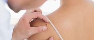
Survey
In order to identify pathological changes in the formation, an optical device is used - a dermatoscope. By applying the device to an area of skin, the doctor can examine in detail the structure of the formation and understand whether it poses a danger to the patient . Based on this, the specialist determines which method of removing the formation is suitable for the patient.
People often come to Melanoma Unit to eliminate cosmetic defects associated with moles, warts and papillomas. For this purpose, an individual method of removing formations is selected, with maximum cosmetic effect.
Our doctors - oncosurgeons, dermatologists - remove moles, papillomas, warts using surgical, radio wave, and laser methods.
All procedures are virtually painless, safe and can be performed in one clinic visit.
Care after mole removal
Within 10-14 days after laser removal of a mole, you should pay attention to your health and follow all doctor’s recommendations.
What not to do:
- Always cover the scab with a bandage or plaster - this can slow down the regeneration process.
- Expose the skin to heat - do not go to the bathhouse or sauna, sunbathe in the sun, or make warm compresses.
- Be in direct sunlight - even after the crust falls off, you must apply sunscreen before going outside (if it is a visible part of the body).
- Play sports - regardless of the location of the mole, it is necessary to exclude physical activity for 1-2 weeks.
- Take a hot bath - on the first day after the procedure, you should not wet the mole with water at all. Next, hygiene procedures must be performed with water at room temperature. The ideal option is a shower. When taking a bath, there is a risk that after steaming the protective crust will fall off and an unaesthetic scar will appear in its place. You should also not rub the mole with a washcloth.
If a mole has been removed from the surface of the palms or soles, any physical activity should be reduced as much as possible. To treat the damaged area, use only medications recommended by your doctor. If you notice swelling, redness or pain in the area where there used to be a mole, do not self-medicate, but immediately consult a doctor.
The essence of the occurrence of moles
You have probably noticed the appearance of new moles where there were none before. In fact, moles form throughout a person’s life. Their secret is quite simple: these small and large dots on the body represent an excessive local accumulation of melanocytes under the skin - cells responsible for the production of melanin pigment. Each mole is fundamentally different from the other in size and shape.
Each mole has its own life cycle. As a rule, the first nodules appear in infancy, and by 7-10 years they acquire their final form, although they may change over time. First, a nevus appears - a flat tiny spot of a dark shade; then you can notice how it grows, becomes voluminous and convex. The intensity of the color and shape of the surface directly depends on the concentration and location of melanocytes - the fewer there are and the deeper the layer of their localization, the lighter the mole. If multiple cells are located in the upper layers of the skin, the mole has a distinct contour, a “puffy” shape and a dark color.
Dangerous moles
Most often moles degenerate:
- Purchased. Which appeared over time. Melanoma affects birthmarks much less frequently from birth. Important! The more a person sunbathes, the more moles appear - the risk of getting cancer increases significantly.
- Flat and located in areas where there is no hair. On the feet, palms, inner shoulders and thighs.
- Borderline. They look like a fried egg - there is a dark circle in the center, and a lighter shade around it.
- Often injured. Those who are constantly irritated by a belt, bra clasp,
underwear straps, jewelry. Advice! Dermatologists recommend removing such moles. But it is better to do this in the fall. At this time, the healing process is faster.
Signs of melanoma
Run to the doctor if you find that a mole:
- Asymmetrical. Mentally divide your education into two parts. If they are unequal, that's bad.
- With jagged edges. The contours are jagged and torn.
- Changed color. It became darker or lighter. White, blue or red areas appear.
- Big size. Diameter is more than 1 cm.
- It began to grow quickly. Within a month and a half it increased sharply in size.
- Causes pain. There are crusts, compactions, peeling, bleeding.
- I was left without hair. They shouldn't fall out. Hairiness, on the contrary, is a sign that the mole is not dangerous!
Contraindications
Depending on the method of removal, there are various contraindications to removing formations.
If during the examination the doctor reveals a pathologically altered formation , it is immediately subject to surgical removal, since there is a huge threat to the health and life of the patient. In this case, this is a contraindication for other methods of tumor removal.
If the formation does not pose a threat to the patient’s health, but there are concomitant diseases associated with poor blood clotting, diabetes mellitus and other diseases that may affect the healing of the postoperative wound, it is also worth refraining from the removal procedure.
The medical clinic is equipped with modern, expensive medical equipment, which allows for a high-quality examination of the skin before the removal procedure, such equipment includes:
- Dermatoscope “Heine Delta 20” (Germany) - Leader in the market of hand-held dermatoscopes
- Digital video dermatoscope “FotoFinder ATBM Master” (Germany) - the latest innovative equipment for early diagnosis of malignant skin diseases
- Radio knife “Surgitron” (USA) - used to remove formations using the radio wave method
- Co2 Laser AcuPulse Lumenis (USA-Israel) - used to remove formations using the laser method
Make an appointment
What are the risks of melanoma?
Melanoma and mole are very similar in appearance. They consist of identical melanocyte cells. These cells produce melanin. A pigment that protects the skin from high doses of ultraviolet rays. However, when a mole becomes injured or burned, it becomes dangerous. Melanocytes begin to multiply rapidly. They crowd out healthy cells.
A tumor forms. From the surface of the skin it quickly makes its way deeper. At the same time, it affects all layers of the skin. It is important to stop the tumor in time! Otherwise, metastases may appear - cancer cells that separate and affect internal organs (brain, liver, lungs, etc.).