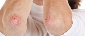
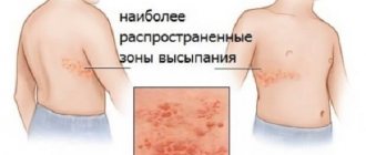
In children after birth, rashes are often physiological; they should not cause much concern to parents. The skin adapts to its new habitat - without water, which is often accompanied by some problems. As a result, the influence of any negative factors can cause a skin rash.
The first children's medical center offers the services of qualified pediatric dermatologists in Saratov. The Center’s specialists aim not only to rid your child of the external symptoms of the disease, but also to find and eliminate the cause that caused it. This is very important, because if the cause of the disease is not eliminated, it can recur again and again, taking a chronic course.
What diseases do staphylococci cause?
The content of the article
The main causative agents of purulent-septic diseases are staphylococci (from the Greek "staphyli" - clusters, "kokkos" - kernels, balls, which corresponds to the appearance of these microbes under a microscope) and streptococci (from the Greek "streptos" - chain).
We owe to these microorganisms such diseases as boils, carbuncles, pyoderma, abscesses, phlegmons, panaritiums and other suppurations of the skin. When the mucous membranes are damaged, conjunctivitis, stomatitis, otitis, and enteritis develop.
Severe forms of the disease are pneumonia, osteomyelitis (bone tissue lesions) and septic - generalized inflammatory processes in internal organs when staphylococci and other microorganisms circulate in the blood (blood poisoning) are especially dangerous.
After purulent infections, various complications arise, including those that can lead to a chronic course of the disease and disability (myocarditis, heart defects, thrombophlebitis, peritonitis - inflammation of the peritoneum).
Losses associated with loss of ability to work only from staphylococcal diseases exceed those caused by all infectious diseases, with the exception of influenza. The number of newborns and adults who die from staph infections is more than twice the number who die from other contagious diseases.
Pustular skin diseases
Currently, pustular skin diseases are the most common dermatoses. Often the development of pyodermatitis (pyon-pus, derma-skin) is caused by staphylococci, streptococci, less often by Proteus vulgaris, Pseudomonas aeruginosa, mycoplasmas, Escherichia coli, etc. When examining the normal microflora of the skin, the greatest contamination with staphylococci is revealed. In this case, the skin of the folds, subungual spaces, mucous membranes of the nose and pharynx is most contaminated, which can serve as a source of endogenous infection.
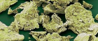
Today staphylococci have been studied quite well. They are cells of a regular spherical shape, with a diameter of 0.5-1.5 microns. Staphylococci are gram-positive and do not form spores. During their life, staphylococci secrete an exotoxin that has the ability to lyse human red blood cells. The pathogenicity of staphylococcal cultures is always associated with coagulase activity. Coagulase exoenzyme is easily destroyed by proteolytic enzymes and inactivated by ascorbic acid. Coagulase-positive and coagulase-negative pathogens can be found in pyoderma. Coagulase-negative pathogens, in addition, are currently considered among the most likely causative agents of gram-positive sepsis. It should be noted that changes in the etiology of sepsis are associated with the selection of resistant gram-positive pathogens as a result of the widespread use of antibacterial therapy. When transformed into L-forms, their reproduction function is inhibited while growth is preserved. Cells in the L-form state have reduced virulence and may not cause inflammation for a long time, which creates a misleading impression of inflammation. Probably, the formation of bacilli carriage and chronic forms of pyoderma, the appearance of atypical forms of bacteria, and drug resistance are due to the transformation of staphylococci into L-forms.
When developing therapeutic and preventive measures, it is necessary to take into account that staphylococci have a high degree of survival in the external environment. They tolerate drying well, are preserved in dust, and spread with air flow. The routes of transmission of staphylococci are very diverse: transmission by airborne droplets, transmission by contaminated hands, objects, etc. is possible.
Carriage of streptococci is much less common. Streptococci have a spherical shape. Facultative anaerobes form endo- and exotoxins and enzymes. Exotoxins have cytotoxic, immunosuppressive and pyogenic effects, erythrogenic activity, and suppress the functions of the reticulohistiocytic system. Streptococci produce deoxyribonuclease, hyaluronidase, streptokinase and other enzymes that provide optimal conditions for the nutrition, growth and reproduction of microorganisms.
In the pathogenesis of pyodermatitis, a decisive role is played by a decrease in local and general antibacterial resistance of the body. The integrity of the stratum corneum and the presence of a positive electrical charge between bacterial cells and the skin provide a mechanical barrier to the penetration of pyococci. The secretion of sweat and sebaceous glands with a high concentration of hydrogen ions (pH 3.5-6.7) has bactericidal and bacteriostatic properties. This “chemical mantle” is regulated by the autonomic nervous system and endocrine glands.
Among the most significant exogenous factors contributing to the development of pyoderma are skin pollution, dry skin, exposure to aggressive chemical agents, temperature irritants, etc.
Endogenous factors include overwork, unbalanced nutrition, especially leading to hypovitaminosis, chronic intoxication, diseases of the gastrointestinal tract, foci of chronic purulent infection, immune imbalance, endocrine diseases. In particular, it is known that pyoderma occurs most severely and torpidly in patients with diabetes mellitus.
There is no single generally accepted classification of pyodermatitis. In this work, we used the most common working classification. It should be noted that the proposed division into superficial and deep pyodermatitis is conditional, since superficial lesions can spread deeper. On the other hand, streptococcus can be cultured from the surface of a staphylococcal pustule, and, conversely, staphylococci are sometimes isolated from the surface of a streptococcal lesion.
The classic division into staphylococcal and streptococcal lesions is based on a number of typical features. Thus, staphylococcal lesions are characterized by a connection with the hair follicle, sweat or sebaceous gland, deep spread, predominantly conical shape, local, sometimes in combination with general, temperature reaction, thick creamy yellow-green purulent contents. Streptococcal pustule is located on smooth skin, lies superficially, has a round or oval shape, transparent or translucent purulent contents.
The most superficial form of staphyloderma is ostiofolliculitis. A pustule appears at the mouth of the follicles, the size of a pinhead to a lentil grain. It has a hemispherical shape, permeated with hair. The cover of the pustule is dense, the contents are purulent. There is a small hyperemic corolla along the periphery. The bottom of the pustule is located in the upper parts of the outer root sheath of the hair follicle. The purulent exudate shrinks into a crust. After three to four days, the element resolves without scar formation.
Folliculitis is an acute purulent inflammation of the hair follicle. Unlike ostiofolliculitis, it is accompanied by infiltration and severe pain. The pustule opens with the release of pus and the formation of erosion or shrinks into a crust. The element resolves by resorption of the infiltrate or with the formation of a scar. The duration of the disease is five to seven days.
Deep folliculitis differs from ordinary folliculitis by its significant spread into the dermis, resolves exclusively with the formation of a scar, and the duration of the disease is seven to ten days.
A boil is an acute purulent-necrotic lesion of the follicle, sebaceous gland and surrounding subcutaneous fat. The development of a boil from ostiofolliculitis or folliculitis is often noted. The growth of the pustule is accompanied by the spread of sharply painful infiltration. After opening the pustule and separating the pus, the necrotic core is clearly visible, gradually separating along with the pus. An ulcer forms at the site of the detached necrotic core. As the necrotic core is opened and separated, the pain decreases, the symptoms of general inflammation subside, the infiltration resolves, the ulcer granulates and heals.
The duration of the evolution of the boil depends on the reactivity of the tissues, localization, state of the macroorganism, etc. When localized on the face or scalp, there is a danger of developing sepsis or thrombosis of the superficial and deep veins that have direct anastomoses with the cerebral sinus.
The carbuncle is characterized by purulent-necrotic lesions of several hair follicles. The inflammatory infiltrate increases not only due to peripheral growth and the possible involvement of new follicles in the process, but also as a result of its spread deep into the underlying tissues. On palpation, sharp pain is noted. Gradually, deep skin necrosis occurs in several places around the hair follicles located in the central part of the lesion. The lesion acquires a slate-blue or black color and “melts” in one or several places (the name “carbuncle” comes from carbo - coal). At the next stage, multiple holes appear, from which purulent-bloody fluid flows. An ulcer with uneven edges is formed, shallow at first, greenish-yellow necrotic rods are visible at the bottom, which are rejected much more slowly than with single boils. After rejection of the necrotic masses, a deep, irregularly shaped ulcer is formed, with bluish, flaccid, undermined edges. The ulcer is gradually cleared of plaque, granulated and healed within two to three weeks.
Furunculosis is a recurrent form of boil. Conventionally, a distinction is made between local furunculosis, when rashes are observed in limited areas, and disseminated, when elements appear on different areas of the skin. As a rule, the process develops against the background of a pronounced immune imbalance, for example in HIV-infected people, patients with diabetes mellitus, etc.
Vulgar sycosis is a chronic recurrent inflammation of the follicles in the growth zone of short thick hair. Most often, the disease occurs in men with signs of imbalance of sex hormones and is localized in the area of beard and mustache growth. Infiltration of the foci is pronounced, ostiofolliculitis and folliculitis appear. After resolution of the elements, scars do not form, but when attempting to forcefully open the folliculitis, scarring is possible.
Hidradenitis is a purulent inflammation of the apocrine sweat glands, observed in young and adulthood. In children before puberty and the elderly, the disease does not develop because their apocrine sweat glands do not function. Most often, hidradenitis is localized in the axillary areas, sometimes on the chest around the nipples, navel, genitals, and anus. The disease develops slowly and is accompanied by discomfort, pain in the affected area, and in some cases itching, burning, and tingling in the affected area. At the beginning of the disease, the surface of the skin has a normal color. Subsequently, the affected area increases to 1-2 cm, the surface of the skin becomes bluish-red. Hidradenitis is characterized by the formation of conglomerates protruding above the level of surrounding healthy areas (popularly the disease is called “bitch udder”). Upon opening, one or several fistulous tracts are formed; necrotic rods are not formed. With regression, retracted scars remain. People with immune imbalances often experience relapses of the disease.
Staphyloderma of early childhood differs in a number of features. The elements do not have the typical properties of a staphylococcal pustule (there is no connection with the hair follicle, sebaceous or sweat gland, the elements are located superficially, have transparent or translucent contents). In newborns, the most common condition is vesiculopustulosis, which is a purulent inflammation of the mouths of the eccrine sweat glands. With adequate management of such patients, the process does not spread deeper and is not accompanied by infiltration. The duration of the disease does not exceed seven to ten days. Epidemic pemphigus of newborns is more severe. Superficial elements quickly spread throughout the entire skin, the resulting erosions are bordered by a fringe of exfoliating epidermis. In a malignant course, erosions merge with each other with peripheral growth of blisters and detachment of the epidermis. The severity of the condition is directly proportional to the affected area. The child's condition is assessed as serious; staphylococcal pneumonia, otitis, and sepsis are developing. The most dangerous form of epidemic pemphigus of newborns is exfoliative dermatitis. Blisters with a flabby tire quickly enlarge and open, forming erosions bordered by exfoliated epidermis. Skin rashes are accompanied by severe fever, weight loss, often diarrhea, pneumonia, otitis media, etc.
Staphylococcus aureus can also be detected in acne vulgaris, acting in association with Propionbacterium acne. Hyperandrogenemia predisposes to increased secretory function of the sebaceous glands. On the skin of the face, scalp, chest and interscapular area, the skin becomes oily, shiny, uneven, rough with enlarged openings of the hair follicles. The thick form of oily seborrhea, which is more often observed in men, is characterized by dilated openings of the sebaceous glands; When pressed, a small amount of sebaceous secretion comes out. The liquid form of oily seborrhea is more common in women and is characterized by the fact that when pressure is applied to the skin, a translucent liquid is released from the mouths of the ducts of the sebaceous glands. Mixed seborrhea is somewhat more often observed in men, while the symptoms of oily seborrhea appear in the facial skin, and dry seborrhea - on the scalp, where fine-plate peeling is expressed, the hair is thin and dry. Acne develops in individuals suffering from oily or mixed forms of seborrhea. Among the patients, adolescents (somewhat more often boys) and women with ovarian cycle disorders as a result of long-term use of glucocorticoid hormones, bromine, iodine preparations, and prolonged work with chlorine-containing substances predominate.
The most common form of the disease is acne vulgaris, localized on the skin of the face, chest, and back. After the pustules resolve, dried yellowish crusts form. Subsequently, an increase in pigmentation is observed or a superficial scar is formed. In some cases, after acne resolves, keloid scars (acne-keloid) appear. If the process proceeds with the formation of a pronounced infiltrate, then deep scars (phlegmonous acne) remain at the site of acne resolution. When the elements merge, acne confluens is formed. A more severe form of the disease manifests itself in the form of acne conglobata, accompanied by the formation of a dense infiltrate of nodes in the upper part of the subcutaneous fat. Nodes can form into conglomerates with subsequent formation of abscesses.
After the ulcers heal, uneven scars remain, with bridges and fistulas. Acne fulminans is accompanied by septicemia, arthralgia, and gastrointestinal symptoms.
Streptoderma is characterized by damage to smooth skin, superficial location, and a tendency toward peripheral growth. In clinical practice, the most common phlyctena is a superficial streptococcal pustule.
Let's look at a few examples.
Streptococcal impetigo is highly contagious and is observed mainly in children, sometimes in women. Phlyctens appear on a hyperemic background, do not exceed 1 cm in diameter, have transparent contents and a thin flabby cover. Gradually, the exudate becomes cloudy and shrinks into a straw-yellow and loose crust. After the crust falls off and the epithelium is restored, mild hyperemia, peeling, or hemosiderin pigmentation temporarily persist. The number of conflicts is gradually increasing. Dissemination of the process is possible. Complications in the form of lymphangitis and lymphadenitis are common. In weakened individuals, the process may spread to the mucous membranes of the nasal cavities, mouth, upper respiratory tract, etc.
Bullous streptococcal impetigo is localized on the hands, feet, and legs. The size of conflicts exceeds 1 cm in diameter. The cover of the elements is tense. Sometimes the elements appear against a hyperemic background. The process is characterized by slow peripheral growth.
Zaeda (slit-like impetigo, perleche, angular stomatitis) is characterized by damage to the corners of the mouth. Painful slit-like erosion appears on a edematous, hyperemic background. Along the periphery one can detect a whitish rim of exfoliated epithelium, sometimes a hyperemic rim, and infiltration phenomena. Often the process develops in people suffering from caries, hypovitaminosis, atopic dermatitis, etc.
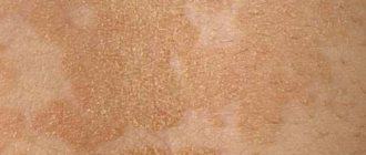
Lichen simplex most often occurs in preschool children in the spring.
Round pink spots, covered with whitish scales, appear on the skin of the face and upper half of the body. With a large number of scales, the spot is perceived as white.
Superficial paronychia can be observed both in people working in fruit and vegetable processing plants, in confectionery shops, etc., and in children who have the habit of biting their nails. The skin of the periungual fold turns red, swelling and pain appear, then a bubble with transparent contents forms. Gradually, the contents of the bubble become cloudy, the bubble turns into a pustule with a tense tire. If the process becomes chronic, deformation of the nail plate is possible.
Intertriginous streptoderma (streptococcal diaper rash) occurs in large folds and axillary areas. Conflicts that appear in large numbers subsequently merge. Upon opening, continuous eroded, weeping surfaces of a bright pink color are formed, with scalloped borders and a border of exfoliating epidermis along the periphery. Painful cracks can be found deep in the folds. Screenings often appear in the form of separately located pustular elements in various stages of development.
Syphiloid papular impetigo is observed mainly in infants. Favorite localization is the skin of the buttocks, genitals, and thighs. Characteristic is the appearance of quickly opening conflicts with the formation of erosions and a slight infiltrate at their base, which was the reason for choosing the name “syphiloid-like”, due to the similarity with erosive papular syphilide. Unlike syphilis, in this case we are talking about an acute inflammatory reaction.
Chronic superficial diffuse streptoderma is characterized by diffuse damage to large areas of the skin, legs, and less often, hands. The lesions have large scalloped outlines due to growth along the periphery, are hyperemic, sometimes with a slight bluish tint, somewhat infiltrated and covered with large lamellar crusts. Under the crusts there is a continuous wet surface. Sometimes the disease begins with an acute stage (acute diffuse streptoderma), when acute diffuse skin damage occurs around infected wounds, fistulas, burns, etc.
A deep streptococcal pustule is ecthyma. The inflammatory element is deep, non-follicular. The disease begins with the formation of a small vesicle or perifollicular pustule with serous or serous-purulent contents, which quickly dries into a soft, golden-yellow convex crust. The latter consists of several layers, which served as the basis for the now textbook comparison with Napoleon cake. After the crust falls off or is removed, a round or oval ulcer with a bleeding bottom is discovered. There is a dirty gray coating on the surface of the ulcer. The ulcer heals slowly, over two to three weeks, with the formation of a scar and a zone of pigmentation along the periphery. In severe cases of vulgar ecthyma, a deep ulcer (ecthyma terebrans - penetrating ecthyma) can form, with symptoms of gangrenization and a high probability of sepsis. Mixed pyoderma is distinguished by the presence of both staphylococcal and streptococcal pustules (in fact, in addition to staphylococci and streptococci, other pathogens can be detected).
Let's look at a few examples.
Vulgar impetigo is the most common. Mostly children and women are affected. Favorite localization is the skin on the face around the eyes, nose, mouth, sometimes the process spreads to the upper half of the body and arms. Against a hyperemic background, a bubble with serous contents appears. The vesicle cover is thin and flaccid. The contents of the vesicle become purulent within a few hours. The skin at the base of the pustule becomes infiltrated, and the rim of hyperemia increases. After a few hours, the lid is opened, forming erosion, the discharge of which shrinks into “honey crusts”. On the fifth-seventh day, the crusts are torn off, a slightly flaky spot remains in their place for some time, which later disappears without a trace.
Chronic deep ulcerative-vegetative pyoderma has a predominant localization on the scalp, shoulders, forearms, axillary areas, and legs. On an infiltrated bluish-red background with clear boundaries, sharply different from the surrounding healthy skin, irregular ulcerations appear in place of the pustules. On the surface you can find papillomatous growths with verrucous cortical layers. When compressed, purulent or purulent-hemorrhagic contents are released from the openings of the fistula tracts. With regression, the vegetation gradually flattens, and the separation of pus stops. Healing occurs with the formation of uneven scars.
Pyoderma gangrenosum often occurs in patients with chronic inflammatory infectious foci. Changes in the skin develop against the background of chronic inflammatory infectious foci, connective tissue diseases, and oncological processes. Blisters with transparent and hemorrhagic contents, deep folliculitis quickly disintegrate or open with the formation of ulcers that expand along the periphery. Subsequently, a lesion with an extensive ulcerative surface and uneven, undermined edges is formed. Along the periphery, these edges appear to be raised in the form of a roller, surrounded by a zone of hyperemia. Bleeding granulations are found at the bottom of the ulcers. The ulcers gradually increase in size and are sharply painful. Scarring of different areas does not occur simultaneously, i.e., when one area is scarred, further growth of another may be observed.
Chancriform pyoderma begins with the formation of a vesicle, after opening which remains an erosion or ulcer of round or oval shape, the base of which is always compacted. As the name implies, an ulcerative surface of a pinkish-red color with clear boundaries is subsequently formed, resembling a chancre in appearance. Certain difficulties in differential diagnosis may also be due to the similar localization characteristic of these diseases: genitals, red border of the lips. Unlike syphilis, a pronounced infiltrate is palpable at the base of the lesion, sometimes painful when pressed. Repeated negative tests for the presence of treponema pallidum and negative serological tests for syphilis confirm the diagnosis.
For the treatment of superficial pyodermatitis, alcohol solutions and aniline dyes are used. If necessary, the cover of phlyctena and pustules is aseptically opened, followed by washing with a three percent solution of hydrogen peroxide and lubrication with disinfectant solutions. Ointments containing antibiotics are applied to widespread multiple lesions.
In the absence of effect from external therapy, deep lesions on the face, neck (furuncle, carbuncle), pyoderma, complicated by lymphangitis or lymphadenitis, parenteral or oral use of broad-spectrum antibiotics is indicated. For chronic and recurrent forms of pyoderma, specific immunotherapy is used (staphylococcal toxoid, native and adsorbed staphylococcal bacteriophage, staphylococcal antiphagin, antistaphylococcal g-globulin, streptococcal vaccine, streptococcal bacteriophage, autovaccine, antistaphylococcal plasma).
In severe cases, especially in weakened patients, the use of immunomodulatory agents is indicated.
In the case of chronic ulcerative pyoderma, courses of antibiotics can be supplemented by the administration of glucocorticoids in a dose equivalent to 20-50 mg of prednisolone per day for three to six weeks. In the most severe cases, cytostatics are used.
Prevention of pustular skin diseases, including compliance with hygiene rules, timely treatment for intercurrent diseases, adherence to diet, etc., should also be carried out at the national level (increasing the standard of living of the population, introducing methods of protection against microtrauma and contact with chemicals at work , solving environmental problems, etc.).
I. V. Khamaganova , Doctor of Medical Sciences, Russian State Medical University, Moscow
Antibiotic resistance of staphylococci
The more we attack, for example, pathogens of purulent-septic infections with antibiotics, the more often antibiotic-resistant microorganisms appear.
Under the influence of antibiotics, the inhibitory effect on staphylococci of other microbes - common inhabitants of our body - is disrupted. Moreover, sometimes antibiotics simply stimulate the development of staphylococci and streptococci. Therefore, without medical prescription, the independent use of antibiotics (and other antibacterial agents) in the event of purulent skin lesions should be excluded.
Any treatment for staphylococcal skin pathologies should be prescribed by a dermatologist! Diseases of other organs are treated by appropriate specialists - gynecologists, urologists, therapists.
Why is staphylococcal infection dangerous?
Staphylococcus is especially dangerous in association with viruses and fungi, as well as with concomitant childhood droplet infections that reduce the overall reactivity of the body. It is not without reason that in many diseases local or general complications caused by staphylococci are observed.
Thus, during influenza epidemics, staphylococcal pneumonia is not uncommon, which most often turns out to be the cause of a child’s long-term illness and is the most common prerequisite for death.
Staphylococcal and streptococcal toxins have pronounced sensitizing properties, causing allergies and toxic damage to the heart muscle, kidneys and other important organs. Unlike a number of pathogens of childhood infections, the site of action of which is limited to certain areas of body tissue, staphylococcus and streptococcus are “omnivorous”.
They can cause inflammation of the gallbladder (cholecystitis), toxic dyspepsia and gastroenteritis, inflammation of the joints (arthritis), and genitourinary tract (urethritis and endometritis). In newborns, staphylococcal and other purulent inflammatory phenomena begin with the umbilical wound or other skin lesions that are invisible at first glance (scratches, abrasions).
It must be emphasized that up to 90% of cases of sepsis in young children are associated with staphylococcus.
Features of children's skin
At birth, babies have very thin skin. The skin of a newborn is almost half as thick as the skin of an adult. The outer layer thickens with age.
The skin of newborns is red or purple in color due to the close proximity to the upper layer of blood vessels and an insufficient layer of subcutaneous cellular tissue, as a result, the skin appears “transparent”. This phenomenon is especially pronounced if the baby is frozen - the appearance of a marbled vascular network on the body is observed.
Moisture evaporates faster from the skin of newborns. Children are more susceptible to bacteria, viruses, fungi and mechanical influences. Thickening of the skin begins at 2-3 years of age and ends by 7 years.
Staphylococcus in pregnant women is a threat to the life of newborns
The source of staphylococcal infection in maternity hospitals, children's hospitals and at home can be purulent lesions of the skin and mucous membranes of adults - the mother, others, and staff. Therefore, it is impossible to separate contagious skin diseases from other human infectious diseases.
Under normal conditions, the skin has protective properties in the fight against microbes, in particular, its secretions contain antibacterial substances. However, nutritional disorders, general diseases, and microtraumas of the skin can contribute to the development of pathogenic microflora on the child’s skin.
Active immunization of pregnant women with staphylococcal toxoid, as practice has shown, does not have a harmful effect on either the course of pregnancy or the development of the fetus, while at the same time reducing the incidence of purulent infections in mothers and newborns by 3-5 times.
At the same time, the mother develops active and the child develops passive immunity to pathogens of such infections as otitis media, pneumonia, skin suppuration, tonsillitis (tonsillitis), laryngotracheitis, sinusitis and other inflammatory diseases of the nasopharynx.
Staphylococcal diseases in young people and children
Pustular skin diseases are called pyodermatitis, which, depending on the cause of occurrence, are divided into strepto and staphyloderma. Their mixed form is often observed - streptostaphyloderma.
Streptoderma most often affects children and young men and is usually localized around natural openings - the nose, mouth, ears, that is, in areas subject to irritation by the discharge of these cavities (saliva, mucus). These lesions are very contagious and quickly spread to healthy areas of the skin. Streptoderma is transmitted through toys, diapers, clothes, and underwear.
A fairly common streptococcal skin lesion in newborns is impetigo (from the Latin “impetus” - sudden, attacking). With this disease, flat cavities with a flabby folded covering are formed, filled with serous-purulent contents - phlyctenas.
Merging, they form continuous foci with tortuous inflammatory outlines. Exposed parts of the body are predominantly affected, especially the skin of the face. Nearby lymph nodes often swell, and as the infection spreads, phenomena of general intoxication are observed.
Another form of purulent lesions is neonatal pemphigus. This type of pyoderma is characterized by the formation of large blisters that reach the pigeon egg and are filled with purulent contents. When they rupture, a bare bright red surface (erosion) is formed, which is easily subject to additional infection, resulting in a sharp deterioration in the child’s condition.
Streptococcal pemphigus is extremely contagious, contagious and, in the absence of proper preventive measures, can cause massive outbreaks of pyoderma in maternity hospitals and children's hospitals.
When a child moves home from the maternity hospital, a vesiculopustular form of pyoderma often occurs in places that are most often irritated by sweat (on the back of the head, neck, forehead, groin). Small bubbles with transparent contents (vesicles) the size of a pinhead appear. Then the contents of the vesicles become purulent (pustule) and are surrounded by a halo of hyperemia.
After two to three days, the pustules burst or undergo reverse development and superficial crusts form in their place. Despite the relative ease of this infection, its complications can be very insidious. They are expressed in widespread and severe phlegmon, occurring with significant swelling and subsequent necrosis (melting) of the subcutaneous tissue.
In infants with low nutrition, in improperly bottle-fed children, as well as in older children with weak protective reactions (due to tuberculosis or other types of intoxication), streptococcal infection is not limited to superficial skin lesions, but invades deeper tissues, forming deep ulcers - ecthyma .
They occur on the most frequently injured parts of the body (lower back, lower legs) and are characterized by a sluggish, long-lasting course. After they are healed, scars remain.
Speaking about streptococcal pyoderma in children, we cannot ignore erysipelas, which especially often affects the face (hence the name “erysipelas”). The skin of the face becomes red and swollen. The disease is accompanied by an increase in temperature and sometimes a severe general condition requiring emergency treatment measures.
The so-called “zaeda” is also known - streptococcal phlyctena (a type of impetigo) in the corners of the mouth, which opens and forms a long-term non-healing wound, constantly renewed in connection with facial movements of the facial muscles and food intake. With insufficient sanitary control, seizures can cause real outbreaks in children's groups.
Pemphigus of newborns can also be caused by staphylococci. In the most severe form of this disease, serous fluid accumulates in large areas under the stratum corneum of the skin and detachment of the skin occurs to such an extent that the child gives the impression of being “scalded.”
With staphyloderma, hair follicles are often affected, for example on the head, which leads to a small pustular rash, which disappears without a trace after treatment.
Staphylococci cause not only local, limited skin lesions, but also sore throat, severe pneumonia, inflammatory foci in various organs and tissues (liver, spleen, kidneys), resulting from the spread of microbes through the blood and lymphatic tracts. A generalized staphyloccal infection - sepsis - poses a mortal threat.
Staphylococci can cause real epidemic outbreaks in maternity hospitals and children's hospitals. Due to them, the vast majority of skin and septic lesions in newborns and purulent diseases in mothers occur. Postpartum sepsis, mastitis, pneumonia, meningitis, inflammation of the birth canal, conjunctivitis are a real disaster. Of course, they are especially dangerous for women in labor and newborns.
Causes of pustular diseases and their prevention
Pustular diseases (pyoderma, pyodermitis) are caused by microorganisms that penetrate the upper layers of the skin. Intact skin is impermeable to microorganisms. In most cases, the causative agents of pyoderma are strepto- or staphylococci or combinations thereof, less commonly Escherichia coli or Pseudomonas aeruginosa, gonococci, pneumococci, etc. All pustular diseases are divided into streptoderma, staphyloderma and mixed streptostaphyloderma. Although the main elements of the rash in these diseases are an abscess or acute inflammatory nodules and nodes, more than 20 types of the disease are distinguished. The diagnosis is established mainly by eyes, based on existing rashes, anamnesis (history of the disease). Analysis to determine the pathogen is not necessary because streptoderma and staphyloderma have their own characteristics, known to specialists. Streptococci and staphylococci are permanent inhabitants of human skin and cause diseases only by penetrating the skin. With a decrease in local or general immunity of the human body, they become more aggressive, as experts say, they become more pathogenic, i.e. capable of causing disease. In such cases, the patient can become a source of infection for other family members or organized groups, especially among children. Considering that the skin is a barrier between the internal and external environment of the body, all causative factors of pustular diseases are divided into external (exogenous) and internal (endogenous). Often a combination of both is important. External causes include microtrauma of the skin, contamination, overheating or hypothermia of the body (in children this occurs due to imperfections in their thermoregulation). Microtraumas include abrasions, abrasions, scratches, pricks, scratches, insect bites, microcracks that occur when the skin becomes dry due to daily showering or other general water procedures (this removes the fatty lubricant produced by the sebaceous glands of the skin), etc. Skin contamination can occur in at home and at work. Industrial (occupational) pollution is caused by building materials (cement, lime, etc.), lubricating oils (for example, oil folliculitis of the thighs occurs when work clothes, in particular trousers, are contaminated and not washed regularly), earthen, peat dust, coal dust, etc. d. In everyday life, especially among children, skin contamination occurs due to poor hygienic care. Irregular changes of linen and clothes, rare baths lead to increased proliferation of microorganisms on the surface of the skin, the breeding ground for which are exfoliated epidermal cells, sebum, food debris, etc. Overheating of the body contributes to increased sweating and sebum secretion and an increase in the presence of microorganisms. In young children, this leads to inflammation of the sweat glands (various types of heat rash). Hypothermia causes vasoconstriction, which disrupts its nutrition and reduces its resistance to microbes. Internal factors or causes of pyoderma include disorders of carbohydrate metabolism (diabetes mellitus), endocrine disorders (for example, insufficient activity of the thyroid gland), nutritional disorders (deficiency of proteins, excess carbohydrates, fats), hypovitaminosis (especially lack of vitamins A and C), physiological conditions ( for example, hidradenitis occurs in mature people, before puberty and in old age when the function of the gonads fades, it does not occur), acute and chronic diseases, for example, chronic alcoholism, cancer, chronic infectious diseases, etc., taking medications that reduce the body's defenses (for example, corticosteroids, hormones, including their local use). In children, the development of pyoderma is facilitated by increased looseness, humidity, tenderness of the stratum corneum of the epidermis, and its easy vulnerability. When you eat fruits, such as apples, their juice helps soften the epidermis around the mouth. If the skin of the face is not washed after eating, the juice dries out and becomes a breeding ground for microbes; in addition, contaminated skin is accompanied by itching, which leads to disease. At first, single pustules appear, most often in the form of purulent crusts with a rim of inflammation around them, but very quickly the child’s entire face is covered with such elements of the rash. This disease is called impetigo (translated as “suddenly occurring”) and causes fear among parents. Based on the above, in order to prevent pustular diseases, it is necessary: 1) promptly treat skin microtraumas with antimicrobial agents, such as a 1-2% alcohol solution of brilliant green; fucorcin, 3-5% tincture of iodine 1-2 times a day; Sometimes a single treatment is enough, sometimes it is necessary to use them until complete healing, for example, an abrasion, a cut or burns, etc. 2) compliance with the rules of hygiene, cleanliness of skin, linen and clothing, etc. When taking a shower or other water procedures every day, it is necessary to take care of restoring the fatty mantle of the skin with the help of emollients (for example, Trixer cream) and special cosmetics for use after a shower. 3) avoid overheating and hypothermia of the body (for example, wearing clothing appropriate to certain weather conditions). 4) rational, nutritious nutrition consistent with a healthy lifestyle (do not overeat, get up from the table a little hungry, do not abuse flour and sweets, try to eat fresh vegetables and herbs at every meal, eat fruits, for example 1 apple on an empty stomach daily ). 5) eliminate alcohol abuse. 6) treatment of diseases that reduce the skin's resistance to infections (for example, diabetes). 7) when pustules appear, washing the skin, bathing, etc. is prohibited. water and the use of washcloths, rubbing with a towel lead to softening of the stratum corneum of the epidermis and the spread of microbes to other areas of the skin. For hygienic care, it is recommended to wipe the skin around the pustules with 2% salicylic alcohol and wipe the fingertips of sick children with the same solution to prevent the disease from being transferred to other areas by hand. With the start of treatment, it is necessary to change underwear, bed linen, towels, and clothes that come into contact with the skin rashes in order to prevent re-contamination of the skin with pyogenic material. Infected items of clothing and linen must be washed with detergent and ironed. Wash sick children's toys with soap. 9) rational and timely treatment of diseases accompanied by itching and scratching (pediculosis, scabies, fungal diseases, atopic dermatitis, eczema, etc.). Head of Far Eastern Branch No. 3 Work phone 372 73 67 Gumbar Slavamir Anatolyevich
