Risks of the appearance and development of melanoma
One of the main factors in the development of melanoma is an increase in the duration of exposure to ultraviolet radiation in people whose skin is not genetically adapted to it. As a rule, this type includes people with white skin that is not prone to tanning. Representatives of dark and dark skin, on the contrary, suffer from melanoma much less frequently (10-20 times less often). In addition to genetic predisposition, there are other risk factors:
- Physical – ionizing radiation, trauma, excessive insolation, fluorescent lighting;
- chemical – contact with nitric acid, benzene, coal and pharmaceuticals;
- biological - with alcohol and certain medications (for example, estrogen-containing hormonal drugs).
External biological characteristics of the body that increase the risk of developing melanoma also include: the first two skin phototypes (in which the skin is light and prone to sunburn), the presence of freckles and lentigo, a family history, and immune disorders. Based on the above factors, 3 risk groups are distinguished, which are more likely to undergo screening examinations according to indications.
Factors contributing to changes in nevus color
There are many reasons that influence the darkening or lightening of a mole. The leading factor is genetics. Neoplasms are often congenital. If relatives have malignant ones, then there is a high risk that the child will also develop melanoma.
Another reason why a mole becomes light is due to exposure to ultraviolet radiation. People with many moles on their body should not stay in the open sun for a long time or visit a solarium.
Regardless of the reason for the appearance of a nevus, any changes in it indicate the development of a certain process in the skin, and how dangerous it is should be determined by a doctor.
Changes in the color of a mole also occur for other reasons:
- Exposure to chemicals. If the nevus is constantly exposed to toxic agents, this leads to its modification and irritation.
- Damage. Banal scratches, scratches, wounds or insect bites lead to the degeneration of birthmarks. This causes inflammation of the skin and leads to the production of substances that increase cell growth.
- Hormonal imbalances. A nevus may acquire a heterogeneous consistency, change size and shade due to hormonal imbalance. Most often, dysfunction occurs in pregnant women, adolescents and people with diseases of the endocrine system.
Diagnostics
There are a number of signs of malignant degeneration of a mole:
- in the area of the mole there are signs of a violation of the skin pattern, peeling;
- the color of the mole changes from black to pale pink;
- uneven coloring of the mole;
- the formation of an inflammatory corolla around the mole;
- changing boundaries;
- increase in size, thickening of the mole;
- vertical or horizontal growth.
The appearance at the base of a mole of nodes with areas of decay, cracks, ulcers, bleeding, the presence of itching, burning, tingling should also be alarming.
The first signs of melanoma are an increase in the size of the mole. Discoloration, bleeding, ulceration and pain appear much later.
Diagnosis also consists of a thorough examination of the skin, including the armpits, scalp, interdigital folds, genitals, perianal area and oral mucosa, as well as palpation by the doctor of the lymph nodes and liver.
For unambiguous confirmation, an ultrasound examination (to determine the thickness and depth of invasion), ultrasound of the lymph nodes and abdominal organs, chest x-ray, biopsy and computed tomography (if metastases are suspected) are performed. A biopsy is indicated in some cases - when the thickness of the tumor is from 1 to 4 mm and if the entire range of diagnostic measures did not make it possible to verify the diagnosis.
Danger signs requiring medical advice
When a mole changes color, the worst consequence is the development of melanoma. This is a cancerous tumor that develops from nevi or epidermal cells. The exact causes of malignancy have not yet been established. It is known that birthmarks of heterogeneous color degenerate on the lower extremities in women and on the torso in men.
In order to promptly identify melanoma and seek medical help, you need to know what dangerous signs indicate the initial stage of its development. Symptoms that should alert you:
- the mole has changed in size;
- suppuration;
- the nevus has become lighter or darker;
- ichor or blood is released from the formation;
- change in shape.
Medical classification of melanomas
To determine the stage of melanoma, the Unified Staging System is quite informative, which distinguishes 4 stages - according to the thickness of the tumor and the depth of its invasion according to Clark:
- 1 – tumor thickness up to 1.5 mm with invasion level 2-3;
- 2 – tumor thickness up to 4 mm and invasion level 4-5;
- 3 – regional presence of metastases about 3 centimeters in diameter;
- 4 – distant metastases.
Melanoma can occur in both cutaneous and extracutaneous forms (10-20%) - with localization on the mucous membrane of the esophagus, rectum, genitals, eye mucosa, etc. The proportion of cutaneous forms is incomparably greater - 70-80%, but poses no less a threat for a large number of people around the world.
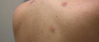
Why does a mole have uneven coloring?
If you look closely at the moles on your body with a magnifying glass and a bright lamp, you can see interesting things. Not a single mole is 100% evenly colored. Some have darker areas in the center, others on the periphery, and others generally have lighter fragments.
You might unwittingly think that you have lived with melanoma all your life and only now noticed it...
Again, this is most likely not the case. Please look at this photo - there is a mole on it with 10x magnification:
Like any part of the human body, a mole does not have perfectly straight lines, identical proportions, or 100% symmetry. In the photo you can see how the pigment melanin, which is produced by the cells of the mole, forms a pigment network. It can be uniform by definition.
In a living organism that is constantly changing, such a situation is impossible in principle. Therefore, the pigment in some moles may be produced unequally, and our eye will see an uneven color.
Preventing and reducing the risk of melanomas
The postulates in the prevention of skin melanoma are: screening, limiting damaging factors, removing altered pigment formations.
Screening includes non-invasive diagnostic methods - dermatoscopy or epiluminescence microscopy annually, and for risk groups at least 2 times a year. Eluminescent microscopy is a non-invasive method for studying skin formations in a special immersion environment that can save a digital image, enter it into a database and track changes by comparing it with existing and subsequent images.
Primary prevention is to avoid the damaging effects of ultraviolet radiation, chemicals and other external factors. Secondary prevention is the removal of all moles suspicious for malignancy.
Tips that can prevent the development of melanoma are quite simple and effective. Will help reduce the risk of mole degeneration:
- wearing dark, thick clothing;
- eating vitamin D;
- removal of altered or injured moles.
The widespread use of screening and preventive measures will have a beneficial effect on the timely diagnosis and treatment of this dangerous disease.
Is uneven coloring of a mole a sign of melanoma?
Yes, this may be one of the signs of a malignant mole. However, to correctly answer this question, we need to analyze more than just one sign that caught our attention. It is necessary to comprehend all the information about the mole. Long-term existence without any significant changes, absence of asymmetry, bleeding without trauma will be arguments in favor of benignity. If the mole has a slightly uneven color, most likely everything is fine.
On the other hand, the presence of uneven coloring and, for example, rapid growth (more than 1-2 mm per year) or pronounced asymmetry will make any oncologist wary.
Summary or briefly about the main thing:
A very small number of unevenly colored moles turn out to be melanoma. The likelihood of a malignant mole tends to zero if there are no other signs of melanoma. If doubts persist, be sure to see an oncologist and have a dermatoscopy done. If you are in doubt whether to go or not to go, it is better to go. Communication with a living doctor who is responsible for his every word, as a rule, dispels all fears.
If you still have questions, the following will help you:
- In-person appointment with an oncologist
(St. Petersburg) - Mole removal
with histology (St. Petersburg) - My online consultation (from anywhere in the world)
Moles, neoplasms, ultraviolet radiation and HIV: an interview with a dermatologist
How can you spot a suspicious mole yourself, distinguish it from a normal one, and manage to remove it before it’s too late? Do you need to examine moles yourself at home? Is it true that sunbathing is strictly forbidden, or is it still possible a little? How are skin diseases related to HIV? Dermatologist-oncologist at the Rassvet clinic, Tatyana Kuzmina, answers these and other questions.
— How often should I undergo a preventive examination with a dermatologist if nothing bothers me?
— It depends on the results of the initial examination by a dermatologist. I examine all formations on the skin, and if there are suspicious ones, I highlight them, measure them and draw up a map of nevi (moles). If there are formations that require dynamic monitoring, then we invite patients to an appointment once a year. Also, once every six months or a year, you need to come for an examination to a dermatologist for those who have many formations (in this case, only a nevus is considered a formation) on the skin, more than fifty to one hundred. Such people are classified as patients with dysplastic nevus syndrome: they develop new moles all the time and are more likely to change towards dysplasia (a condition that is a precursor to melanoma).
We also invite adults over forty for examination once a year - if at this age a new nevus appears on the skin, then there is a higher risk that it will turn out to be something malignant. If a person is under forty and at the initial appointment there are no formations that would cause suspicion, and there is no tendency that something suspicious may appear in the future, then the appointment will only need to come once every three years. But this is provided that the person at home independently observes nevi and conducts self-examination at least once every six months.
— What should you pay attention to at home and how to conduct independent examinations between visits to the doctor?
— At home, from time to time you will need to check the parameters of the tumor map. If you see any dynamics: growth, changes in color, coloring, then you need to come to the appointment earlier. For example, if a mole is actively growing (by more than two millimeters per year), this should alert you. Suspicious formations also include nevi larger than a centimeter, asymmetrical, with uneven coloring and borders. If such formation begins to change, it is better to come and show it to the doctor.
But we also need to keep an eye on new formations. Until the age of forty, a person may develop new nevi, this is normal, but if they are black, then one should be wary. When one appears, regardless of its size, you need to consult a doctor. If you are not sure whether the lesion is black or not, take a black object, such as a phone, apply it to the nevus and compare. If the lump is dark brown in color, it is most likely benign.
Tatyana Kuzmina, dermatologist-oncologist at the Rassvet clinic.
— What does a doctor do if, during an examination, he reveals something suspicious?
— If a nevus seems suspicious, we always suspect a malignant pigment formation, melanoma in situ, and suggest removing it. Depending on the visual assessment, we determine what it could be: a benign nevus that has just begun to change, or an already changed formation.
In the first case, we remove the nevus with a certain indentation, in the second - a more serious intervention; additional examinations need to be carried out to find out whether the process is only in the skin or has already spread beyond its borders. If this formation turns out to be melanoma in situ or severe dysplasia, then everything ends with surgical removal, and the materials after the operation are sent for histology. If the histology results indicate stage 1 or 2, then this is a good prognosis, and in the future you can count on a complete cure. In the third or fourth stage, treatment must be continued, but final recovery is unlikely.
Therefore, it is very important to visit a doctor at the first signs of changes in the skin, when color or size has just begun to progress. Melanoma skin cancer develops quickly, a small dark spot of two millimeters may appear on the skin, this will already be melanoma, and active measures will need to be taken to remove it. No need to wait. If the patient manages to see a doctor within six months, then, as a rule, the formation does not have time to move to the stage when we cannot help him and additional treatment in addition to surgery will be required.
— How can you reduce your risk of getting skin cancer? Don't sunbathe at all? Sunbathing only with photoprotection?
— Melanoma is triggered by sunburn. The more active the sun, the easier it is to get a sunburn. The activity of the sun increases as one approaches the equator - in countries close to it, for example in Thailand, where people now often travel for several months, ultraviolet radiation is of higher intensity (the activity index on a scale from 0 to 10 reaches its maximum values, the sun's rays damage the genetic apparatus of cells more strongly skin). Such radiation can provoke melanoma without visible sunburn.
No protective equipment can completely resist radiation, but with photoprotection and photoprotective clothing, you can avoid skin damage and reduce the risk of developing melanoma. However, even with protective equipment, ultraviolet radiation will accumulate - we call it chronic ultraviolet radiation, which increases the risk of developing non-pigmented skin cancers, in particular basal cell carcinoma. This is a type of tumor that develops from non-pigmented skin cells of the epidermis (keratinocytes) rather than from pigment cells (melanocytes).
How to figure it out?
If after reading this material you still have doubts, the oncologist should say the last word on the mole during a face-to-face consultation. Not a dermatologist, not a surgeon, and certainly not a cosmetologist. It is optimal if the doctor knows the technique of dermatoscopy and has this device at hand.
Very often I hear the following objection regarding a visit to the doctor about a mole: “I don’t want to go to the oncologist, in case he says that I have cancer!!!” Unfortunately, this position borders on absurdity. After all, if there is cancer, it needs to be diagnosed and treated, and not “hidden in the sand.” This is especially true for melanoma, which at stage 1 is completely curable in almost 100% of cases.