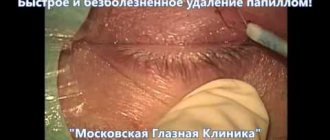
Conditionally benign neoplasms often appear on the skin, which do not pose a direct threat to human life and health, but are a cosmetic defect. Tumors are often damaged during water procedures and can bleed and become inflamed. Keratoma is one of these neoplasias. No specialist can exclude the risk of spontaneous malignancy of a benign neoplasm. Therefore, when a keratoma appears, it is better to immediately contact a dermatologist or dermatologist-oncologist.
Patients in most cases mistake a keratoma for a large mole. But any neoplasm, especially one prone to active growth and darkening of tissue, requires examination by a dermatologist. Only a specialist can say exactly what to do with a certain type of keratoma. At LASER+ clinics in St. Petersburg you can make an appointment with a dermatologist and remove skin tumors of any size
What is a keratoma
Keratomas can be single or multiple. Some types of neoplasms are classified as borderline neoplasias, which can degenerate into malignant tumors under the influence of unfavorable factors. Externally, the keratoma resembles a lumpy brown mole. It protrudes above the surface of the skin. Since it is useless to treat the tumor conservatively, it is better to remove it with a laser. The method is the least traumatic and at the same time affordable. The most common type is senile or senile keratoma. Over time, it grows along the periphery, is capable of transforming into a cutaneous horn and is prone to magnetization. Seborrheic, follicular, solar, and horny keratomas are also found. All of them are potentially dangerous and require removal.
Why you may need to remove a keratoma
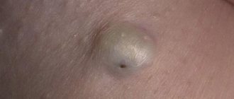
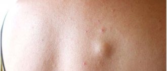
Keratoma is a benign neoplasm on the skin, which consists of epidermal cells. It can be either small in size (a few millimeters) or quite large (up to 5 centimeters in diameter). Keratomas are usually slightly darker than the natural color of the skin or have a brown color. Over time, the surface of the keratoma becomes rough and rough.
The neoplasm is formed from human skin cells, which gradually layer on top of each other. At the same time, the upper layer becomes horny, skin scales die and fall off under mechanical stress, for example, during washing, but the deeper layers of the dermis also become keratinized. The process of continuous cell renewal leads to the fact that the keratoma does not disappear on its own, but can cause inconvenience for a long time.
Under normal conditions, the number of dead and new cells on human skin is balanced in such a way that the epidermis has time to renew itself in time and maintain a healthy state. If this balance is disturbed and the cells become keratinized at a faster rate than they are renewed, then various growths appear on the skin, including benign ones, such as keratomas.
Benign neoplasms on the skin are not dangerous to human health, since they consist of normal, unchanged cells. However, there is a fairly high probability that these formations will degenerate into malignant ones when exposed to a number of negative factors. These include, for example, excessive sun exposure or contact with toxic chemicals.
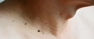
Keratomas can occur on any area of the skin. They can be single or form groups. Most often, keratomas are located on the neck, shoulders and arms. Less common on the lower body.
Currently, the reasons for the appearance of these formations on the skin are not fully understood. Nevertheless, scientists have proven that prolonged exposure to the sun and visiting a solarium promotes the growth of keratomas.
Where do keratomas most often form?
Keratomas can appear on any part of the body, as well as on the face. The tumor is a cosmetic defect. When squeezed by clothing and damaged, it becomes inflamed, bleeds, and provokes a secondary infection.
- Keratomas on the body Keratomas appear mainly on open areas of the body. They most often grow slowly, but can become injured and cause inflammation. Capillary keratoma on the leg or angiokeratoma is dark red or almost black in color. Outwardly, it resembles an irregularly shaped knot with unclear boundaries. Seborrheic keratomas on the arm are common, especially in patients over 50 years of age. The favorite location is the shoulders. In men, due to pronounced hair growth, keratomas appear on the hands more often. In this case, there is a dysfunction of the sebaceous glands with the formation of a large number of loose scales.
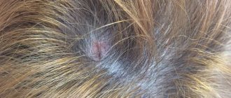
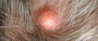
- Keratomas on the head Seborrheic keratomas form on the head. The disease begins with the appearance of yellow spots of small diameter. Gradually they become covered with scales, which grow and transform into crusts with cracks on the surface. Also, a follicular keratoma may appear on the head, resembling a flesh-colored nodule with a small depression in the center. Scales rarely appear on the surface of such a neoplasm. Magnetization of the tumor is unlikely.
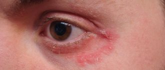
- Keratomas on the face Solar and horny keratomas grow on the face. They are considered precancerous skin hyperplasias. Solar keratoma of the eye begins with the formation of small pink papules that are prone to peeling. After some time, they turn into brown plaques with a characteristic rim along the periphery. Solar keratoma on the eyelids and face is capable of spontaneous magnetization. Much less common on the face is keratoma keratoma or cutaneous horn. Outwardly, it resembles a convex tumor that rises above the surface of the skin
Diagnostics
An appointment with a dermatologist begins with taking an anamnesis. The doctor clarifies how long ago the formation was, asks the patient about sensations - painful manifestations, itching, and studies the symptoms. Next, the skin is examined to determine the size and location of the keratoma. The instrumental method - dermatoscopy - allows you to clarify the diagnosis and exclude other skin diseases.
A special device allows you to identify the formation, examine the structure of its tissues, and the depth of its occurrence. The dermatoscope magnifies the image many times, so errors are excluded. And if there is a risk of the keratoma degenerating into a tumor, the doctor will determine this. In some cases, histology is recommended - the formation is sent to the laboratory to determine the cellular composition.
Methods for removing keratomas
There are several ways to remove keratomas. Dermatologists and oncodermatologists can use laser therapy, radiosurgery, surgical excision with a scalpel with a small capture of healthy tissue, electric knife, and cryodestruction. But laser treatment is considered the most effective method.
Benefits of using laser:
- bloodlessness;
- high safety of the method;
- excellent cosmetic effect;
- fast recovery.
Seborrheic keratomas
Seborrheic keratoma affects areas of the body with hair and may appear on the scalp. The top of the tumor is covered with loose scales. As the keratome grows, it becomes brown, thickens with the formation of small cracks, and may bleed.
Causes of keratoma formation
Keratomas have a multifactorial etiology. First of all, age-related degeneration of skin cells is important. In addition, the following factors are important:
- Excess ultraviolet radiation.
- Age-related decrease in immune defense. As a consequence, intensive growth of epidermal cells and the formation of areas of hyperkeratinization and keratinization occur.
- Neuroendocrine pathology.
- Vitamin A deficiency.
- Endocrine disorders and sexual involution (menopause).
- Long-term use of certain medications.
- Skin exposure to chemicals.
Where to remove keratoma
malignant skin keratoma can become malignant at any time. An effective and safe treatment method is laser removal. The skin keratoma is evaporated layer by layer, without affecting healthy tissue.
IN LASER REMOVAL CLINICS, experienced dermatologists and oncodermatologists treat skin tumors. After laser removal of a keratoma, a small crust forms, which falls off on its own after a few weeks. There will be new healthy skin under the scab. It is recommended to protect it from UV rays for several months using sunscreen.
Treatment
Treatment includes drug therapy and surgical removal. Drug therapy involves the local use of cytostatics or other antitumor drugs selected according to an individual regimen. Treatment may require hospital stay.
Surgical methods for removing keratomas are varied:
- Traditional excision within healthy tissue using a scalpel. Used in cases of suspected cancer.
- Curettage of the hair follicle - the keratoma is scraped out with special instruments with sharp edges.
- Laser removal - using laser radiation, the tumor is evaporated layer by layer. In its place, a crust remains, which disappears after healing, and “young” skin remains in its place.
- Electrocoagulation - the tumor is excised under the influence of high-frequency electric current. It leads to local heating of tissues and their evaporation. At this point, they separate and form a cut. After removal, a wound with a crust remains in place, healing occurs, as after exposure to a laser.
- Radio wave surgery is a non-contact method of removal using radio waves. A modern, effective method with minimal risk of the formation of rough scars.
- Cryodestruction is the effect of liquid nitrogen on the keratoma, which leads to its immediate freezing and necrosis. After a few days it disappears on its own.
Surgical removal is performed on an outpatient basis using local anesthesia. After the procedure, the patient can leave the clinic almost immediately.
Our clinic has all the necessary equipment for keratoma removal, and experienced specialists will offer treatment based on specific clinical features.
Book a consultation around the clock +7+7+78
Indications and contraindications for the procedure
A distinctive feature of keratomas among most skin tumors in humans is that not every tumor requires mandatory medical intervention and removal. Typically, doctors recommend that if a patient has neoplasms of any kind on the body, periodically visiting a dermatologist for preventive purposes, for example, once or twice a year. This measure is enough to prevent the transformation of benign tumors into malignant ones, or, if the process has already begun, to detect it in time.
As for the indications for laser removal of keratomas, the main one among them is keratoma, which is dangerous, that is, it has the likelihood of degenerating into a cancerous tumor. The main symptoms that should alert a person:
- peeling;
- itching;
- pain in the keratome;
- darkening of its surface;
- falling off of the keratoma, leaving a bleeding wound.
In such cases, you should immediately contact a dermatologist or oncologist.
It should be noted that laser removal, along with surgical and radio wave, is the most preferred method of treating malignant keratomas, in contrast to cryodestruction or electrocoagulation, since the latter do not provide adequate efficiency in removing cancer cells.
In addition, laser destruction can also be carried out in cases where the keratoma does not pose any threat to the patient’s health, but at the same time represents an obvious cosmetic defect, that is, removal can be prescribed at the request of the patient.
Contraindications to the procedure may be:
- pregnancy;
- general serious condition of the patient;
- acute infectious or inflammatory processes in the body;
- some psychiatric diseases.
If a person has an acute inflammatory or infectious lesion, it is necessary to first remove the aggravation, after which it will be possible to perform an operation to remove the keratoma.
Pregnant women are traditionally advised to avoid any interference with the body while they are carrying their baby. Of course, if the situation is critical and cannot be delayed, the procedure for removing a tumor with a laser can be prescribed as an exception, however, if possible, it is better to postpone it until the child is born.
Wound surface care techniques
After laser destruction of the keratoma, a characteristic dark brown crust forms in its place. It covers the surface of the wound, and underneath it the tissue healing process is actively occurring. It is strictly forbidden to rip off or comb the scab, as this opens the door for pathogenic microorganisms to enter the wound until the need for it disappears - then it disappears on its own. For the first two days, it is not recommended to wet the wound site.
Postoperative care includes the following rules: once or twice a day, the procedure site must be washed with water and baby soap, after which it is necessary to treat the surface with antiseptic drugs and healing ointments. Next, a sterile gauze bandage is applied to the wound. If possible, it is better not to use an adhesive plaster, as it restricts air flow into the wound, thereby slowing down its healing.
Complete skin restoration occurs after 4-6 weeks. During this entire time, it is prohibited to visit the bathhouse, sauna, or swim in pools or open reservoirs. Exposure to ultraviolet rays is also undesirable, and not only during postoperative recovery, but also in the next 6-8 months, in order to avoid recurrence of keratoma, so it is better to forget about beaches and solariums for a while, and go outside in clothes or a hat that covers the affected area and treat it with sunscreen with a high UV filter.
Possible complications and consequences of laser intervention
Laser surgery of neoplasms can leave scars - small and unnoticeable, or larger ones if the keratoma was initially large. In addition, patients note the appearance of numbness in the tissue at the site where the tumor was removed.
Such manifestations are natural consequences of the procedure.
It is necessary to pay attention to some dangerous symptoms of poorly carried out destruction of the tumor, for example, the appearance of redness around the crust formed after removal, severe itching or rash at the site of the keratoma.
If the crust has fallen off on its own, and underneath it an ulcer in the form of a red spot with an uneven surface is found that has not healed for a long time, you need to visit a dermatologist as soon as possible.
In addition, allergic reactions or dermatitis may develop around the procedure site due to constant wearing of a bandage, as well as the use of various ointments on the skin. At the first manifestations of dermatitis or allergic rashes, you must stop treating the area with any medications and also consult a dermatologist.