Dermatoscopy is an instrumental non-invasive study in which the doctor examines moles and other formations on the skin using a special device that creates multiple magnification. In oncology, dermatoscopy helps to distinguish harmless formations from skin cancer and identify melanoma in the early stages.
- Indications for dermatoscopy
- Why are moles dangerous?
- How is dermatoscopy performed?
- Equipment for dermatoscopy at Euroonco
- How to assess the malignancy of a mole
- Advantages of dermatoscopy using the FotoFinder system
- Prices for dermatoscopy at Euroonko
Indications for dermatoscopy
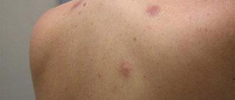
- the appearance of any red, brown or black formation on the skin;
- injury to a mole; uneven change in its color;
- an increase in the total number of moles and age spots;
- itching and tingling in the area of education;
- increase in size of the mole.
For preventive purposes, dermatoscopy is recommended:
- fair-skinned people with moles and age spots;
- with a family history of melanoma (if it was diagnosed in close relatives);
- people who have many moles and freckles on their body;
- when taking oral contraceptives for more than 1 year;
- when working in hazardous industries;
- with regular trips to hot countries;
- For dysplastic nevi, it is recommended to undergo dermatoscopy every six months to a year.
No special preparation is required for dermatoscopy. It is not recommended to use cosmetics or topical medications on the day of the examination. To obtain a clearer image, a small amount of gel is sometimes applied to the test site to reduce the reflection of light from the surface of the skin.
Modern digital dermatoscopes have many advantages over conventional ones. The device tube is connected to a computer monitor, on which the image is displayed. Digital dermatoscopy serves as a monitoring method for patients at high risk for developing melanoma. It also allows you to map moles over the entire surface of the body and observe their changes in dynamics, comparing the results with previous ones. The technique of digital dermatoscopy is simple, safe and fully automated. Within 3 minutes you can get a detailed analysis of all neoplasms.
It should be remembered that dermatoscopy does not allow a definitive diagnosis of melanoma; this requires a biopsy and histological examination of a tissue sample. [1,3]
When to remove a mole
Education is removed in the following cases:
- studies showed the beginning of cell degeneration and atypia;
- the mole is in the way or is injured;
- there is a cosmetic defect;
- the mole hurts, itches, becomes inflamed and grows;
- studies showed a suspicious dysplastic nevus, and the patient had a family history of melanoma.
It is pointless to cut out all moles, because each such impact has an impact on the body. In one session, a maximum of five large or eight small formations are eliminated.
Early detection and removal of melanomas saves the lives of patients in 80% of cases. In later stages, survival does not exceed 20–50%, depending on the type of tumor. Therefore, it is better to play it safe once again - if you notice a suspicious mole on yourself, immediately go to a dermatologist!
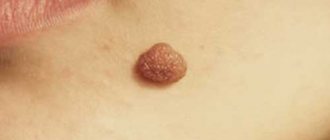
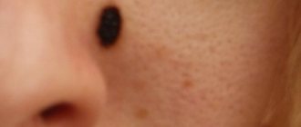
Why are moles dangerous?
Moles (pigmented nevi) are benign neoplasms, but some of them can transform into an aggressive, dangerous malignant tumor - melanoma. Risks are increased in the following conditions:
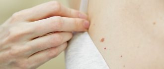
- A large number of moles on the body.
- Dysplastic nevi are special moles that are large in size, uneven edges, and uneven in color.
- Congenital melanocytic nevi are moles that are present on a child’s body from birth. Usually children are born without moles - they appear throughout life. The most dangerous are giant congenital nevi with a diameter of more than 10 cm. They degenerate into melanoma with a probability of 30%.
Important to know: Scientific research shows that only 30% of melanomas develop from pre-existing pigmented nevi. In 70% of cases, a malignant tumor occurs on unchanged skin, where there were no moles. [2]
Melanoma symptoms
There are five types of melanoma, differing in appearance, growth rate and metastasis:
- Superficial. 70% of cases of the disease belong to this form. The tumor grows in the upper layer of the skin and does not metastasize for a long time. The cancer appears as a flat spot with uneven borders and heterogeneous coloring, most often occurring on the lower half of the body, legs and back.
- Lentigo maligna is similar to superficial melanoma. Developing for a long time in the upper layers of the skin, the tumor does not metastasize. Forms on the ears, face and upper half of the body. When it grows deep into the skin it becomes lentigo-melanoma.
- Acral lentigenic melanoma appears as black or dark brown spots on the palms, soles, and under the nails. The disease progresses, growing deep into the skin layers.
- The nodular form is extremely malignant, grows upward, metastasizes early and leads to death after 6–18 months. This dangerous type accounts for 10–15% of cases of the disease.
- Amelanotic melanoma is light pink or flesh-colored. This is a type of nodular form that, due to its unusual color and rarity (less than 5% of cases of the disease), is difficult to diagnose. Due to aggressive growth and metastasis, most cases of the disease have an unfavorable outcome.
How is dermatoscopy performed?
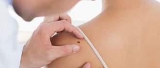
Classic dermatoscopy is carried out using a special instrument resembling a magnifying glass - a dermatoscope. Using it, the doctor examines the skin, assesses the size and appearance of the detected tumors.
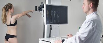
A more modern technique, which is used at Euroonco, is videodermatoscopy using the German PhotoFinder. The device takes pictures of the entire surface of the patient's skin, creates a “mole map” and stores the images in a computer. The procedure is absolutely painless. [3]
Hardware diagnostics of melanoma
The doctor examines suspicious moles using a special device - a dermatoscope. This device provides a tenfold magnification, which allows you to examine the skin area in detail, identifying pigment inclusions and heterogeneity of the structure.
With confocal laser scanning microscopy (CLSM), the doctor takes images of “slices” of the skin without damaging it. It is a painless and quick alternative to a biopsy. The result is confirmed in 97% of cases.
A biopsy for suspected melanoma is rarely used, as it can provoke the degeneration and growth of malignant cells. The method is more often used on distant tumors. When the diagnosis is confirmed, the nearest lymph nodes are punctured.
Equipment for dermatoscopy at Euroonco
At Euroonko, dermatoscopy is performed using a modern PhotoFinder installation from the German company FotoFinder Systems GmbH. This manufacturer has been producing high-tech turnkey solutions for visualizing skin tumors and image analysis for more than 20 years. The device takes pictures of the entire surface of the body and loads them into a computer, where they are saved and processed by a special program.
How to assess the malignancy of a mole
During dermatoscopy, a number of characteristics are assessed, such as size, its elevation above the skin, shape, symmetry, state of the borders, color, nature of the surface, and the presence of ulcerations. However, the assessment of these indicators is quite subjective. In this regard, various methods for determining the malignancy of a neoplasm have been developed. The most commonly used are the following:
- Scale of 3 signs. Asymmetry, the typical pigment network and the presence of blue-white structures are assessed.
- Scale of 7 signs (G. Argenziano). The main criteria (atypical pigment network and vascular pattern, blue-white veil) and additional ones (atypical branches, pigmentation, spots, as well as areas of regression) are assessed.
- 11 signs scale (S.Menzies). Within this system, positive criteria are identified (black dots along the edge of the formation, a blue-white veil, many brown dots, the presence of more than 5 colors in the formation, many blue and gray dots, etc.), as well as negative ones (symmetry of shape, the presence of one color ).
- ABCD system (W. Stolz). This algorithm takes into account asymmetry, unevenness and clarity of boundaries and color, as well as the presence of dermoscopic structures.
Each of the described systems has demonstrated high practical significance and has its own counting algorithm. For the presence of one or another attribute, a certain number of points are added. If their summation exceeds the threshold value, the neoplasm is considered suspicious, it is biopsied or immediately removed as a malignant neoplasm. [4]
In the PhotoFinder system, a computer program automatically analyzes images, thereby achieving greater diagnostic accuracy.
"Pearls" on the Internet.
We will not analyze in detail the “pearls” that can be found on the Internet on the topic of dermatoscopy.
Just follow the first link for this request:
The author suggests that we urgently do dermatoscopy immediately after traumatization of a mole. In order not to waste precious time, suddenly the mole has already become malignant...
The article has a signature, but it is impossible to understand from it whether the doctor wrote it or not.
I am sure that any oncologist will agree with me - a person who gives such recommendations, at best, is far from diagnosing skin cancer. At worst, it has nothing to do with medicine at all.
Very often I have to deal with manifestations of depression in people who have read such publications.
But we only examined one of them...
Advantages of dermatoscopy using the FotoFinder system
- The diagnostic accuracy is 99%.
- While a dermatologist using a conventional dermatoscope does not examine the entire surface of the skin and may miss pathological neoplasms in hard-to-reach places, the FotoFinder system provides the most complete scanning of the body surface.
- The ability to detect melanoma and other small skin tumors in the early stages.
- If videodermatoscopy is performed regularly, the doctor is able to assess the condition of the skin over time, and this further increases the accuracy of diagnosis.
- The system can automatically analyze images.
- The study is completely safe, painless, and has virtually no contraindications. [5,6]
Why should the removal of skin tumors be performed by an oncologist?
Moles, nevi, warts, papillomas and other neoplasms often become an unpleasant cosmetic defect. Removing undesirable effects on the skin can solve the problem not only from an aesthetic point of view, but also prevent a malignant disease. However, in order for the operation to proceed without consequences, it is necessary to contact an oncologist.
Moles and other neoplasms can pose a potential health hazard from an oncological point of view. Therefore, they cannot be removed in the office of a regular surgeon, dermatologist or cosmetologist. Only related specialists—oncologist surgeon and dermatologist-oncologist—can accurately distinguish “good” from “evil.” At the same time, excision of tumors is carried out according to certain rules and strict indications.
Patients often come to us with benign formations - viral warts, raised moles, nevi, pedunculated papillomas. Primary consultation is provided by oncologists of two directions - a surgeon or a dermatologist. They examine not only the “problem” area on the skin, but also the patient completely. By the way, this is also an important aspect - an ordinary surgeon, cosmetologist or dermatologist may not pay close attention to all the patient’s skin tumors. Recently a woman came to me with a traumatized nevus, and upon examination she discovered melanoma on her eye.
After the examination, the doctor may prescribe cytology - a test for the presence of cells with signs of malignant degeneration. If there are no malignant cells, then removal can be done using a laser or radio wave device. If the result shows “evil,” then the oncologist surgeon excises the formation within healthy tissue and sends the material for histological examination to his own laboratory of the National Medical Research Center of Oncology named after. N. N. Petrova.
An ordinary cosmetologist does not conduct a cytological examination, which can lead to dire consequences. Sometimes patients come to us who have had a tumor excised incorrectly or incompletely and have experienced a relapse. And this is an additional risk of cancer.
There is no need to be afraid of the oncologist's office - now specialists can help the patient in one-day surgery, easily and painlessly fix the problem and provide qualified assistance.
Prices for dermatoscopy at Euroonko
- Initial consultation with a clinical oncodermatologist—RUB 5,100.
- Consultation with professor, doctor of medical sciences — 10,500 rub.
- Examination of the skin under magnification (dermatoscopy) - 4,600 rubles.
- Screening using FotoFinder - 13,400 rub.
Book a consultation 24 hours a day
+7+7+78
Bibliography:
- Bakulev A. L., Konopatskova O. M., Stanchina Yu. V. Dermatoscopy in the diagnosis of pigmented skin nevi. Bulletin of dermatology and venereology. 2019;95(4):48–56. https://doi.org/10.25208/0042-4609-2019-95-4-48-56
- V.G. Polyakov, R.V. Shishkov. Clinical guidelines for the diagnosis and treatment of melanoma in children and adolescents. All-Russian Union of Public Associations. Association of Oncologists of Russia.
- Recommendations for performing dermatoscopy of skin tumors, protocol for dermoscopic examination: a textbook for doctors. - Ekaterinburg: SV - 96, 2022. - 23 p.
- Korovin S.I., Litus A.I., Litvinenko B.V., Kukushkina M.N., Palivets A.Yu. Dermatoscopy of skin melanoma, applied significance and prospects. Clinical Oncology, No. 8 (4), 2012
- Julia K. Winkler, Katharina Sies, Christine Fink. Melanoma recognition by a deep learning convolutional neural network – Performance in different melanoma subtypes and localisations. European Journal of Cancer 127 (2020) 21-29 https://doi.org/10.1016/j.ejca.2019.11.020
- D.V. Sokolov, N.N. Potakaev, L.V. Demidov. Experience in automatic recognition of skin melanoma based on digital epiluminescence dermatoscopy. Clinical dermatology and venereology 3, 2010.
Self-examination
It is useful for people of any age to regularly examine their skin for new growths. Of course, it is not necessary to spend a lot of time and run to doctors with every mole that you find. But there are some symptoms that will help you pay attention to changes occurring in the tissues when examining moles. A mole that is not yet going to turn into melanoma:
- symmetrical;
- has clear outlines;
- evenly colored;
- retains its size, does not increase or decrease;
- It does not itch and does not cause any discomfort.
Accordingly, if the nevus begins to change somehow (be it a blurring of the border or a change in color), itches, hurts or causes any other unpleasant sensations, then it’s time to consult a doctor - this is a mole that needs to be observed professionally. Remember that a healthy organ does not remind you of itself.