Pigment spots on the face and body for a woman often become a cause of psychological discomfort and self-doubt. But in modern cosmetology there are many effective methods for treating age spots that help a woman save her nerves and preserve her image.
Types of age spots
There are several types of age spots:
- Freckles are the most common and safest age spots; they arise due to the inability of a young body to distribute sun tan evenly throughout the skin. During the cold season they disappear and appear again in the spring. With age, the sun tan usually begins to spread evenly and freckles disappear completely.
- Lentigines are age spots on the face, arms, back and shoulders. They appear after 40 years, as a rule, in those places where there were freckles in youth. They reveal their real age, which is why women try to get rid of them.
- Melasma (chloasma) is pigmentation during pregnancy, which manifests itself as a result of hormonal imbalance in the body as a result of bearing a child. Once the hormonal balance is restored, the spots disappear on their own.
- Birthmarks (moles) are age spots that can be congenital or acquired with age. The reason for their appearance is a disruption in the functioning of the skin and the accumulation in one place of cells overflowing with pigment. Moles are considered benign tumors, which under unfavorable circumstances can become malignant.
- Vitiligo is pinkish or milky-white pigment spots that can form at any age. Their exact origin has not yet been clarified by science; it is believed that their appearance is provoked by a failure of the endocrine system.
Why do brown spots appear?
Brown spots on the skin are a result of increased production of melanin in the skin of the body. When the level of melanocytes increases, it leads to pigmentation disorders. This reaction is usually caused by an imbalance in metabolic processes and contributes to the development of skin disease.
An imbalance of pigments is associated not only with internal abnormalities in the functioning of organs, but is also provoked by stress and environmental factors.
The defect is caused by the following reasons:
- Intensive production of melanin under the influence of ultraviolet radiation, especially in fair-skinned people.
- Hormonal disorders, pregnancy, PMS, childbirth. Areas of skin may darken during pregnancy in the area of the nasolabial folds on the face. The deviation is caused by hormonal changes in the body and usually disappears after childbirth in a few months.
- Genetic factor (hereditary transmission).
- Age-related changes associated with deterioration of body systems, decreased tissue regeneration, and slower metabolism.
- Stress and depression disrupt the production of hormones and cause pigmentation.
- Chemical and thermal burns, wounds and blisters leave a brown mark on the body.
- Advanced form of acne, boils.
- Deficiency of microelements and vitamins (especially vitamin C, copper).
- Uncontrolled use of hormonal medications.
- Cosmetic procedures including acid peels, Botox, hyaluron, chemical cleansing, mesotherapy. Activities can injure the skin layer and stimulate increased melanin production.
- Diseases of the liver, stomach, kidneys, gallbladder, adrenal glands.
- Allergic reactions to cosmetics, certain foods.
Skin mastocytosis
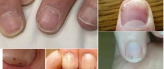
With such a lesion, spots with black dots appear on the person’s body. The disease occurs due to the pathological proliferation of mast cells (they are responsible for the state of human health) and their accumulation in large numbers in the epithelium. Doctors divide the disease into a skin form, in which dark spots, nodules and spider veins appear on the human body, and a systemic form (the spots spread to internal organs).
Mastocytosis in a child occurs in the first years of his life. The most common form of mastocytosis is cutaneous. The disease goes away on its own as you grow older.
In elderly and adult people, the disease spreads not only to the skin, but also to internal organs (heart, kidneys, liver, spleen).
There are the following types of disease:
- Maculopapular - multiple dark rashes appear on the skin, which, when scratched, turn into blisters and hives. This type of mastocytosis is also called urticaria pigmentosa.
- Nodal form. With this condition, a person is diagnosed with small blisters ranging in size from 7 to 10 millimeters. They can be pink or light brown, often merge and form large plaques.
- Solitary form. In this case, one large dark spot with a size of 5 to 6 centimeters appears on the body. It is usually localized on the shoulders, abdomen, back and neck. If you accidentally touch the lesion, it will turn into a blister and cause severe itching.
- Erythroderma - yellow-brown dense spots. They have no boundaries, are easily deformed and provoke the formation of cracks and ulcers. Most often, the spots spread to the buttock folds and hollows.
- Telangiectasia is a large number of dark spider veins in the neck and chest. In most cases, such formations are formed in women.
Treatment of any type of mastocytosis should be comprehensive and include the use of hormonal agents, cytostatics, antioxidants and antiallergic medications. If black spots appear on the body in a single quantity, then it can be eliminated surgically.
Where can brown spots be located?
Pigmented areas can be located throughout the body. The nature of the disease depends on the location of the defect.
Localization of spots is as follows:
- Face and area around the nose, chin and mouth. It manifests itself mainly in women experiencing reproductive disorders.
- Knee joints, toes. An indicator of thyroid imbalance. More often seen in men.
- Area of the palms, arms and wrists. Caused by senile changes in which metabolism is disrupted. The marks often have a bluish tint.
- Neck and shoulder area. At a young age, the pigment appears as a consequence of tanning; at an older age, after the age of 45, it indicates lentigo.
What are pigment spots on the back?
Such formations on the back are areas on the skin of various shapes and sizes. Flaws are rarely single, often multiple. Outwardly, it looks like a large number of minor formations or one continuous spot.
The reasons for the appearance of spots of different pigmentation on the back are usually heredity, hormonal and immune disorders, allergies, excessive ultraviolet radiation, and vitamin deficiency.
Diagnosis is carried out by a dermatologist to determine the cause of the spots.
If the need arises, the stains are removed using cosmetic procedures: cryotherapy, laser therapy, mesotherapy, peelings; Keratolytics are used locally.
Their shade is dark or light, like a discolored area, with a smooth or rough surface. To exclude any adverse effects, you should consult your doctor.
Safe Types of Brown Spots
Formations of a brownish tint can be benign; such spots are not dangerous to health.
The category of safe pigmentation includes:
- Lentigo. The pigment spot resembles a mole, is a benign formation, and is located in the area of the legs, legs, upper torso and face. Pathological changes appear at different ages, the disease has a slow pace of development. It is extremely rare for a spot to degenerate into a malignant tumor. Provoking factors: sun rays, AIDS, hormonal imbalance. The spot has a uniform color with clear boundaries. Children during prenatal development are most susceptible to the disease, but adults can also get sick.
- Freckles. Light brown marks are more often observed in the face area. This type of pigmentation is associated with hereditary transmission, as well as with the intensity of sunlight during spring and summer.
- Chloasma. Brown areas vary in color and size. Chloasma varies in appearance depending on its location on the torso, chest, face or genitals. Formations are more often manifested in women as a sign of sexual disharmony, ovarian failure, menopause or reactions to pregnancy.
- Moles. They can appear from birth or during life. Localization of spots occurs throughout the body. Dark areas can be smooth or rough, merge with the general skin or protrude above it.
Seborrheic warts
Brown or black raised lesions that appear on any part of the skin. They are quite easy to recognize: their surface is loose, and the base is relatively small - it looks as if the wart is stuck to the skin.
In the early stages of development, a wart can be confused with a mole, but over time it noticeably changes its color and size.
The number of warts also increases with age. From a medical point of view, they are absolutely harmless, but for aesthetic reasons they can be removed.
Other types of hyperpigmentation
After the age of 25, the skin of the eyelids in women may become hyperpigmented. Pathological manifestations are associated with the action of sunlight or hormonal imbalance, the cause of which is improper functioning of the thyroid gland, adrenal glands, ovaries, or poor genetics.
Hyperpigmentation of the eyelids can result from improper use of oral contraceptives, a certain category of medications, or the use of cosmetics containing citrus fruits.
Secondary hyperpigmentation can appear after suffering from neurodermatitis, lichen, inflammatory skin processes, which include burns, ulcers, and secondary syphilis.
How to recognize dangerous brown spots on the skin.
At a more mature age, you can see senile manifestations of lentigo. Spots of this type are characterized by round, oval or irregular shapes. The color of the pigment ranges from light to dark brown. The manifestation is caused by a decrease in the number of melanocytes.
It is believed that their level decreases by 8% every decade after the age of 30. Another reason is due to a decrease in the supply of pigment to cells. Sunlight activates the density of pigment cells, which manifests itself as photoaging of the skin.
Freckles on the skin
Almost every person has a certain number of freckles on their body. Sometimes they spread to the hands, back, and chest. Freckled rash appears in humans as a result of heredity. It is formed due to the fact that melanin in the skin is distributed unevenly.
The peak prevalence of freckles on the human body occurs in the spring and summer. Freckles are numerous small rashes (size 2 to 3 millimeters). Their color ranges from light yellow to dark brown.
The freckled rash may become darker when exposed to sunlight. Experts believe that the skin of people who are susceptible to the formation of hemp is particularly sensitive.
Freckles do not need treatment. Many people even like such rashes on the body. If a person wants to eliminate them, then he can use a specialized whitening cream. To prevent the appearance of rashes, it is important to have the right diet, start taking a complex of vitamins, and also protect your skin from the rays of the sun.
What diseases cause brown spots?
Brown spots on the skin may be a sign of internal organ disease. The greatest imbalance of pigment is caused by liver diseases due to cirrhosis or other serious pathologies.
A significant proportion is caused by endocrine disorders that affect metabolic processes (hormonal imbalance, diabetes mellitus). Diseases such as tuberculosis provoke a cachectic form of melanosis. Kidney dysfunction can cause melanosis of the uremic type.
The following types of pigmentation are common:
- Toxic melanosis. Brown formations occur due to close and prolonged interaction with a chemical substance - coal, oil, oil. Inattention to this moment leads to pigmentation and malfunction of the internal systems of the body. As a result, chronic ailments develop.
- Becker's nevus. Formations on the body are presented as small yellow-brown spots with jagged edges. Localization occurs on the limbs and upper body. The disease most often affects males during puberty from 10 to 15 years. The cause of the spots has not been fully studied. Experts agree that the manifestations are caused by surges in hormones.
- Arsenic form of melanosis. The development of brown spots is associated with taking medications containing arsenic elements. Usually present in people working in manufacturing who have close contact with the chemical.
- Dubreuil's melanosis. Darkening of small areas of skin with jagged edges is an oncological manifestation and indicates the development of skin cancer. Neoplasms are located in the upper part of the body, in the initial stage they look like a mole, slightly protruding above the skin. In a short period of time, the spot grows to global proportions, resembling a geographical map. The color of the formations varies from light yellow to deep brown. Over time, the spot thickens, nodules and papules appear on the surface. Nearby skin turns red and begins to itch. At the final stage, the spot peels off, adjacent tissues acquire brown marks similar to freckles, as evidenced by the degeneration of the affected areas into a malignant form.
- Acanthosis nigricans. Characterized by the formation of black spots on the body. The disease is rare and can have a malignant or benign form. You can see akanatosis nigricans in places where there are folds - armpits, groin area, neck, buttocks. The causes of development may include hormonal drugs, thyroid dysfunction, and genetic inheritance.
Diagnostics
A dermatologist provides consultations on skin diseases. When examining pigmentation, he will pay attention to details such as:
- size of education;
- surface of the stain (smooth, rough, etc.);
- color intensity;
- number of rashes and location;
- accompanying symptoms (itching, peeling, etc.).
This will help the doctor make a preliminary conclusion about what it might be. For a more accurate diagnosis, you will need to take blood tests - general and hormone tests, and possibly do an ultrasound of the liver and thyroid gland. To examine the nature of brown spots on the body, a dermatoscope will be used and scrapings will be taken from the surface of the affected areas. If necessary, the dermatologist will prescribe the patient a consultation with an endocrinologist, gastroenterologist, gynecologist and other specialists.
Brown spots in a child
The skin of children has a thin stratum corneum, and the tissues are intensely saturated with blood vessels. All this, combined with a weak immune system, can lead to the formation of pigmented areas on the body. Hyperpigmentation often manifests itself as freckles, nevi (giant, flaming, melanocytic, non-cellular), in the form of hemangioma, congenital coffee spots.
Brown spots on the skin are caused by a hereditary form of the disease.
These types include:
- Neurofibromatosis type 1. Initially, café-au-lait spots are small in size, on the surface of which a cluster of freckles forms. The disease often occurs at an early age. The neglected process leads to oncological complications, on the basis of which diseases develop: respiratory tract cysts, growth retardation, formation of voids in the spinal cord, narrowing of the renal vessels, enlarged mammary glands.
- Peutz-Jeghers disease. Pigmentation of this type affects large areas of the body, mucous membranes and leads to polyposis of internal organs. The formations are localized around the eyes, around the lips, in the anus and palms.
- Poikiloderma. Hereditary spots are observed in infants from 3 to 5 months. Initially, hyperemia occurs in the limbs, face, and neck. Then atrophic pigment changes form on the affected area.
- Urticaria pigmentosa. This form of pigmentation can trigger the development of mastocytosis. A small dark red spot leads to the formation of vesicular inflammations filled with fluid. At the completion stage, the bubbles open, and in their place brown areas remain, which disappear after a few months. If left untreated, urticaria can cause disability or death. Pathology can be triggered by climatic conditions, stress, exposure to ultraviolet radiation, weak immunity, infectious and inflammatory processes.
Older children are susceptible to mastocytosis, allergic pigmentation, intradermal and borderline pigmentary nevus, as well as impaired melanin production (hypomelanosis). Deviations can manifest themselves in the form of diseases: albinism, vitiligo, Ito hypomelanosis.
A diagnostic examination is carried out using a dermatoscope, pigmentation is examined under ultraviolet radiation (Wood's lamp). In serious cases, computer diagnostics and biopsy tissue analysis are performed, followed by histological examination.
Treatment of pigment spots in children does not involve the use of lightening and whitening preparations containing hormonal components. At the age of 6-7 years, it is allowed to treat pigmented areas with parsley, lemon, and cucumber juice. If removal of the formation is required, then surgical method, laser therapy, electrocoagulation or cryotherapy are used.
Treatment methods
If the spots are red or pink, then the cause is an internal infection. In this case, medications are prescribed that contain keratolytics and fungicides. Efficiency differs:
- Clotrimazole - taken three times a day;
- ten percent sulfur ointment - applied twice a day to the affected skin;
- ointment with salicylic acid - used as a replacement for sulfuric ointment.
We recommend reading
- Treatment with folk remedies for age spots on the face
- The danger of pigment spots on the skin
- The effectiveness of Melanativ cream against age spots
Freckles can be removed with cosmetic procedures in the salon. The accumulation of pigments can be observed in different layers of the skin: in the epidermis (upper layer) and dermis (deep layer). Cosmetologists recommend removing only those pigments that are located on the surface of the skin. Treatment methods:
- chemical peeling – carried out using acids. This method lightens pigments and cleanses the epidermis of the stratum corneum. Peeling is recommended to be done in winter, as the skin becomes too sensitive to light;
- photo-treatment – removal of pigments using light rays. Photorejuvenation is painless, but the skin takes a long time to recover;
- cryomethod – treatment of freckles with liquid nitrogen;
- laser – reduces the brightness of pigments gradually. The use of laser beams is the safest.
In addition, doctors recommend an integrated approach that uses:
- professional procedures;
- use of creams with lactic acid;
- proper nutrition - eating fresh fruits and vegetables, a balanced supply of all necessary substances to the body;
- traditional methods of treatment - the use of masks with lemon juice, parsley and fermented milk products.
Hyperpigmentation due to fungal disease
Brown spots on the skin can be caused by a fungus. Neoplasms can affect any part of the body, depending on the type of fungal disease.
The following types are distinguished:
- Keratomycosis. Small pigmentation caused by a fungus (lichen versicolor, Piedra's disease). In the case of deprivation, in the normal state of the body, neoplasms are not dangerous. Piedra's disease is localized in the head and throughout the body, becoming chronic.
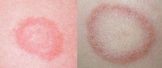
- Trichophytosis. The fungus is transmitted through animals and contact with infected people. There are several forms - superficial (pink spot, peels off, becomes crusty), infiltrative (the rash contains lymphatic fluid with the presence of blood), suppurative (located on open areas of the body).
- Mycosis. Pigmentation begins from the feet, hands, and crotches of the fingers. Sometimes the fungus spreads to the head and face area.
- Candidiasis. Pathological changes develop not only on the body, but also on the mucous membranes and internal organs. The symptom is manifested by burning and dryness of the skin layer.
Fungal pigmentation spots are light brown in color and may crack and peel.
Types of pigmentation and age spots
Dermatologists distinguish two types of pigment disorders:
- Type 1: Cutaneous, affects the top layer of skin.
- Second type: Epidermal, disorders in the deep layers of the skin.
The first type of pigmentation is much easier to solve. With proper treatment, the marks disappear after a couple of months.
Freckles
These spots are completely harmless. This is an aesthetic flaw that many even like. They most often appear on light skin under the influence of ultraviolet radiation. Freckles appear on the surface of the skin and are easily removed using cosmetic procedures (for example, chemical peeling). How many of these procedures will be needed depends on the condition of the client’s skin.
Nevi
Moles, called nevi, are highly pigmented areas associated with benign growths on the skin. These congenital spots are divided into two categories:
- Limited focal dysplasia is a disorder of peripheral nerve development. It is characterized by a change in the appearance and color of the dermis around the problem area.
- Focal proliferation of melanin cells is benign (nevoid tumor).
It is important to monitor moles that are located in areas of constant friction (back, groin, armpits) or on open areas of the body. Doctors advise removing moles from such places in a timely manner so as not to damage them. If you experience changes in the area of the spot, you should immediately consult a doctor.
If you are watching:
- Soreness
- Color change
- The appearance of satellites (moles around the “mother”)
- Changes in the structure of the educational sector
- Bleeding; Increase in size
- Itching
These symptoms indicate the beginning of degeneration. Only an oncologist can decide whether to remove moles or not; touching some of them is strictly prohibited. Burning moles with anything at home or in a beauty salon is strictly prohibited. Such actions can cause ultra-rapid tumor growth. All actions with moles are carried out only after receiving the biopsy result. And only an oncologist surgeon can perform such operations.
Chloasma
This type of pigmentation is most typical for women. Chloasma is uneven growths. Chloasma can affect most of the body and almost blend into the skin tone. Most often, such darkening is concentrated on the surface of the face, on the neck or ears. They very rarely reach the forearms and chest.
Chloasma appears against the background of hormonal disorders in women:
- While taking hormonal medications
- In the early days of the cycle
- During menopause
- While pregnant or breastfeeding
In most cases, such defects disappear on their own after the birth of a child or after completing a hormonal course of therapy.
Melasma
It develops according to disorders similar to chloasma - against the background of hormonal imbalances in the body. Uneven grayish or dark brown, usually located symmetrically to each other. The defect increases under the influence of ultraviolet radiation.
Diseases are caused by the following disturbance factors:
- thyroid gland
- gastrointestinal tract
- liver
- gonads
- pituitary gland
In women, spots disappear when hormone levels in the body normalize. In men they rarely turn pale because... are formed in youth.
Lentigo
This is age-related pigmentation , but it can appear by the age of forty. Lentigines are round spots of dark brown or tan color that protrude above the surface. The manifestation is associated with a natural slowdown in metabolism. In females, such pigmentation is aggravated due to hormonal imbalance during menopause.
They appear on:
- Face
- Brushes
- Neckline areas
- Upper back
Vitiligo
This is a disease, the main indicator of which is white pigment spots on the body of different sizes. It most often occurs in young people under the age of 20, but can appear at any age. These spots appear on different parts of the body.
This pathology also disrupts hair pigmentation. The system of development of this disease has not been fully studied, but the cause of this depigmentation has been identified (death or cessation of functioning of cells that produce melanin).
Doctors have identified three types of such pigmentation:
- Generalized is when the white spots are located symmetrically to each other (for example, on two arms or two legs);
- Segmental - affects only one part of the body. Usually appears in young people, progresses in the first year, then stops spreading;
- Localized - manifests itself in single fragments on the body.
Color variation begins on exposed skin on the arms, legs and face and gradually covers the rest of the body. The speed of this process cannot be predicted. Sometimes the spots stop appearing on their own without any treatment and even disappear. But in most cases they cover almost the entire surface of the body. This type of pigmentation develops in people of any skin color, but discolored spots are more noticeable on dark skin.
This disease is not contagious, does not harm health, but significantly reduces the quality of life due to the development of complexes in humans.
Brown spots due to liver disease
Brown formations in the facial area may indicate liver disease. Hepatic pigmentation (chloasma) indicates chronic disturbances in the functioning of the organ. The spots are localized in the cheek area with a transition closer to the neck and do not have clear boundaries. Another important indicator is the vascular network.
Liver spots are located not only on the face, but in the entire body in the form of a rash, red and yellowish spots, plaques, and stars. But more often the defect is localized in the neck, arms, shoulders, and face.
Brown pigments appear on the skin with age-related changes after 40 years. But if there are too many spots, and their color has become grayish or bronze, this may be a signal of an unhealthy liver.
Preventive actions
To prevent the development of spots on the body, the following recommendations should be followed:
- in sunny weather, it is mandatory to wear a hat;
- eat foods with plenty of vitamin C, and also limit your intake of vitamin A;
- Before going outside, you should use sunscreen;
- wash your face with fermented milk products (kefir, yogurt).
Burning and itching during pigmentation indicates liver disease. When scratching the affected area of the body for a long time, the skin becomes yellow. Eliminating the root cause of dark spots will help get rid of the problem and prevent metastasis.
In case of adrenal gland dysfunction
Improper functioning of the adrenal glands provokes the development of excess pigmentation. Changes occur due to the production of certain hormones. There are different types of hyperpigmentation of this type, which include: primary (congenital or acquired), secondary (post-infectious), local and widespread type.
Pigmented formations caused by improper functioning of the adrenal glands can be represented by:
- Bronze tint of the skin and mucous membranes. The pathology is often caused by Addison's disease. At first, only open areas of the skin are affected, then the entire body.
- Chloasma. The symptom often appears during pregnancy and is called the “mask of pregnancy.”
- Vitiligo. Predominance of lightened skin lesions.
- Itsenko-Cushing syndrome. Spread of red streaks like stretch marks in the thighs and abdomen.
Causes
The smallest disruptions in the functioning of internal organs can affect a person’s condition and appearance. Pigment spots of red, pink or black color can form on the back under the influence of various circumstances.
Pigmentation changes due to improper care of the skin after sunbathing, in particular in the abdomen and back, chest and shoulders.
After a rash, various marks also form on the back. The most popular provoking factors are:
- Genetic and hereditary disposition leads to disorders in the production of melanin, which, in particular, is characteristic of people with fair skin, who are very susceptible to ultraviolet radiation.
- rashes in childhood and any traumatic procedures (scratches, aggressive influence of cosmetic products), which provokes the loss of the skin’s ability to regenerate.
- Hormonal and immune disorders, as well as hidden diseases of the body (diseases of the thyroid gland, liver, intestines). In addition, prolonged use of antibiotics or oral contraception can be a factor in hormonal imbalance.
- Allergy of the body.
- Excessive use of ultraviolet treatments.
- Skin diseases .
- Age-related changes within the body.
- Lack of vitamins, in particular C, A and group B.
Having established the root cause of pigmentation, it is possible to select effective therapy for the disease.
How to treat brown spots
It is not always possible to cure pigmentation with medication, but modern means can make the spots less noticeable. You can purchase several types of whitening preparations at the pharmacy, some of them are presented in the table.
| Type | Name |
| Antifungal agents | Sulfur or zinc ointment, Clotrimazole |
| Antibacterial agents | Salicylic and syntomycin ointment |
| Exfoliates and reduces melanocyte production | Salicylic-zinc paste, folic, azelaic, kojenic acid. |
| Cream | Melanativ, Achromin |
| Cosmetical tools | Bodyaga forte, Boro plus cream, Clearwin, Biocon cream, Vitex mask. |
The desired result can be achieved by using the listed remedies for about 4-8 weeks.
Symptoms
Pigmented spots on the back are skin formations that have a brown tint. The size of the spots may vary. In some cases, a continuous pigment spot is formed, and in other situations a large number of small spots are formed.
To the touch, such formations on the back can be smooth or rough. In order to make sure that the defect is directly a pigment spot, as well as in order to identify the cause of its formation, you need to find out the doctor’s recommendations.
Salon treatments for hyperpigmentation
Salon therapy sessions are recommended as the fastest way to eliminate pigment disorders.
Cosmetologists can offer several ways to remove stains:
- Laser grinding. It is considered a highly effective procedure. The session is carried out mainly for the face. Under the action of a laser beam, melanin is destroyed, the problem area turns red, and then the skin becomes its normal color.
- Chemical peeling. It will help radically solve the problem of brown spots using an acidic composition (salicylic or triacetic). After 3-10 sessions, a significant result is noticeable.
- Phototherapy. Thanks to pulsed waves, the skin is lightened, the result is noticeable from the first use. Infrared radiation destroys melanin and does not damage the epithelium.
- Mesotherapy. The procedure consists of subcutaneous administration of a drug according to an individual prescription. In addition to the brightening effect, the technique rejuvenates and is considered less traumatic than laser resurfacing.
Despite the advantages of the procedures, there are contraindications that should be reviewed during consultation with a specialist.
Removing pigment spots
The most effective and fastest ways to eliminate age spots are offered by beauty salons. The most popular procedures in this area are laser removal of age spots, phototherapy, cryotherapy, chemical and ultrasound peeling and mesotherapy.
Phototherapy
Phototherapy is a procedure for removing age spots of any size, color and texture. The method is one of the most effective in combating discoloration from various sources. Broadband pulsed light acts on the necessary target cells: oxyhemoglobin and melanin.
Unlike laser treatments and chemical peels, all other components of the skin are not damaged. Removal by phototherapy is indicated for the following types of spots:
- Melasma
- Chloasma
- Lentigo
- Keratomas
- Post-traumatic hyperpigmentation (for example, caused by chemical peels in sunny seasons)
- Freckles (partially)
The phototherapy procedure can get rid of stagnant age spots on the face after acne, infiltrates, wounds and cuts. Sometimes one procedure is enough to significantly lighten age spots. If the spots are too large and too colorful, the doctor may prescribe a course of three to five sessions.
Long-term phototherapy regimens are also relevant for those who pursue the following goals:
- Skin thickening and tightening
- Narrowing and smoothing of skin relief
- Elimination of vascular forms
- Refreshing complexion
Phototherapy is not currently the best solution for removing age spots.
Cryotherapy
Helps completely eliminate unwanted pigmentation, warts and various formations. Cryotherapy or freezing can only be performed by a qualified specialist with medical education.
There may be some mild discomfort during the procedure , but no anesthesia is required as the sensations are tolerable. If the pain does not go away after the procedure, you can use painkillers. Very rarely, the procedure for removing age spots with nitrogen leads to swelling of the lower extremities and the appearance of large blisters. This is a short-term phenomenon that quickly disappears.
Chemical peeling
Used to renew skin over large areas. This procedure is based on the effect on the upper layers of the epithelium using special chemicals. Chemical peeling is one of the most effective products designed specifically for removing stains:
- eliminates pigmentation
- removes scars and small scars
- brightens the skin and eliminates freckles
The disadvantages of chemical peeling include the risk of complications , including redness of the skin, allergic reactions and the occurrence of dermatological diseases. This should not be done during pregnancy and breastfeeding, in the presence of non-healing rashes, skin ulcers and in people with epilepsy.
Ultrasonic peeling
A unique procedure that allows you to restore youth and purity of your skin after the first session. This is done using mineral water or ultrasound. Both options are completely painless. The main advantage is that one session is enough to remove small pigments. If necessary, the process can be repeated after 2 weeks. Not recommended for neuralgia, tumors, pregnancy and the postoperative period.
Mesotherapy
The mesotherapy technique was developed in the middle of the last century by French doctors. Features of the procedure:
- Treatment involves introducing various biologically active substances into the body through injections.
- The choice of suitable substances is decided by the cosmetologist.
- They are based on vitamin complexes, amino acids and active substances for certain molecules.
- The downside is that the first effects do not appear immediately.
To completely eliminate skin pigmentation, up to 15 sessions every 7-10 days will be required. Contraindications: individual intolerance to the selected injection drugs, pregnancy, tendency to epilepsy and allergies.
Folk remedies for brown spots
At home, homemade whitening masks, lotions, serums and creams help a lot.
Some popular recipes:
- Finely grind a slice of lemon and mix with sour cream in a 1:1 ratio. The composition is applied to the brown spot, left for half an hour, then washed off with lukewarm water. Actions are performed twice a day.
- Gauze should be soaked in hydrogen peroxide and applied to the pigmented area. Once the fabric is dry, the process is repeated. Thus, they arrive within 15 minutes. The procedure is performed once a day, for no more than two weeks in a row. After a week of break you can repeat.
- You should prepare a mask of white clay and chamomile infusion. To do this you need to mix 2 tbsp. l. clay with chamomile decoction – 100 ml. Keep the prepared composition for half an hour, then wash off with warm water without soap.
- Rubbing with curdled milk or kefir, the juice of parsley, lemon, cabbage, raspberries and viburnum helps well in the fight against pigmented areas.
- A carrot mask will help lighten the brown spot. To prepare, mix a medium spoon of sour cream, yolk and a spoon of carrot juice. Wait 5 minutes, then apply the mask for 15 minutes. Rinse off only with cool water.
To restore normal color to the skin and prevent the reappearance of a brown tint, the use of cosmetics and salon procedures is not enough. It is advisable to regularly protect the skin from direct exposure to ultraviolet radiation, monitor the diet and condition of the body, then unaesthetic spots on the body will no longer bother you.
Author: Semenova Elena
Acanthosis nigricans
Acanthosis nigricans also leads to the appearance of dark pigmentation on the human body. This is a rare type of skin dermatosis, which manifests itself in thickening of the stratum corneum of the skin, age spots and papillomas.
In most cases, pigmentation spreads in the folds of the skin: in the armpits, under the knees, near the neck, under the breasts, in the groin area and thighs.
Why do black spots appear on the body? The main factors leading to the onset of the disease are not precisely known. Doctors believe that acanthosis may indicate diseases of the endocrine system or the presence of malignant and benign formations in the body.
Pigmentation in acanthosis nigricans can be light brown or dark in color, do not have a clear boundary, and also spread over large areas of the body. The skin in the affected areas becomes thicker and is often covered with a large number of small papillomas. This form of rash does not affect the patient’s condition in any way, but only brings cosmetic discomfort.
To get rid of acanthosis nigricans, it is first important to eliminate the root cause of the lesion. To do this, the doctor prescribes the use of immunostimulants, vitamin complexes and cosmetic gels. You can see the black spots on the body in detail in the photo.
