Demodicosis is a dermatological lesion of the skin of the eyelids by the Demodex mite . This arthropod, if hygiene is not observed and the immune system is weak, can settle at the base of the eyelash follicles, causing a burning sensation and itching.
And as a result of this progression, without appropriate treatment, a person’s eyelashes fall out.
For your information! Treatment of such a disease is long and complex, and the sooner it is started, the greater the chances of getting rid of the tick quickly and without consequences.
The cause of demodicosis is the acne iron mite
The demodex mite (D. follicullorum) belongs to the genus Demodex, family Demodicidae, suborder Trombidiformes, order Acariformes.
A little history
- The parasite was first discovered in 1841 by two independent researchers Henle (identified the parasite on human skin) and F. Berger (identified the parasite in earwax).
- In 1842, C. Simon identified mites in hair follicles and described the parasite for the first time. Between 1917 and 1923, acarologist Hirst identified 21 species and several subspecies of demodex mites in animals.
- In 1963, L. Kh. Akbulatova described a new species of iron mite, Demodex folliculorum brevis.
Spreading
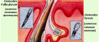
Demodex mites are the most common parasites living on the surface of the body of humans and animals. They live in the mouths of hair follicles and the excretory ducts of the sebaceous and meibomian glands. Of the 100 known species of Demodex mites, 2 species live on human skin: Demodex folliculorum longus (long) and Demodex folliculorum brevis (short). Contamination of the skin occurs in childhood through contact. The maximum incidence occurs in females aged 20–40 years. At older ages, tick parasitism is registered in 80–100% of the population.
Acne iron mites live in places with high humidity; the parasites do not live long outside the host, are distributed everywhere, and are recorded in all races and in all geographical zones.
General characteristics of parasites
Acne gland mites live in the mouths of hair follicles and excretory ducts of the sebaceous and meibomian glands of the skin of the face (T-shaped zone), in the ears, and less often on the back and chest. There is an atypical localization of parasites.
Demodex mites feed on epidermal cells and sebum.
At night, ticks crawl to the surface to mate. Ticks move along the surface of the skin at a speed of 8 - 16 mm/hour.
At the head end (gnathosoma) there is a mouth opening equipped with sharp chelicerae that facilitate the absorption of food (epidermal cells and sebum). Digestion of food occurs under the influence of lytic enzymes.
Rice. 2. Photo of demodex mites under an electron microscope.
Reproduction and development
Ticks crawl onto the surface of the skin at night to mate. That's when they end up with other hosts. The spring-summer period is the most favorable for parasites.
The life cycle of parasites is 14 - 18 days and consists of 5 developmental phases. After fertilization, the females return to the hair follicles and sebaceous gland ducts and lay diamond-shaped eggs, from which larvae emerge after 60 hours. In the developmental cycle, larvae transform from eggs, protonymphs and nymphs into adults. The appearance of a large number of parasites coincides with the first rashes on the skin.
Rice. 3. The photo shows (from left to right) the developmental stages of the demodex mite from egg, larva, protonymph and nymph to adult.
Features of Demodex folliculorum longus (long mite)
The tick of this species is long (0.3 - 0.4 mm), females and males are of the same size. The body is worm-shaped. The head end (gnatosoma), thorax (subsoma) and abdomen (opisthosoma) are well differentiated. The chelicerae of the oral opening are sharper.
The subsoma has setae. The covering (cuticle) is transparent. There are very short three-membered legs. Transverse striations of the posterior part of the body are noted. They live in hair follicles in groups.
Features of Demodex folliculorum brevis (short mite)
This species of tick has a short body (0.18 - 0.19 mm). The opisthosoma is short with a pointed cone-shaped end. The head end is short and flattened. Males are smaller than females and die after fertilization. The breast is wide and lacks setae. The cuticle is less transparent. They live in the mouths of the excretory ducts of the sebaceous and meibomian glands singly, which makes them difficult to detect. They rarely rise to the surface, making treatment difficult.
Demodex folliculorum longus is found 4 times more often in men and 10 times more often in women than Demodex folliculorum brevis.
Rice. 4. On the left are long demodex mites (Demodex folliculorum longus), on the right are short mites (Demodex folliculorum brevis).
Rice. 5. In the photo there is a short and a long demodex mite.
Causes of demodex
Tick reproduction is not associated with any specific climatic zone, but is associated with seasonal peaks, which is due to the biological characteristics of the pathogen. At the same time, the appearance of clinical symptoms of demodicosis often leads to disturbances in the central nervous system with the occurrence of neurotic disorders.
The mite, the causative agent of the disease, is an acariform parasite that lives in the ducts of the sebaceous glands, as well as hair follicles. It mainly affects humans, but is sometimes detected in animals, usually living at home. When a gland or follicle is blocked, the most favorable conditions are created for intradermal reproduction of the parasite. In places of its large accumulation, an inflammatory process is formed, ending in the formation of acne and small abscesses.
Two types of mites can parasitize human skin: short and follicular. It is worth knowing that sometimes both of these types can be detected in one person. A similar situation is observed in approximately 10-27% of cases. In 20-80% of cases, a follicular mite is found as an independent parasite, and a short mite is much less common - in 7-13%. Both types of ticks have favorite places to live. At the beginning of their development, they both can be detected in the skin of the face (forehead, chin, eyelids and nasolabial folds). Extremely rarely, they are detected in the thickness of the skin of the abdomen, back, chest, pubis and ears.
Both follicular and short types of mites have different habitats, so they do not compete. The area affected by follicular demodex is mainly the hair follicles, while its reproduction occurs in the cavity of the pouch. It is there that the largest groups of it are found. Short demodex is usually detected in the ducts of the sebaceous glands and the glands themselves or in the glands of the cartilaginous plate of the eyelids. During the process of mating and reproduction, tick colonies can migrate.
When clinical symptoms occur, special structures are identified in the hair follicles - nests of parasites. In shape, they vaguely resemble a cocoon that is attached to the hair. In these structures, mite eggs and a small amount of tissue of the parasite itself are detected, which remain when their surface covers are replaced.
Depending on the type, the nutrition of ticks also differs. So follicular demodex feeds on the cells of the epithelial layer or the contents of the follicles. The short mite prefers to consume the secretions of sebaceous and cartilaginous glands and the contents of cells. The feeding process of the parasite is also of some interest. So, the tick first penetrates the tissue using a piercing-sucking apparatus. After this, he injects a specific secretion of salivary jelly, under the influence of which the products decompose. In addition to enzymes, this substance contains antigens that cause allergic reactions on the body of the carrier.
The seasonal activity of ticks is explained by the biological cycle of parasites. They are often detected in the summer. At this time, their number on the surface of the skin is almost 10 times higher than their number in winter. Moreover, in summer, as a rule, a large number of young individuals of both sexes parasitize on the skin, while in winter, only males are predominantly found.
People in the age group of 30-50 years are especially susceptible to tick damage. In persons under 20 years of age, cases of Demodex infection are quite rare. Cases of infection in infants have not been described. The incidence of the parasite in the female population is almost twice as high, which is associated with the characteristics of age-related physiological and biochemical processes that occur in the body.
The theory about the connection between the risk of Demodex infection and hormonal levels has become widespread. Thus, when studying the level of estrogen and testosterone in women with demodicosis, it was found that the concentration of the hormone testosterone affects the severity of clinical symptoms. In the case of dermatological manifestations, mites are most often found in individuals with acne - rosacea. In patients with seborrheic eczema, neurodermatitis or eczema, mites are usually not detected or are detected in very small numbers.
In the case of eye diseases, demodex is detected in approximately 62-70%. As a rule, these are episcleritis, blepharoconjunctivitis, keratitis, iridocyclitis, periorbicular dermatitis. Demodectic mange most often affects patients with gastroenterological problems (gastric or duodenal ulcers, enteritis, gastritis, cholecystitis), as well as diabetes mellitus, diseases of the endocrine glands, tonsillitis, focal infection, and rheumatism.
It is worth mentioning that even when examining absolutely healthy people, the presence of a tick is detected in almost 100% of cases. In this regard, some experts are inclined to believe that demodicosis is an opportunistic invasion. In addition, there is research data indicating the role of demodex mites in removing excess biomaterial from the ducts of the human sebaceous glands.
Epidemiology of demodicosis
Source of infection
The main source of demodex mites are sick people or carriers.
Distribution routes
- Iron mites can be transmitted through bedding, underwear, and personal items. The parasites were found in house dust, the source of which was bedding.
- Some researchers suggest that ticks can get on a child during breastfeeding from a sick mother.
Sustainability
Constant humidity, darkness and air temperature within 30-40°C are optimal conditions for iron mites. Outside the host, at room temperature, constant humidity and in the dark, ticks live up to 9 days.
According to the observation of some researchers, parasites remain viable in water for 7 days, and up to 2 weeks in immersion oil.
Rice. 6. Demodex mite under a microscope.
demodex mite
Demodex mite on the eyes is a type of parasite that affects the follicles of the eyelashes.
Two types reproduce in the human body :
- Demodex folliculorum , which reproduces specifically in or near the eyelash follicles;
- Demodex brevis , which is found less frequently and only in the sebaceous ducts of the eyelid.
But both of these species cause demodicosis - an inflammatory disease of the edge of the upper and lower eyelids.
The average size of a demodex mite is about 0.4 millimeters (adult).
The food for such a parasite is not only particles of eyelashes, but also particles of the skin of the eyelid and sebaceous secretions of the glands located here.
The mite reproduces quickly, laying eggs in the follicles, from which new individuals hatch within the next three days.
It is worth noting! The disease worsens not only with an increase in the number of ticks, but also due to inflammatory processes provoked by the decomposition of dead parasites.
How the disease develops
Demodicosis in humans can develop for the first time and occur as an independent disease, or develop against the background of existing skin diseases (acne, perioral dermatitis, rosacea, seborrheic dermatitis, etc.). From 55 to 100% of the population are carriers of iron ticks and do not have any manifestations of the disease, which proves that the parasites belong to the opportunistic flora. Damage to follicles by parasites increases with age.
Factors in the development of the disease
Acne iron mites are an opportunistic infection. They can parasitize a person for many years without causing him any harm. Their activation and active reproduction depends on a number of factors.
- Changes in microbiocenosis, the function of the sebaceous glands and the composition of sebum are the trigger mechanism of the disease. Changes in the composition of surface lipids lead to the development of dysbiosis.
- Demodex mites are carriers of viruses and microbes into the sebaceous glands and hair follicles. When pathogenic pyococci and fungi Pityrosporum spp are introduced, purulent-necrotic inflammation develops.
- The bacillus Bacilluss oleronius found on the surface of the parasite increases the activity of ticks and stimulates staphylococci, streptococci, acne propionobacteria, and fungi of the genus Malassezia to produce anti-inflammatory proteins, which triggers a cascade of immune reactions.
- The presence of foci of chronic infection, disturbances in the functioning of the liver and gastrointestinal tract, endocrine glands, nervous system, and corticosteroid therapy contribute to the activation of ticks.
- The rise in incidence in the spring-summer period is associated with increased insolation. An increase in the production of vitamin D under the influence of ultraviolet radiation leads to an increase in the synthesis of cathelicidins, which support the inflammatory process.
- The likelihood of infestation by Demodex mites increases in individuals with a sharply reduced functioning of the immune system, which occurs with primary and secondary immunodeficiencies, long-term use of corticosteroids and cytotoxic therapy, HIV infection, leukemia, T-cell lymphoma and other malignant neoplasms.
- The mite infestation maintains the activity of the existing inflammatory process. An imbalance in the cytokine cascade develops when the immune response is too strong.
Mechanisms of action of iron mites on humans
The impact of Demodex mites on humans is multifaceted:
- When feeding, the parasites secrete aggressive saliva and mechanically destroy the cell walls of the sebaceous glands and follicles, inflammatory infiltrates and granulomas form in the dermis, and the processes of keratinization and pigmentation develop.
- Substances formed after the death of ticks have antigenic properties, which leads to allergization and increased immune response.
- When the immune system is suppressed, opportunistic flora is activated, which successfully colonizes the host.
Acne iron mites act as carriers of bacteria and viruses, which complicates the course of demodicosis and complicates treatment.
Rice. 7. Histological preparation of a skin section. Arrows indicate the location of demodex mites.
Treatment
To cure eyelash demodex, you need to conduct the necessary research that will show possible complications. It is necessary to begin therapy by observing the rules of personal hygiene. This will further minimize the risk of relapse of the disease. As a result, the effectiveness of treatment will increase significantly.
An integrated approach to the treatment of the disease is the key to successful treatment of demodex. The use of antibiotics, immunomodulators and antihistamines is combined with the use of acaricides and local medications. The duration of treatment is usually about a month, but can reach a year. It depends on various complications and aggravating factors.
It is very important to regularly clean the eyelid from formed scales. To do this, you can use an alcohol infusion of eucalyptus or calendula. You also need to apply an ointment that contains tar, sulfur, ichthyol, yellow mercury, benzyl benzoate or zinc thyol. These acaricidal substances attack the tick, which leads to a decrease in the number of the parasite. Preparations containing metronidazole are used orally.
Demodex Complex is highly effective, helping to cope with ticks in various ways. Also, in some cases, anticholinesterase drugs are used, which paralyze the parasite due to their effect on muscle and nervous tissue. At the same time, it is worth paying attention to normalizing metabolism. To do this, you need to adjust the functioning of the gastrointestinal tract and immune system.
Clinical variants of demodicosis
Demodicosis in humans can develop as a primary independent disease, or it develops against the background of existing skin diseases (perioral dermatitis, rosacea, acne, seborrheic dermatitis, etc.), aggravating their course.
Primary demodicosis
In primary demodicosis, inflammatory skin elements resolve after antiparasitic therapy. About 40% of cases of primary demodicosis develop in patients suffering from rosacea.
Primary demodicosis is characterized by:
- the onset of the disease over the age of 40 years;
- The areas most often affected are around the mouth, eyes and outer ear;
- inflammatory elements are located asymmetrically and are accompanied by itching;
- acne and rosacea are absent;
- high content of mites on the skin;
- positive effect of the antiparasitic treatment.
Secondary demodicosis
Secondary demodicosis develops against the background of diseases accompanied by a sharp suppression of the immune system (HIV infection, leukemia, etc.), long-term use of corticosteroids and cytotoxic therapy, or develops against the background of acne and rosacea, complicating their course. This form of the disease is registered in 33% of patients with acne.
Secondary demodicosis is characterized by:
- the disease appears at any age;
- lesions are widespread;
- presence of anamnesis and clinical picture of relevant diseases.
Papulopustular rashes on the skin of the face are a reason for diagnosing demodicosis.
Rice. 8. Demodectic mange on a woman’s face.
Signs and symptoms of demodicosis
The disease always occurs gradually and tends to spread and progress.
The main localization of demodex mites
Short mites live in the mouths of the excretory ducts and the secretion of the sebaceous glands of the skin of the face, ear canal, chest and back, sometimes on the scalp, as well as the meibomian glands, which are modified sebaceous glands that open at the edges of the eyelids. Long mites live in hair follicles. Mites were found on the penis, buttocks, and in the area of ectopic sebaceous glands.
Rice. 9. Demodectic mange on the face in the area of the nasolabial folds.
Rice. 10. Demodectic mange on a woman’s face in the chin area.
Rice. 11. Demodectic mange on the skin of the cheeks, nose and around the mouth.
Rice. 12. Demodectic mange on the face of women on the skin of the forehead, cheeks and nasolabial folds.
Rice. 13. Demodectic mange on the face of women.
Rice. 14. Demodectic mange of the eyelids.
Characteristics of the rash
- Initially, erythematous (red) spots appear around the mouths of the follicles. The rash is located asymmetrically and is accompanied by lamellar peeling.
- The appearance of the rash is accompanied by a feeling of heat, burning, crawling and distension.
- Very quickly, pink or red papules with a cone-shaped top and gray scales appear in the reddened area. Papulovesicles (a vesicle is a blister filled with clear fluid) or papulopustula (a pustule is a blister filled with purulent fluid) often appear.
- Inflammatory infiltrates and granulomas form in the dermis. The skin over the affected areas thickens and its elasticity decreases. A feeling of tightness appears. The affected areas are covered with serous, serous-sanguineous or purulent crusts. Over time, hyperpigmentation develops: the skin becomes brownish or yellowish-brownish in color.
- With the massive development of a pyogenic bacterial infection, large follicular pustules (purulent elements) appear at the site of inflammatory infiltrates. Over time, they spread into the deeper layers of the dermis, which leads to facial disfigurement.
- The clinical picture of demodicosis for different types of mites is somewhat different from each other. Thus, when Demodex folliculorum is affected, erythema and peeling (desquamation of the epithelium) are more often recorded; when Demodex brevis is affected, papulopustular elements are often recorded, which are often located symmetrically.
- With long-term (chronic) demodicosis, thickening of the skin, loss of elasticity, and a feeling of tightness appear. Bloody-purulent crusts are located against the background of hyperemic skin. When a pyogenic infection is attached, pustules and microabscesses appear. Dense infiltrates form in the deep layers of the dermis, disfiguring the face.
Rice. 15. Demodectic mange on a woman’s face, severe.
Deterioration in appearance creates aesthetic discomfort for the patient and leads to the development of neuroses and depression.
Forms of demodicosis in humans
Depending on the nature of the manifestations on the skin, several forms of demodicosis are distinguished, the symptoms of which are characteristic of the corresponding diseases. Not to be confused with these diseases!
Acneform form
The acneiform type of demodicosis is characterized by the appearance of papules and pustules on the facial skin, which are similar to acne.
Rice. 16. Demodectic mange of the face in women. Acneform form of the disease: papular (photo on the left) and papulopustular (photo on the right).
Rosacea-like form
The rosacea-like type of demodicosis is characterized by the appearance of papules against a background of diffuse erythema. Demodex brevis (short mite) causes the appearance of inflammatory infiltrates and granulomas in the deep layers of the dermis, which are similar to the granulomatous form of rosacea.
Rice. 17. Rosacea-like form of demodicosis: in the form of diffuse erythema (photo on the left), granulomatous form (photo on the right).
Seborrheic (erythemo-squamous) form
The seborrheic form of demodicosis is characterized by the appearance of redness and rash on the facial skin, accompanied by lamellar peeling.
Ophthalmic form
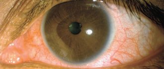
The ophthalmic form of demodicosis occurs with damage to the eyeballs and eyelids. Redness, dryness, irritation, sensation of a foreign body in the eye, fatigue are the main symptoms of eye demodicosis. Itching of the eyelids, redness and swelling of their edges, the appearance of scales or crusts on the eyelids are the main symptoms of demodicosis of the eyelids.
Read in detail in the article “Demodicosis of the eyelids and eyes”
Rice. 18. Demodectic mange of the eyelids. Lamellar peeling is clearly visible (photo on the left). On the ciliated edge of the eyelid, scales form a “collar” around the eyes (photo on the right).
The role of demodex mites in the formation of androgenetic alopecia
There is an assumption that the presence of mites in the hair follicles aggravates the development of androgenetic alopecia. During inflammation, T-lymphocytes are activated, which induce collagen synthesis, resulting in fibrous degeneration of hair follicles.
Rice. 19. Demodicosis of the scalp.
Symptoms of demodicosis of the eyes
With demodicosis of the eyelids, symptoms characteristic of this disease occur. Often the patient complains that even after minimal strain his eyes get tired. In addition, photosensitivity, burning and irritation of the eyes, itching, and heaviness of the eyelids appear.
Manifestations of demodicosis bring great discomfort to the patient. In this case, the edge of the eyelid usually becomes red, inflamed, accompanied by swelling, viscous and foamy discharge, which are localized in the corners of the eyes. Externally, scaly formations can be distinguished between the eyelashes.
If complications arise against the background of demodicosis of the eyelids, then the growth of ciliary hair is disrupted and atrophy occurs. Sometimes eyelashes simply fall out and their growth is disrupted. In addition, with this disease, ulcers, ulcers, plaque, and papillomas form along the edge of the eyelids. The eyelashes themselves are covered with sticky crusts.
Demodectic mange against the background of rosacea
Demodicosis complicates the course of rosacea in 88.7%. The disease is more common in women over 30 years of age. The phymatous form of the disease is more common in men. The pathological process is characterized by redness of the skin of the face, associated with the expansion of small superficial vessels, the appearance of papules and pustules. Papules increase in size over time and merge, forming dense infiltrates. The sebaceous glands become hyperplastic. Fibrosis develops. Persistent redness and fibrous lumps on the nose are called rhinophyma.
There are many reasons for the development of diseases. Factors contributing to the development of the disease can be divided into endogenous and exogenous.
Rice. 20. Erythematotelangiectatic form of rosacea. Multiple telangiectasias (dilated subcutaneous arterioles) are visible on the skin.
Rice. 21. The photo shows the papulopustular form of rosacea. Against the background of erythema (redness), multiple rashes are visible, which are papules with thin scales on the surface. The affected areas are infiltrated and swollen.
Rice. 22. Demodex brevis (short mite) causes the appearance of inflammatory infiltrates and granulomas in the deep layers of the dermis, which are similar to the granulomatous (phymatous) form of rosacea. The photo shows the phymatous (papular-nodular) form of rosacea.
Rice. 23. Severe form of rosacea.
Rice. 24. The ocular form of rosacea is registered in 50% of patients. Inflammation of the eyelids, redness of the conjunctiva, iritis and keratitis are the main manifestations of the disease.
Demodicosis on the background of acne
Among acneiform dermatoses, demodicosis accounts for 10.5%. Acne (acne vulgaris) is a chronic, relapsing disease that affects the sebaceous glands and hair follicles. Most often, acne occurs in males during puberty between the ages of 14 and 16 years. The cause of juvenile acne is propionobacteria acne, epidermal staphylococci, pityrosporum ovale and orbitale, which constantly live on the skin of the face. Many factors contribute to the development of acne.
Rice. 25. Demodicosis is registered in 10.5% of patients with acne. The photo on the left is papular acne. When a staphylococcal infection is attached, pustules (ulcers) and microabscesses develop - pustular and abscess acne (photo on the right).
Rice. 26. With the indurative form of acne, inflammatory infiltrates appear in the deep layers of the dermis. The skin has a bumpy appearance.