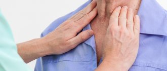
Often, patients with rashes on the skin of the face or other parts of the body and limbs, for a number of reasons, resort to treatment on their own, purchasing creams or ointments at the pharmacy recommended by a pharmacist, a friend, or found on the global Internet. These funds may be useless, but they are not always harmless. It’s good when the drugs helped and did not have any side effects. However, long-term use without the advice of a doctor of external products, mainly on the skin of the face, which contain a hormonal (or more correctly, glucocorticosteroid) component, can lead to the development of such complications as perioral dermatitis, when the rashes are located around the mouth. With the correct prescription and mode of use, there is no need to be afraid of hormonal (glucocorticosteroid) drugs; today they have a high level of safety.
Perioral dermatitis
Perioral dermatitis on the face is a disease that affects the skin.
It appears in the form of small pimples that form in the area of the lips and mouth. It is accompanied by unpleasant itching, redness and negative consequences, which can be eliminated through a medical examination and properly selected treatment. In each specific situation, based on the results of the consultation, doctors recommend the patient medications that are relevant to his case. These include specialized pastes, ointments, sometimes hormonal drugs, as well as therapy that quickly eliminates burning and other symptoms. For perioral dermatitis, medications (ointment, tablets, cream) are selected based on medical diagnosis and tests. You can purchase the necessary medications for treatment on our website “Pharmacy 911”.
Treatment of skin infections in atopic dermatitis in children
In the international consensus document on atopic dermatitis (USA, 2002), experts note that atopic dermatitis is typically an inherited disease often associated with asthma, food allergies, allergic rhinitis and recurrent skin infections. Infection can significantly change the course of the disease. If a patient develops a bacterial, fungal or viral skin infection, it is necessary to quickly identify this infection and begin specific therapy to eliminate it [1].
This approach is due to the fact that viruses, bacteria and fungi can be triggers of atopic dermatitis and lead to exacerbation of the disease. On the other hand, ceramide deficiency as a result of a pronounced decrease in sphingomyelin levels contributes to dry skin and easier penetration of infectious agents and allergens into the damaged epidermis. In addition, the epidermal skin barrier is disrupted by proteases from house dust mites and Staphylococcus (S.) aureus, which, in combination with other factors, also increases the risk of allergen penetration and supports the inflammatory process in the skin. There are also observations indicating the role of bacterial and fungal colonization in the formation of therapy-resistant and more severe recurrent atopic dermatitis [2, 3]. Finally, secondary infection of the skin or deep scratching can lead to scarring, which is generally not typical for atopic dermatitis itself.
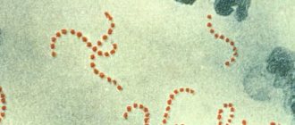
The microflora of healthy skin is usually not characterized by colonization by S. aureus, while the skin of more than 90% of patients with atopic dermatitis is contaminated with this type of bacteria, the number of which in weeping lesions can be over 14 million per square meter. see [2]. Moreover, a high degree of skin colonization with S. aureus is possible even in the absence of clinical manifestations of a bacterial skin infection. Toxins (so-called superantigens) produced by S. aureus on the skin surface stimulate the activation of T lymphocytes and macrophages and thereby enhance the allergic inflammatory immune response of the skin. Studies have shown that S. aureus exotoxins worsen the course of atopic dermatitis, and the level of specific immunoglobulin (Ig) E antibodies to staphylococcal superantigens correlates with the severity of the disease [3]. It is also assumed that superantigens are involved in the development of skin insensitivity to topical glucocorticosteroids and/or a decrease in the therapeutic effect of these drugs [3–5].
Another cause of severe atopic dermatitis, refractory to standard therapy, is considered by some authors to be fungal sensitization [5–7]. In Europe, 30–68% of patients with recurrent severe atopic dermatitis, as well as with a variant of the localization of the disease on the head and neck, have specific IgE antibodies in the blood or a positive reaction to prick or patch tests for the fungi Malassezia furfur (formerly called Pityrosporum ovale) [5].
In rare cases, the skin of patients with atopic dermatitis becomes infected with viruses, most often with herpes simplex virus types 1 and 2, as well as with molluscum contagiosum virus [1, 5, 7]. Herpetic skin infection can occur repeatedly in the same patients, while in others it almost never occurs. It is believed that patients with severe atopic dermatitis or those who have not received proper treatment are at greatest risk. Sometimes eczema herpeticum in patients with atopic dermatitis should be differentiated from a bacterial skin infection such as impetigo. Eczema molluscatum, unlike herpetic eczema, is a harmless complication that is rare.
Scientists believe that one of the important reasons for skin infection in patients with atopic dermatitis is the deficiency of antimicrobial peptides in the skin, which are necessary to protect the body against bacteria, viruses and fungi [5]. It is believed that infectious skin complications in atopic dermatitis are accompanied by certain immunological disorders: in bacterial infections - a deficiency of epidermal antimicrobial peptides and an imbalance of T-helper cells (Th1/Th2) - in the case of viral skin lesions [5]. According to recent data, in patients with atopic dermatitis with a complication such as herpetic eczema, significantly lower expression of antimicrobial peptides was found in skin keratinocytes [5].
We list the skin infections that most often can complicate the course of atopic dermatitis and their causative agents:
- superinfection, folliculitis, furunculosis - Staphylococcus aureus; herpes simplex, herpetic eczema - herpes simplex virus type 1;
- genital warts, common warts, plantar warts - human papillomavirus;
- molluscum contagiosum - molluscum contagiosum virus;
- “head and neck dermatitis” - Malassezia furfur.
However, the possibility of developing such complications should be considered not only in the case of a clinically pronounced stage of exacerbation of atopic dermatitis, but also in the absence of a response to appropriate therapy. High colonization of the skin with S. aureus or a fungal infection can occur in the absence of a clear clinical picture of the disease.
According to M. Furue et al., who observed 1271 patients with atopic dermatitis for 6 months (including 210 young children, 546 children and 515 adults), the following complications were diagnosed: herpetic infections and/or Kaposi's dermatitis in 2.4% young children, 2.5% older children and 3.5% adults; association of atopic dermatitis with molluscum contagiosum in 7% of young children, 9% of children and 0.2% of adults [8].
Domestic experts point to the formation of complicated forms of atopic dermatitis in a much larger number of cases: in 25–34% of children suffering from atopic dermatitis [7].
The participation of microbes and viruses in the development of exacerbations and recurrent severe atopic dermatitis justifies the need for a differentiated approach to complex therapy for such patients. Let us dwell on the main stages of treatment of complicated forms of atopic dermatitis in children. We will talk about external, etiotropic and immunomodulatory therapy [7].
External therapy
Let us recall that the key point in the treatment of any forms and stages of atopic dermatitis is auxiliary basic therapy: regular use of cleansing and moisturizing products of the dermatocosmetic line [1]. The goal of this approach is to limit bacterial growth and restore skin barrier function.
When a child with atopic dermatitis is diagnosed with an active bacterial skin infection, treatment should include proper skin care (cleansing/hydration, antiseptics) and topical and/or oral antibiotics.
- For pustular rashes, it is better to use local baths with the addition of a solution of potassium permanganate until a light pink color is achieved (for infants - 10 ml of a 3% solution per 1 liter of water), lasting 15–20 minutes.
- Pustular rashes should be immediately extinguished with water (for children under 2 years of age) and alcohol solutions of aniline dyes: Castellani liquid, fucorcin, brilliant green solution or other antiseptics.
- Skin cleansing is carried out by taking a short bath or shower with a water temperature of 35–36 ° C and a soft washing base (pH = 5.5) that does not contain alkali (Bodygel, Mustela StelAtopia for bath, Atoderm mousse and etc.).
- After swimming or a bath, it is necessary to dry the skin with a towel without rubbing it, and apply a moisturizing cream (Topicrem, Exomega, Atoderm, etc.), and etiotropic drugs in combination with anti-inflammatory drugs to the areas of skin lesions.
Etiotropic therapy
For bacterial infections with exudation and oozing of the skin, topical antibiotics are used. It has been established that the growth of colonies of microorganisms, which are the most common causative agents of skin infections (including S. аureus, Streptococcus pyogenes, S. epidermidis, Corynebacterium spp.), is most effectively suppressed by fusidic acid [9]. In addition, the concentration of this antibiotic after a single application, in particular fucidin ointment, is 2.2% in the skin (in the epidermis - ~430 µg/ml, in the dermis - 286 µg/ml and subcutaneous fat - ~109 µg/ml) ; for comparison: the penetration ability of erythromycin is 0.3%, tetracycline - 0.28%, mupirocin (Bactroban) - 0.02%. In other words, fusidic acid penetrates the skin 100 times better than mupirocin, and 8 times better than tetracycline and erythromycin. It has been shown that fusidic acid penetrates even into devascularized areas and necrotic tissues, and its bioavailability is 90%.
Since in atopic dermatitis, eczematous skin lesions, in particular areas of exudation and excoriation, represent a favorable environment for the proliferation of S. аureus, it is believed that in the treatment of the disease it is preferable to use a combination of a topical glucocorticosteroid with an antibiotic. This will break the vicious circle: S. aureus reduces the effectiveness of corticosteroids through the production of staphylococcal toxin, while corticosteroids suppress inflammation.
Thus, the pathogenetic role of bacteria in the development and maintenance of allergic inflammation in atopic dermatitis has been proven, which justifies the need for the use of combination drugs containing topical corticosteroids and antibacterial agents. It has been shown that the use of such drugs can more effectively combat the clinical signs of atopic dermatitis than therapy with only one external corticosteroid. Thus, in two randomized clinical trials (from 34 to 207 patients), the effectiveness of topical application of various combination drugs containing antibacterial agents in combination with topical corticosteroids and monotherapy with topical corticosteroids was compared in patients with atopic dermatitis with skin manifestations of secondary infection [10, 11 ]. Clinical improvement was observed in 54–95% of participants, with no statistically significant differences in subjective and objective data depending on the use of various combination drugs. However, in the group using fusidic acid–hydrocortisone acetate (Fucidin G), clinical improvement occurred faster than in the group using miconazole–hydrocortisone [10], and the laboratory confirmed positive effect of treatment in the group using betamethasone valerate–fusidic acid (Fucicort) was higher than in the group where miconazole-hydrocortisone was used [11].
This approach—the topical use of an antibiotic and a cream—the mild corticosteroid hydrocortisone—led to a significantly earlier regression of the symptoms of skin inflammation in mild to moderate atopic dermatitis [12]. The administration of a cream containing an antibiotic and a corticosteroid seems absolutely necessary in the case of clinical manifestations of skin infection (wetting eczema). It is advisable to use such a drug in a short course (up to 1–2 weeks) in order to avoid the development of resistance to S. аureus [13]. In this case, for use on the face, skin folds, armpits, groin area, and open areas of the body, it is preferable to choose a combined drug in the form of a cream—a mild corticosteroid and an antibiotic (for example, fusidic acid + hydrocortisone acetate (fucidin G), 2–3 times per day in the form of light applications). In the case of acute and chronic forms of dermatitis, localized in areas with thick skin (palms and feet), resistant to weak topical corticosteroids, it is more advisable to prescribe an ointment form of a combination drug containing a corticosteroid of medium/high activity (for example, fusidic acid/betamethasone valerate (Fucicort) , celestoderm B with garamycin, etc.) 2–3 times a day.
However, studies have shown that skin colonization with S. aureus reoccurred 2–4 weeks after completion of treatment with a combination of topical corticosteroid and antibiotic. This is explained primarily by the fact that even when highly sensitive antibiotics are used, it is very difficult to achieve complete eradication of S. аureus. The maintenance of skin infection is facilitated by such factors as the possibility of transmission of S. aureus from parents who are in direct contact with a sick child, as well as the content of S. aureus in the nasal mucus of the patient himself.
If topical antibiotics are ineffective, as well as atopic dermatitis complicated by a bacterial infection (for example, impetigo), occurring with fever, intoxication, inflammatory changes in the blood, the administration of a broad-spectrum antibiotic orally (cephalosporins 1–3 generations, macrolides, aminoglycosides) is indicated for 7– 10 days. Taking fluoroquinolones and tetracyclines is not recommended [7].
However, despite the fact that as a result of taking systemic antibiotics, the level of microbiologically confirmed colonization of the skin with S. aureus was significantly reduced, this did not contribute to the clinical improvement of severe atopic dermatitis in children [14].
The ability to significantly reduce the colonization of S. aureus and the frequency of skin infections has also been described in relation to drugs of a new class of non-steroidal topical immunosuppressants - the calcineurin inhibitors tacrolimus and pimecrolimus (Elidel) [1, 4]. Thus, as a result of long-term use of pimecrolimus in children aged 3–23 months of life suffering from atopic dermatitis, significantly fewer cases of skin infections were noted [15]. This fact further confirms the important role of Th2-mediated skin inflammation and bacterial colonization, in which the relationship between cytokines, adhesion molecules and antimicrobial peptides plays a huge role [4, 16]. Another immunosuppressive drug, which also works by inhibiting calcineurin, is cyclosporine. This drug has the ability to reduce the number of S. aureus on the skin of patients with atopic dermatitis, which is most pronounced in patients who simultaneously have a high degree of colonization of the microbe and clinical manifestations of a bacterial skin infection [17]. However, the use of this drug is limited in pediatric practice.
For a fungal infection confirmed microbiologically, the following are prescribed: systemic antifungal agents (fluconazole (Diflucan) from the neonatal period, children over 2 years old can be prescribed terbinafine (Exifin), itraconazole (Orungal), ketoconazole - from 12 years old) for 7-14 days in combination with local therapy.
Exifin inhibits the biosynthesis of ergosterol in the fungal cell, thereby promoting the accumulation of squalene inside the cell and its death. The drug does not affect the metabolism of hormones or other drugs; in low concentrations it has a fungicidal effect on dermatophytes, molds and some dimorphic fungi. Depending on the type of yeast, its effect can be fungicidal or fungistatic.
Ketoconazole and itraconazole inhibit the production of interleukin-4 and interleukin-5 and the synthesis of ergosterol, which leads to a nonspecific anti-inflammatory effect and a decrease in the level of specific IgE to Malassezia furfur [5].
In case of combined skin lesions of patients with atopic dermatitis with a bacterial and/or fungal infection, the most optimal drugs of choice are combination agents: Triderm, Akriderm GK. Triderm in the form of an ointment is applied to the skin of the torso and limbs, and in the form of a cream - on delicate areas of the skin (in the area of folds, forearms). In pediatric practice, Triderm cream is mainly used. The drug is applied in a thin layer to the affected areas of the skin 2 times a day, the course of treatment averages 7–12 days.
As is known, topical corticosteroid therapy is the standard treatment for exacerbations of atopic dermatitis. However, the powerful anti-inflammatory effect inherent in these drugs may increase the skin's sensitivity to bacterial and fungal infections, which is an obstacle to long-term use of topical hormonal agents in the presence of these infections. In addition, children are more sensitive to topical corticosteroids due to their high cutaneous absorption; the latter is also caused by dry skin [1]. However, there are suggestions that the use of only monotherapy with topical antibiotics or fungicidal drugs contributes to skin sensitization and exacerbation of allergic inflammation with the development of candidiasis infection [18].
In all cases, the doctor should individually select medications for patients with atopic dermatitis, taking into account the course of the disease and the condition of the skin.
For herpes infection:
- it is necessary to control the factors that provoke the disease (colds, increased insolation, psychological stress, fatigue, etc.);
- in case of viral skin lesions, the use of topical corticosteroids is contraindicated;
- In pediatrics, only one antiherpetic drug is used - acyclovir (Zovirax). Parents of children with recurrent herpes infections are reminded of the need to use acyclovir immediately when the first symptoms of the disease appear (tingling of the skin);
- patients with atopic dermatitis, suffering from recurrent herpes simplex, herpetic gingivostomatitis, as well as in the case of diagnosing the most severe and life-threatening complication - Kaposi's eczema herpetiformis - are prescribed oral acyclovir. The duration of the course depends on the severity of the child’s condition and is 7–14 days [5, 7]. Patients with recurrent disease require prophylactic therapy with acyclovir. According to the latest data, after starting antiviral therapy, in order to quickly restore the skin barrier function, it is advisable to continue treatment with topical immunomodulators (tacrolimus, pimecrolimus) [5].
For molluscum contagiosum:
- the rashes are opened under aseptic conditions by piercing with a sterile needle (the needle is inserted from the side, parallel to the skin) and extinguished with a 5% alcohol solution of iodine. After the necessary training, the child's parents can perform this procedure at home on their own;
- practice cryodestruction;
- Isolation of a sick child is necessary.
Immunomodulatory therapy
In cases where patients with atopic dermatitis are diagnosed with a recurrent bacterial, fungal and/or viral infection, as well as if they have foci of chronic infection that are resistant to standard therapy, immunomodulatory therapy is indicated, which should be carried out under the control of immunological parameters. In pediatric practice, the most widely used is lycopid, an immunomodulator that has the ability to influence the main populations of cells of the immune system (macrophages, T- and B-lymphocytes), which is probably due to the presence of specific intracellular receptors for glucosaminylmuramyl dipeptide [7, 18]. . In its structure, lycopid is a synthetic analogue of the main fragment of peptidoglycan of the cell wall of all known bacteria (muramyl dipeptide). Activation of macrophages under the influence of lycopid leads to increased synthesis of pro-inflammatory cytokines (interleukin-1, tumor necrosis factor a, colony-stimulating factors), as a result of which phagocytosis, proliferation and differentiation of T- and B-lymphocytes are activated, the synthesis of immunoglobulins is enhanced, cytotoxicity increases, and leukopoiesis is stimulated .
A study by S. Yu. Rezaikina, conducted in patients with atopic dermatitis, revealed a predominant decrease in the functional activity of neutrophil phagocytosis (changes in the oxygen metabolism of neutrophils), which was restored when taking licopid [20]. As a result of the use of licopid in complex therapy of patients with moderate atopic dermatitis, the frequency of exacerbations of the disease significantly decreased and a pronounced correction of altered laboratory parameters was observed.
Likopid is prescribed to children aged 1 to 16 years orally in the form of 1 mg tablets. For the treatment of purulent-inflammatory skin diseases, the daily dose of the drug is 1 mg for 10 days. When treating herpes infection, the drug is prescribed orally at a dose of 1 mg 3 times a day for 10 days. Contraindications to the use of licopid: pregnancy, individual intolerance to the drug. In some cases, side effects may occur such as an increase in body temperature to 37.9°C (this is short-term, goes away on its own and does not require discontinuation of the drug). For adults, Lykopid is prescribed orally 30 minutes before meals (10 mg tablets) and sublingually (1 mg tablets) depending on the severity of the disease.
Isoprinosine (inosine pranobex) is characterized by a dual effect - antiviral (due to suppression of the replication of DNA and RNA viruses) and immunomodulatory (due to the inosine complex, the production of interleukins, the chemotactic and phagocytic activity of monocytes and macrophages is enhanced, the synthesis of antibodies increases and the proliferation of T-lymphocytes increases, T helper cells, natural killer cells). Indications for the use of isoprinosine are all viral skin infections in children over 1 year of age suffering from atopic dermatitis (molluscum contagiosum, herpes infection, vulgar warts, human papillomavirus infection). Isoprinosine is prescribed 1 tablet per 10 kg/body weight per day, in 3-4 doses over 10-14 days - depending on the severity of infectious diseases; If necessary, the course of treatment is repeated.
According to L.V. Luss, the inclusion of another immunomodulator, polyoxidonium, in the complex therapy of patients with atopic dermatitis and bronchial asthma, occurring in combination with signs of secondary immune deficiency and resistant to therapy, allows achieving a high clinical effect [21].
Among children suffering from atopic dermatitis, cases of scabies are common. In such cases, the course of atopic dermatitis is accompanied by severe persistent itching of the skin, and often by infection.
When scabies occurs, treatment includes:
- treatment of persons in contact with the patient with anti-scabies drugs;
- disinfection of things and bedding. The premises are wet cleaned or disinfected by employees of the sanitary and epidemiological station on the day of diagnosis and after completion of treatment. Medical control is carried out for 1.5 months;
- applying an aerosol preparation a-steam to the surface of clothing, furniture, bedding that cannot be boiled or otherwise treated;
- prescribing to the patient one of the following anti-scabies drugs:
– spregal – used in all age groups and has no contraindications; sprayed in the evening onto the patient’s skin from the neck to the soles; The drug is left for 12 hours, after which the child is thoroughly washed. If necessary, it is possible to reuse the drug 4–10 days after the initial treatment;– benzyl benzoate - 20% ointment is applied to the skin on the first, second, fourth day, also with a change of linen, clothes, and cleaning of the room;
– to treat the skin you can also use 33% sulfur ointment, Medifox;
- for atopic dermatitis complicated by scabies, it is necessary to use antihistamines, topical corticosteroids, including those combined with antibacterial drugs.
Thus, microbial colonization and superinfection of the skin (for example, S. aureus, Malassezia furfur) may play a role in exacerbations of atopic dermatitis, which justifies the need for additional prescription of antimicrobial agents to patients. Infectious agents are also involved in the development and maintenance of chronic allergic skin inflammation in atopic dermatitis. As a rule, this course of the disease is characterized by frequent relapses and a severe course. Timely diagnosis of skin infections and prescription of optimal etiotropic therapy are extremely important for achieving clinical improvement in the condition of patients with atopic dermatitis.
Experts on atopic dermatitis emphasize that etiotropic therapy can be stopped only after stopping the infectious process and cleansing the skin [1]. At the same time, treatment with anti-inflammatory drugs should be continued (according to indications, short-course topical corticosteroids, long-term calcineurin inhibitors) along with the continuous use of moisturizers to hydrate the skin [2, 5].
Literature
- Ellis C., Luger T., Abeck D. et al. New clinical data and modern strategies for the treatment of atopic dermatitis // Allergology. 2003. No. 4: 50–58.
- Leung D. Infection in atopic dermatitis // Curr Opin Pediatr 2003; 15: 399–404.
- Leung D. Presence of IgE antibodies to staphylococcal exotoxins on the skin of patients with atopic dermatitis. Evidence for a new group of allergens // J Clin Invest. 1993; 92:1374–80.
- Hank P., Leung D. Tacrolimus (FK506): new treatment approach in superantigen-associated diseases like atopic dermatitis? // J Allergy Clin Immunol. 2001; 107:391–2.
- Boguniewicz M., Schmid-Grendelmeier P., Leung D. Atopic dermatitis // J Allergy Clin Immunol. 2006; 118:40–43.
- Malanicheva T. G., Salomykov D. V., Glushko N. I. Diagnosis and treatment of atopic dermatitis in children complicated by mycotic infection // Russian Allergological Journal 2004. No. 2. P. 90-3.
- Atopic dermatitis and skin infections in children: diagnosis, treatment and prevention: a manual for doctors. M., 2004. 104 p.
- Furue M., Terao H., Rikihisa H. et al. Therapeutics clinical dose and adverse effects of topical steroids in daily management of atopic dermatitis // Br J Dermatol. 2003; 148: 128–32.
- Carr W. Fusidic acid in patients with skin and soft tissue infections// Eur J Clin Res. 1994; 5:87–95.
- Poyner T., Dass B. Comparative efficacy and tolerability of fusidic acid/hydrocortisone cream (Fucidin H cream) and miconazole/hydrocortisone cream (Dactacort cream) in infected eczema // J Eur Acad Dermatol Venereol. 1996; 7 (suppl. 1): 23–30.
- Wilkinson J., Leigb D. Comparative efficacy of betamethasone and either fusidic acid or neomycin in infected or potentially infected eczema // Curr Ther Res. 1985; 38: 177–82.
- Ramsay C., Savoie J., Gilbert M. et al. The treatment of atopic dermatitis with topical fusidic acid and hydrocortisone acetate // J Eur Acad Dermatol Venereol. 1996; 7 (suppl 1): 15–22.
- Menday A., Noble W. Topical betamethasone/fusidic acid in eczema: efficacy against and emergence of resistance in Staphylococcus aureus // J Dermatol Treat. 2000; 11: 143–149.
- Boguniewicz M., Sampson H., Bina P. et al. Effects of antibiotic treatment on S. aureus colonization in atopic dermatitis // J Allergy Clin Immunol. 1999; 103:179.
- Paul C., Cork N., Rossi A. et al. Safety and tolerability of 1% pimecrolimus cream among infants : experience with 1133 patients treated for up to 2 years // Pediatrics. 2006; 117: 118–28.
- Ong P., Ohtake T., Brandt C. et al. Endogenous antimicrobial peptides and skin infections in atopic dermatitis // N Eng J Med. 2002; 347:1151–60.
- Bunikowski R., Mielke M., Brautigam M. et al. Effect of oral cyclosporin A in children with Staphylococcus aureus-colonized vs S aureus-infected severe atopic dermatitis // Pediatr Allergy Immunol. 2003; 14(1): 55–9.
- Wong V., Croce C., Schonfeld S. et al. Use and abuse of topical corticosteroids in infections of the skin and related structures // J Drugs Dermatol. 2003; 2: 268–76.
- Smirnova G.I. Modern approaches to the treatment and rehabilitation of atopic dermatitis complicated by secondary infection // Allergology and immunology in pediatrics. 2004: 1: 34–39.
- Rezaikina S. Yu. Likopid (glucosaminylmuramyl dipeptide) in complex therapy of patients with atopic dermatitis based on a study of opsonophagocytic reactions of neutrophils: abstract. dis. ...cand. honey. Sci. M., 1999. 17 p.
- Luss L.V. Principles of using immunomodulatory therapy in patients with atopic diseases occurring in combination with secondary immune deficiency syndrome // Allergology and Immunology. 2002. No. 3: 159–63.
D. Sh. Macharadze , Doctor of Medical Sciences, Professor of RUDN University, Moscow
Causes of perioral dermatitis
Various factors become prerequisites for the occurrence of the disease. It is generally accepted that the disease can occur in waves, during which the irritation on the skin passes and reappears. Doctors often identify hormonal changes as the cause of perioral dermatitis. Symptoms appear as a result of:
- the use of drugs based on corticosteroids, which provoke the occurrence of rosacea, acne, blackheads and other cosmetic problems
- pregnancy, when the body is at the stage of serious changes and hormonal changes. Dermatitis often worsens before the onset of menstruation.
- using inappropriate decorative cosmetics
- the use of pastes with fluoride, which provoke irritation of the epidermis
- chapping after walking in the cold or sunbathing
- infectious diseases
- problems with immunity. They are usually caused by not getting enough vitamins.
Having discovered a corresponding problem, it is better to immediately go to see a doctor and find out how to treat perioral dermatitis on the face in adults.
Symptoms of the disease
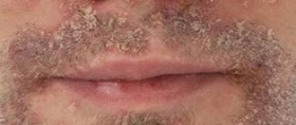
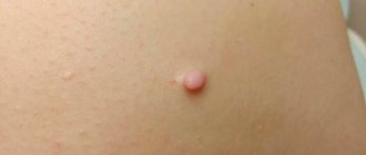
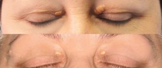
The first sign that you need to undergo an examination is the appearance of small blisters (papules) in the mouth and chin. In severe cases, they can spread throughout the face, reaching the cheeks, nose, eyes and temples. Papules are filled with clear liquid. Color varies from flesh-colored to pink to bright red depending on the stage of the disease. Pimples appear singly or in groups. The skin in these areas becomes rough. Bursting, papules form crusts. If you accidentally touch one, pigmentation will eventually remain on the skin.
Patients do not always experience serious pain. The sooner treatment of perioral dermatitis on the face in children and adults begins, the easier it is to get rid of the disease, and the lower the risk of unpleasant consequences. When you notice the first symptoms of the disease, make an appointment with a dermatologist.
Risk groups and similar diseases
Signs of the disease are most often found in women, but children and adolescents periodically experience them due to weakened immunity. In place of the blisters, ulcers often form. When spread over the entire face, it becomes a threat to vision, which is especially important in the situation with children. Therefore, when checking with a specialized doctor about how to treat perioral dermatitis in children, it is worth paying a visit to an ophthalmologist.
In the case of teenagers, the disease may not be detected immediately, because, experiencing hormonal changes, boys and girls often encounter skin problems. Blackheads and acne affect most people, so it can be difficult to notice papules at an early stage. Patients prone to allergies also rarely accurately diagnose the disease. Perioral dermatitis in children is often attributed to hormonal imbalance.
General notes on therapy
The choice of drug and treatment method for perioral dermatitis depends on the severity and stage of the disease.
For the period of treatment, regardless of the chosen method of therapy, stop using cleansing and moisturizing cosmetics, decorative cosmetics, fluoridated toothpastes, as well as external and systemic drugs containing glucocorticosteroids (when discontinuing systemic drugs containing glucocorticosteroids, it is necessary to take into account the indications for which the drugs were prescribed , the patient should be advised to consult the doctor who prescribed them regarding possible discontinuation of the drug). There are “zero”, external and systemic therapy for perioral dermatitis. For mild perioral dermatitis, “zero” therapy is sufficient, which consists of abolishing all external, including cosmetic, products, especially drugs containing glucocorticosteroids. Improvement occurs on average within 2 weeks. If it is ineffective, drug therapy is prescribed.
Indications for hospitalization
None.
Requirements for treatment results
Regression of rashes.
Prevention
- limiting the use of drugs containing glucocorticosteroids;
- limiting the use of cosmetics.
External therapy
Prescribed for mild to moderate severity of the disease, used as monotherapy, and for severe perioral dermatitis it can be prescribed in combination with systemic therapy:
- Metronidazole, 1% cream 2 times a day externally for 8 weeks
- azelaic acid, 20% cream 2 times a day externally for 2-6 weeks
- pimecrolimus, 1% cream 2 times a day externally for 4 weeks
Note. In the instructions for medical use of azelaic acid and pimecrolimus, perioral dermatitis is not included in the indications for use of the drug.
Systemic therapy
Prescribed for severe forms of the disease, as well as when external therapy is ineffective:
- tetracycline 250-500 mg 2 times a day orally for 4-8 weeks. Prescribed to patients over 8 years of age.
- For tetracycline intolerance in pregnant women, children under 8 years of age and for granulomatous form of perioral dermatitis in children: erythromycin 250 mg 2 times a day orally for 1 to 3-4 months.
Note. In the instructions for medical use of tetracycline and erythromycin, perioral dermatitis is not included in the indications for use of the drug.
If antibacterial therapy is ineffective, isotretinoin 0.1-0.7 mg per kg body weight is prescribed orally once a day for 6-20 weeks.
Note. In the instructions for medical use of isotretioin, perioral dermatitis is not included in the indications for use of the drug.
Diagnosis of perioral dermatitis
Having noticed the first signs of the disease, having felt unpleasant symptoms, you need to make an appointment with a specialized specialist. The initial appointment with a doctor consists of an examination, a description of the medical history based on the patient’s complaints and information about the individual characteristics of his body and possible reactions.
Before treating perioral dermatitis, tests are one of the mandatory procedures. They allow you to identify the prerequisites for the development of the disease and accurately diagnose it, without confusing it with herpes, rosacea and other skin lesions. You will need:
- Dermatoscopy. Gives an assessment of the epidermis and helps assess the course of the disease
- Scrapings. Determine the infectious nature if the symptoms have a corresponding origin
- Allergy tests. Demonstrate the level of skin sensitivity to staphylococcus and streptococcus
As a result of the examination, the doctor explains to the patient how to cure perioral dermatitis, clinical recommendations are entered into the disease record. A course of medications and therapy is prescribed.
Treatment of perioral dermatitis
A confirmed diagnosis usually requires an integrated approach. The treatment regimen involves a complete refusal of cosmetics, as well as corticosteroid drugs. The patient is advised to use antihistamines to relieve burning and itching. Herbal lotions are used to help reduce inflammation. Reflexology is used to stop symptoms by pressing on specific points or placing special needles.
Having weakened the symptoms, you can resort to the use of antibiotics to get rid of the infection if it is diagnosed during the examination. For perioral dermatitis, medications are prescribed by a doctor taking into account age, body characteristics, allergies and pregnancy. Strengthening the immune system and restoring the balance of intestinal microflora are important. The latter may require a specific diet. Avoid fried, spicy and fatty foods, snacks, sweets and fast food. Give up alcohol and cigarettes.
Prevention
Treatment with ointment for perioral dermatitis is not always a panacea. In some cases, the disease can return, so the course of taking medications can be long. Any irritant often leads to the appearance of the disease. Following simple recommendations will help you avoid possible risks:
- Check the composition of cosmetic products and treat their choice with special attention
- Don't neglect personal hygiene
- Strengthen your immunity
- Eat right and monitor your digestive health
- Be aware of allergies
- Monitor hormonal balance during pregnancy, in case of possible changes due to nervousness, illness or other reasons
Perioral dermatitis detected at the first stage is the easiest and fastest to treat, so remain sensitive to the body’s signals and begin the fight against the first symptoms of the disease in a timely manner.
Causes
The exact causes of the development of perioral dermatitis are not known, but trigger factors have been identified, i.e. provoking:
- Deterioration, weakening of immunity;
- Climate change;
- Exposure to ultraviolet rays;
- Use of corticosteroids;
- Fluoridated toothpaste;
- Genetic predisposition;
- The use of low-quality cosmetics against the background of a tendency to allergic reactions to cosmetics;
- Emotional stress.
The most reliable seems to be the development of perioral dermatitis in connection with long-term local use of corticosteroid ointments against the background of other provoking factors. There is a decrease in local immunity, which gives impetus to the development of the inflammatory process.
Bibliography
- Adaskevich V.P. Perioral dermatitis: clinical picture, diagnosis, treatment / V.P. Adaskevich // Dermatology. – 2008. – No. 1. – P. 17–20.
- Grashkin V. A. Diagnostic criteria, epidemiology and substantiation of clinical and pathogenetic types of perioral dermatitis / V. A. Grashkin, M. S. Gromov // Voenmed. magazine – 2010. – No. 10. – P. 32–45.
- Rodionov A. N. Dermatocosmetology. Lesions of the facial skin and mucous membranes. Diagnostics, treatment, prevention / A. N. Rodionov. – St. Petersburg. : Science and technology, 2011. – 912 p.
- Karelina O. Yu. Perioral dermatitis: treatment with azelaic acid / O. Yu. Karelina, Yu. M. Karelin // Klin. dermatology and venereology. – 2006.
Treatment
Perioral dermatitis is a condition that can be treated. The duration of treatment can range from a month or two to six months or a year in severe cases. The main focus of therapy is the normalization of the microflora of the facial skin, including the elimination of pathogenic and opportunistic microorganisms, the diversity and aggressiveness of which increases with this disease. In the absence of success of external treatment and severe disease, doctors currently have effective systemic drugs in their arsenal that provide a high cure rate and, as a result, facial beauty and good mood.
Popular questions about perioral dermatitis
How can you get perioral dermatitis?
Dermatitis is not contagious, as it is an individual reaction of the body to the use of fluoride-containing pastes, inappropriate cosmetics and drugs containing corticosteroids. In the case of diagnosing infectious preconditions for a disease, it is worth finding out its nature, but it will not become a reason to limit communication.
How does perioral dermatitis manifest?
The disease is characterized by the formation of papules, blisters filled with colorless liquid, which spread around the mouth and along the chin, rising to the eyes. Dry skin, itching and burning accompany the disease, but in the first stage patients experience minimal discomfort.
Is it possible to cure perioral dermatitis?
The disease is easily treatable if you follow your doctor’s recommendations, use the necessary medications and avoid products and products that can cause perioral dermatitis. Depending on the extent of the damage, the course can take from several days to several months.
Clinical manifestations of dermatitis
Perioral dermatitis most often affects women, mainly aged 20-30 years. Clinically, it manifests itself as a rash against the background of inflamed skin, most often located around the mouth, and somewhat less often - the nose and eyelids. A distinctive feature of dermatitis is the presence of a light rim of unaffected healthy skin around the lips; in severe forms, swelling may occur. This dermatitis can resolve on its own when the causative factor is eliminated or become chronic, which significantly reduces the quality of life.