Neoplasms on the scalp and neck account for about 5% of benign tumors. They can be located in different areas and have different origins. The medical clinic in Kaluga successfully treats neoplasms on the head and facial skin using modern innovative methods. The treatment method is determined individually after examining the patient and a full diagnosis.
Why does atheroma occur on the head?
Atheromas can be congenital or acquired. In the second case, their development is facilitated by:
- metabolic disorders,
- hormonal changes (high testosterone levels),
- poor hygiene,
- abrasions,
- increased skin oiliness.
Atheromas can be single or multiple, their sizes range from a pea to a chicken egg and larger. Since atheromas develop from the sebaceous glands, they occur more often on those parts of the body where there are many of these glands: on the head, face, neck, and armpits.
Reasons for development
It is still unclear why soft tissue sarcomas occur, but certain risk factors have been identified.
Genetic predisposition
- nevoid basal cell syndrome (Gorlin syndrome);
- neurofibromatosis (von Recklinghausen disease);
- tuberous sclerosis (Bourneville disease);
- Gardner's syndrome;
- Werner's syndrome.
Carcinogens
The risk of developing angiosarcomas increases significantly with previous exposure to phenoxy herbicides (2,4-dichlorophenoxyacetic acid, 2,4,5-trichlorophenoxyacetic acid) and dioxins.
Immunosuppression
The most striking example is Kaposi's sarcoma in people with AIDS, autoimmune hemolytic anemia, and organ transplant recipients.
Most patients come with a complaint of tumor growth after an injury or bruise. There is no connection between trauma and the development of soft tissue sarcomas.
Is scalp atheroma dangerous?
If there are no complications, then the biggest trouble that atheroma of the scalp causes is the lack of hair in the place where it is located. But the situation may change:
- If an infection gets inside the atheroma, it will turn into an abscess. An abscess of the soft tissues of the head will occur.
- Ulcers are dangerous on any part of the body, but especially on the head. The infection may spread deeper to form an intracranial abscess.
- Sometimes (although very rarely, there is such a possibility) atheromas transform into malignant tumors.
If an incomprehensible lump appears on the scalp, you should not wait for complications. Contact our doctors, they will remove it quickly and carefully!
Treatment of papillomavirus infection
Papillomas are only a symptom, not a disease.
These symptoms are caused by the papilloma virus.
To prevent formations from reappearing after removal, treatment for papillomavirus infection is required.
It is carried out using:
- immunomodulators
- nonspecific (general action) antiviral agents
The drugs are used both locally and systemically.
They can be injected into the area where papillomas are located.
Systemic immunocorrection is also required.
It is usually performed by an immunologist under the supervision of an immunogram.
After normalization of immunity, the papillomavirus usually leaves the body within several months.
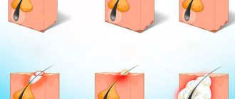
How are atheromas removed?
No amount of burdock, plantain or compresses with alcohol help with atheroma. It needs to be treated surgically. You should visit a doctor as early as possible. If the atheroma has festered, you need to run to the surgeon.
The most senseless way to remove scalp atheroma is puncture, when a needle is inserted into the cyst and its contents are pumped out with a syringe. No normal doctor does this (except perhaps to obtain the contents of the cyst and send it for a biopsy). As long as the walls of the atheroma are preserved, it will fill up again and again. In order to rid the patient of a cyst, its walls must be completely removed. The classic operation looks like this:
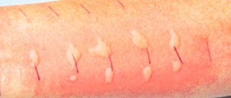
- Two bordering incisions are made on the skin in the area of atheroma.
- A blunt instrument (most often closed scissors) is placed under the atheroma.
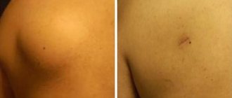
- The atheroma is peeled off, and stitches are placed on the wound.
The intervention is performed under local anesthesia. Usually the operation is performed on an outpatient basis, but if the atheroma is large, hospitalization in a hospital may be required. However, scalpels are becoming a thing of the past. Surgeons now have more efficient tools. For example, lasers. With their help, atheroma on the head less than 5 mm in size can be evaporated without a trace; for larger ones, additional small incisions will have to be made.
An even more advanced way to remove atheromas of the scalp is radio wave and laser surgery. These are the methods that doctors at the ProfMedLab clinic use. Laser or high frequency radio waves work instead of a scalpel. They cause little tissue damage, healing occurs faster, and the cosmetic effect is better.
Clinical picture
The cyst is a soft lump with rounded contours, located subcutaneously. Even small atheromas are clearly visible if you part your hair and carefully examine the changed skin.
Non-inflammatory atheromas are painless and do not cause discomfort, but if an infection occurs (which almost always happens with an accidental injury), the symptoms become more obvious. Suppuration of the cyst leads to its increase in size, intense pain, redness of the skin, and local swelling. The skin in the area of the inflamed atheroma is hot, and pus may be released from the outlet of the sebaceous duct.
Recurrence of atheroma after removal
Relapse is one of the possible complications after surgery to remove atheroma. Most often, the reason is trivial and related to the human factor: the doctor did not completely remove the capsule. We will consider the option when the surgeon did everything “perfectly”, and there was no trace of atheroma left on the head. Can it happen again? Maybe if the same thing happens to another sebaceous gland. Can this be prevented? Following three simple recommendations will help reduce risks:
- Wash your hair regularly and thoroughly.
- Use special shampoos that help fight oily scalp.
- Try to exclude fatty, sweet, and spicy foods from your diet.
If atheroma does appear, do not hesitate to visit a doctor. Make an appointment with a surgeon at the Medical Center: +7 (495) 120-08-07.
Diagnostics
The diagnosis is made based on histological examination of tumor tissue.
There are two methods for obtaining material for research:
- core needle biopsy;
- open biopsy.
The biopsy should be performed in the location that during the operation will be included in the area of excision of the tumor formation.
Diagnostic algorithm:
- Examination: A lumpy, rounded yellow or gray nodule may be observed. It can have different densities and consistencies. Soft nodes - with liposarcomas, dense - with fibrosarcomas, jelly-like formations - with myxomas;
- Complete blood count with calculation of leukocyte formula and platelet count;
- Biochemical blood test with determination of liver and kidney function indicators (including electrolytes);
- Coagulogram;
- MRI of the head and neck area;
- CT scan of the lungs;
- Ultrasonography;
- Radioisotope study of skeletal bones (for myxoid liposarcomas);
- CT scan of the brain (for alveolar soft tissue sarcoma and hemangiopericytoma).
Important: for the purpose of initial examination and clarification of the stage, routine PET scanning is not recommended.
Treatment for a common process
Chemotherapy is the mainstay of treatment for advanced disease because the drugs injected enter the bloodstream and reach cancer cells throughout the body.
The most common chemotherapy drugs used for soft tissue sarcomas are doxorubicin, trabectedin, gemcitabine, docetaxel, and paclitaxel. These drugs may be prescribed alone or in combination.
Chemotherapy in patients with progressive disease should be based on doxorubicin or epirubicin, both drugs belong to the anthracyclines. In patients with angiosarcoma, paclitaxel or docetaxel may be offered instead of doxorubicin.
Adding another drug(s) to doxorubicin or epirubicin may enhance the effect of systemic chemotherapy in some patients. This choice primarily depends on the histological type of cancer.
If the first chemotherapy does not give the expected result, then another chemotherapy may be offered. The choice of one or more drugs will depend on previously used drugs, as well as on the histological type of the tumor. Drugs that may be considered include ifosfamide, trabectedin, gemcitabine, docetaxel, and paclitaxel.
Targeted therapy
This treatment works by binding to a specific protein or structure involved in tumor growth and progression. Side effects differ from traditional chemotherapy and depend on the mechanism of action of the drug. Targeted drugs approved for use in soft tissue sarcomas in Russia are:
- pazopanib – for soft tissue sarcomas other than liposarcomas;
- imatinib – for dermatofibrosarcoma, when systemic therapy is required.
Radiation therapy
Radiation therapy may be used to relieve symptoms or prevent complications, such as bone metastases.
Surgery
Surgical treatment of metastases may be considered depending on their location and medical history. For example, when lung metastases appear long after initial treatment and when, in the opinion of the surgeon, they can be completely removed.
Types of bumps behind the head depending on the reasons
Masses on the back of the head can occur for a variety of reasons, sometimes accompanied by pain and sometimes by deformation of the scalp. Some of the reasons are given below.
Mass due to head injury
If for any reason the blow is struck from behind, a slightly hard mass and bleeding (hematoma) may form at the site of the blow and under the skin. These bumps usually heal gradually over two weeks.
Cold compresses can be used to reduce swelling for minor injuries. Using painkillers is also a great help in reducing pain.
But in the event of a severe injury, a person may suffer a concussion. Symptoms such as headache, nausea, vomiting, blurred vision, extreme tiredness or drowsiness, and memory loss are important and should be treated by a doctor.
Scalp diseases: names and causes
Common skin diseases are considered to be:
- diseases of a dermatological nature (including seborrhea, eczema and hyperkeratosis);
- autoimmune diseases of the scalp (including psoriasis and scleroderma);
- infectious (formation of pustules, appearance of lice, etc.);
- fungal rashes.
Whatever the cause of the disease, it must be eliminated in time. It is important to start treatment as early as possible, because wasted time can have serious consequences.
Symptoms of bruises
Bruises are accompanied by the appearance of the following clinical signs:
- redness, swelling, or swelling;
- hemorrhage (hematoma) or bruise (appears at the site of redness, the area of appearance is subcutaneous fatty tissue, intermuscular and subfascial space);
- severe or aching pain (the intensity of the pain depends on the location, force of the blow and the shape of the traumatic object).
You should pay attention to prolonged pain; it may indicate a fracture, bleeding in internal organs, or ligament ruptures.
1 Treatment of bruises and hematomas
2 Treatment of bruises and hematomas
3 Treatment of bruises and hematomas
Where are the lymph nodes located?
| Part of the body | Where are they located? |
| Upper limbs | - under the arms, - on the elbows. |
| Head | - in the area of the ears, - under the jaw. |
| Rib cage | - in the area of the trachea and bronchus, - near the sternum, - between the ribs. |
| Neck | - in the front of the neck, both on the surface and deep. |
| Pelvis | - in the area of the sacrum, - the ilium. |
| Lower limbs | - in the groin, both on the surface and in depth, - under the knees. |
| Abdomen | - in the area of the liver, - stomach, - internal genital organs in women. |