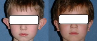
Otoplasty results
Almost any surgical intervention associated with violations of the integrity of soft tissues and cartilage is accompanied by various consequences. Otoplasty is no exception. The consequences, for the most part, manifest themselves in the form of:
- periodic pain;
- swelling;
- hematoma;
- small scars or scars.
After the operation is completed, the patient is given an anesthetic, but discomfort may continue for several days. Swelling and hematomas appear in 9 out of 10 cases, but disappear on their own after 10-14 days. The formation of scars or scars is possible due to the individual characteristics of the patient’s skin or the low qualifications of the plastic surgeon. You should always carefully choose the person you trust with your health and beauty. In order to avoid negative consequences after otoplasty, you must strictly follow all the doctor’s recommendations.
What is tympanoplasty
Tympanoplasty
is a surgical intervention for chronic purulent otitis media, an operation aimed at restoring damaged structures of the middle ear, eardrum and improving hearing.
Unfortunately, chronic suppurative otitis media is a common disease. Its development is facilitated by improper and untimely treatment of acute purulent otitis media, which can result in perforation of the eardrum (hole in it). Pathogenic bacteria enter the tympanic cavity through the perforation, so ear discharge may persist for many years.
Untimely treatment of purulent otitis leads to serious complications and can cause diseases such as meningitis, cerebral thrombosis, and brain abscess.
The inflammatory process is not limited to the mucous membrane of the tympanic cavity, but also leads to the destruction of bone structures - the walls of the tympanic cavity, the auditory ossicles. Over time, not only progressive hearing loss occurs, but there may also be severe purulent complications - meningitis, cerebral thrombosis, brain abscess
.
Early postoperative period
The most crucial moment is rehabilitation after otoplasty, because the extent to which the desired result is achieved depends on how it proceeds. After this operation, rehabilitation is divided into two types: early and late. The duration of the early rehabilitation period after otoplasty takes about 10 days and is accompanied by pain and swelling. Immediately after the operation is completed, a compression bandage is applied to the head. It is very important to follow the recommendations for wearing this bandage, because it performs such important functions as:
- fixing the ears in the desired position;
- protection of ears from accidental injuries;
- containment of swelling and spread of hematoma.
The success of otoplasty and the rehabilitation period largely depend on wearing a compression bandage, which is removed only on the recommendation of the plastic surgeon.
Progress of the operation
There are several techniques for performing otoplasty. They can be divided into three groups.
The first group of operations (suture) to correct the auricle uses sutures on the cartilage.
The second group of operations (resection) involves removing sections of cartilage and then stitching them together.
The third group is a combination of suture and resection techniques.
The choice of surgical technique to correct protruding ears depends on the correct preoperative analysis.
Auricular plastic surgery is performed on an outpatient basis and does not require hospitalization. The operation is performed under local anesthesia and usually lasts about an hour, sometimes a little longer.
The surgical field is treated and covered with sterile surgical linen. A skin incision up to 4 cm long is made behind the auricle. After hemostasis, the cartilage is modeled with incisions and sutures.
Sutures are applied with non-absorbable threads in such a way as to form the correct shape of the auricle and get rid of protruding ears.
The skin incision behind the ear is often sutured with threads that dissolve on their own.
Limitations after ear surgery
There are some limitations for the early period of rehabilitation after otoplasty. Until the stitches are removed, which is about 7-10 days, washing your hair is not recommended to avoid infection and is strictly prohibited for the first 72 hours after the end of the operation. After the doctor removes the stitches, it is better to start washing your hair with baby shampoo, as it contains fewer chemical components that can cause irritation.
It is best to sleep on your back, and to reduce swelling and reduce the likelihood of it spreading to surrounding tissues, the sleeping position should be semi-sitting.
The rehabilitation period for otoplasty is also accompanied by the treatment of sutures with antimicrobial agents. This allows you to significantly speed up the process of cell regeneration. In addition, do not forget that physical activity can cause sutures to separate and cause scars to appear.
Otoplasty: pain after surgery
When preparing the patient for discharge, an injection of pain medication is given - this is standard practice. For home care, the surgeon prescribes analgesics that easily relieve moderate pain. If its intensity increases sharply, is accompanied by bleeding, discharge of pus, an increase in the size of the ears, or high fever, you should contact your doctor.
In rare cases, the individual reaction of the body is such that even 2 weeks after otoplasty, pain after the operation persists. In the absence of complications, violations of the rehabilitation regime, or doctor errors, this is a variant of the norm.
Late postoperative period
The main task of the late period of rehabilitation after otoplasty is to create the most favorable conditions for the healing of the ears.
Rehabilitation ends 25-30 days after surgery and consists of the following:
- adherence to dietary nutrition;
- limiting showering and bathing;
- restriction of physical activity;
- maintaining a comfortable temperature regime.
The late period of rehabilitation after otoplasty is characterized by partial loss of sensitivity, a feeling of discomfort and slight swelling. All this indicates that the tissues are not ready to perform their functions in full.
Complications after otoplasty
Aesthetic otoplasty is a frequently performed operation. There are many different ways to correct protruding ears, and each can have complications. The plastic surgeon performing otoplasty must be aware of possible complications, their cause and how to eliminate them.
Asymmetry
Asymmetry may exist before otoplasty. If asymmetry occurs after surgery, correction can be avoided if the asymmetry is minor.
Surgical correction will depend on the cause of the asymmetry (i.e., too narrow or too weak a suture on one side, too much or little skin excision on one side, etc.).
Bleeding and hematoma
Bleeding after otoplasty may be caused by inadequate hemostasis, hypertension, vasodilation due to the use of local anesthetics, postoperative trauma, or existing coagulopathy.
Surgery should be performed as early as possible to stop the source of bleeding and evacuate clots. Untimely removal of a clotted hematoma can lead to tissue necrosis or chondritis.
Cartilage relief disorders
Palpable and visible cartilage edges or irregularities may be caused by cutting the cartilage along the anterior surface. To avoid this, it is necessary to use cartilage-sparing techniques.
Cartilage necrosis
This may occur due to a hematoma causing excessive pressure on the cartilage. The necrotic area must be removed. And this in turn can lead to deformation that will require correction.
Chondritis and perichondritis
Typically, clinical signs of infection (pain increasingly resistant to analgesics, swelling, redness of the ear) begin 3–5 days after surgery.
Chondritis may require drainage and debridement of the affected cartilage. The use of antibiotics during and after surgery and the placement of a drain can prevent the development of chondritis.
Granulomas
Braided nonabsorbable sutures are more likely to cause granulomas than monofilament nonabsorbable sutures.
To eliminate a granuloma, it is necessary to remove the sutures and the granuloma itself. Non-absorbable sutures, such as nylon or polypropylene, can cut through the skin, causing fistulas or granuloma.
Hypersensitivity
Postoperative hypersensitivity is the result of irritation of the nerve endings of the cutaneous nerves.
It usually subsides when there is no pressure on the ear and is quickly eliminated with mild painkillers.
Infection and suppuration
Infection is usually diagnosed by the presence of excessive pain, erythema, fever, or purulent discharge.
The festering wound must be opened, sanitized and thoroughly washed with an antiseptic solution. Oral antibiotics are also prescribed for at least 7 days. Failure to treat the infection at an early stage can lead to perichondritis, chondritis and cartilage necrosis.
Hypercorrection
Excessive correction of the middle third of the ear leads to a “telephone handset” deformity.
Excessive correction of the upper and lower concha leads to a “reverse telephone ear” deformity.
Narrowing of the external auditory canal
Surgical deformity resulting from anterior displacement of the pinna can cause obliteration (narrowing) of the ear canal.
This can be prevented by careful preoperative planning and adherence to a protruding ear plan.
Pain
Excessive postoperative pain may be a sign of a developing hematoma. The dressing should be removed and the wound examined for swelling indicating a hematoma.
Treatment consists of finding the source of bleeding, electrocoagulating it and evacuating the hematoma.
Recurrence of deformity (protruding ears)
Recurrence of the initial ear deformity can be caused by trauma, insufficient weakening of the cartilage, dehiscence or cutting of the suture.
Rough scars
Hypertrophic or keloid scars may occur in approximately 2-5% of patients.
The earring hole can be a source of keloid scars. Excessive tension in the suture area can cause the scar to expand.
Careful and conservative skin resection can reduce the formation of rough scars.
About 8% of hypertrophic scars resolve without treatment, but 80% of keloid scars recur.
Treatment may include
- use of hormonal drugs (triamcinolone), alone or in combination with 5-fluorouracil or bleomycin,
- removal with or without radiation,
- cryotherapy with or without steroid injection and
- compression therapy.
Skin necrosis
Skin necrosis is usually the result of postoperative compression bandages being too tight or it may be caused by excessive electrocoagulation of the skin. This can lead to rough scars.
Seam extrusion (cutting seams)
Suture cutting can occur as a result of permanent sutures being placed too close to the skin and placing excessive tension on the cartilage. Also, the development of infection can lead to cutting of stitches.
Telephone ear deformity
This deformity is created by the uneven distance of the pinna, where the upper and lower poles of the ear are still convex in relation to the middle third of the ear.
The reason is due to inadequate correction of the middle pole, which is excessively tightened by the suture, as well as due to excessive removal of skin in the postauricular area, soft tissue or cartilage in the middle third of the ear.
Prevention includes
- tightening the middle seam last to avoid potential overcorrection,
- avoiding excessive cartilage removal.
Treatment requires removal of incorrectly placed sutures and application of new ones.
Insufficient correction
Insufficient correction of the upper helix and lobule results in a relatively noticeable upper third of the ear called a “telephone deformity.”
Insufficient correction of an overdeveloped bowl can result in “reverse telephone ear.”
Unrealistic patient expectations
The surgeon must detect the patient's unrealistic expectations before ear surgery. In such cases, it is better to refuse the operation.
Indications and contraindications
Surgical intervention is necessary in the following cases:
- central or marginal perforation of the eardrum, that is, there is damage to it, which leads to displacement of the auditory ossicles;
- perforation of the eardrum without significant inflammation of the middle ear;
- fibrosis of the middle ear;
- tympanosclerosis;
- middle ear polyps;
- chronic inflammation of the middle ear with severe suppuration.
An absolute contraindication to surgery is:
- severe general condition;
- exacerbation of chronic diseases;
- purulent complications and sepsis;
- complete deafness;
- adhesive otitis media;
- persistent disturbances in the patency of the auditory tube - these can be either congenital anomalies or the formation of adhesions and scars as a result of inflammation.
Relative contraindications:
- epidermization of the tympanic cavity - absolute replacement of the mucous membrane of the tympanic cavity by the epithelium of the external auditory canal;
- functional (temporary) obstruction of the auditory tube;
- upper respiratory tract diseases;
- allergic diseases in the acute stage;
- acute inflammatory process in the middle ear.
The doctor assesses the required volume of surgical interventions and always informs the patient about possible risks.
Prohibitions
In order for the wound healing process after surgery to proceed without complications, it is necessary to strictly follow all the recommendations of the plastic surgeon. But in addition to the doctor’s advice, there are a number of prohibitions, compliance with which guarantees the successful completion of the operation and the recovery process.
The following factors are strictly prohibited:
- Smoking and drinking alcohol.
- Eating pickles, marinades, as well as fatty, hot and spicy foods.
- Participation in certain sports that involve contact with an opponent and the possibility of injury to the ears (boxing, wrestling).
- Going to the beach or solarium and exposure to direct sunlight should be limited.
- Use of shampoos and other detergents for washing your hair; you can only use baby shampoo.
- Removing the bandage, stitches and peeling off the crusts from the scar yourself.
In addition, wearing glasses is prohibited for two months. Women are not recommended to wear earrings or other jewelry on their ears.
The rehabilitation period for eliminating defects in the ears does not last that long, so you need to be patient and endure all these inconveniences and discomfort in order to achieve the desired result from the operation without bad consequences.
Full recovery occurs only after six months, then all restrictions are lifted if the correction of the ears was carried out efficiently.
After otoplasty, the ears are tightly pressed to the head. Should I worry?
Don't rush to conclusions, this may be a temporary phenomenon. After six months you can judge the result. Recovery is a complex physiological process that does not occur as quickly as we would like. If, after otoplasty, the ears are pressed tightly against the head, this causes anxiety and discomfort, the procedure may be considered for a second time.
Asymmetry is an indication to operate again. Pathology occurs if the operation was performed on one ear. However, the appearance of a defect occurs when both organs are operated on. In this case, repeated surgery involves reconstructive correction.
If it is impossible to achieve an ideal result the first time, plastic surgery is carried out in two stages. The surgeon warns immediately about a repeat intervention.
It is difficult to understand the causes of the defect. You cannot determine on your own whether additional correction is needed or whether the problem will disappear after some time. A conversation with a doctor will help you decide whether a repeat procedure is advisable. You can sign up for a free consultation by calling the number listed on the website.
Types of tympanoplasty
Myringoplasty
– restoration of the integrity of the eardrum. This type of tympanoplasty is indicated for patients who have preserved and mobile all the auditory ossicles in the tympanic cavity; they only need to close the perforation of the eardrum. The material for myringoplasty is an autograft - a flap of skin with underlying layers is taken from behind the patient’s ear. Own tissues take root well and are not rejected by the body, which is their huge advantage as opposed to foreign materials.
Tympanoplasty with ossiculoplasty
– reconstruction of not only the eardrum, but also the destroyed auditory ossicles.
In chronic purulent otitis media, the auditory ossicles (there are three of them - the malleus, the incus and the stapes, their connections to each other resemble a microscopic joint) can be damaged due to a constant inflammatory process. Without them, it is impossible to conduct sound to the perceptive nerve endings of the inner ear. Therefore, the missing elements of these ossicles are replaced by artificial prosthetic hearing ossicles made from Tiflon, bioceramics and other synthetic materials. Our clinic uses modern titanium prostheses. If the chain of auditory ossicles has minimal destruction, then it is possible to do without artificial materials - the preserved elements are somewhat rebuilt, while maintaining their mobility. The final stage of surgery is myringoplasty
.
In the case of fusion of the two surfaces of the middle ear after complications with otitis, it is possible to restore the tympanic cavity using the remnants of the eardrum and grafts connecting the tissues to each other.
All types of tympanoplasty are performed using a modern microscope for otosurgery.