A fistula always comes suddenly, when the inflammation has already been cured or the surgical wound has long healed. Its appearance cannot be predicted, although its formation does not occur without symptoms, but it is impossible to guess that a fistula will occur in this place. But it is very difficult to confuse an emerging fistula with something else.
- What is a fistula?
- Types of fistulas
- Causes of fistulas
- Symptoms of appearance
- Diagnostic methods
- Methods for treating fistulas
- Prevention of fistulas
What is a fistula?
A fistula is an anastomosis formed as a result of a pathological process between the hollow organs of the gastrointestinal tract and/or genitourinary system. A fistula is also a convoluted tubular passage from an organ, passing through soft tissues with access to the skin.
A more euphonious name for a fistula, borrowed from Latin, is “fistula,” but this medical concept is broader, since it also includes artificially formed anastomosis, such as a surgically created fistula between a vein and an artery in dialysis patients. In one form, a fistula is a fistula; in another, it is an artificially created anastomosis; a fistula is always a miraculous pathology.
A fistula always has a beginning - an internal opening, localized in the primary focus of inflammation with suppuration. The beginning of a fistula can be a non-healing wound from injury or surgery, which is especially typical for organs of the gastrointestinal tract that produce secretions: gastric, intestinal or pancreatic juice, bile.
The external opening of the fistula can open into another organ or onto the skin, as in the case of a rectal fistula, but this opening is not necessary - some fistula tracts blindly end in soft tissues, muscles or fiber, forming purulent cyst-like cavities there - leaks.
How does a staphylococcal infection manifest?
Symptoms, its duration and intensity depend on the strain of the pathogen, the general physical condition of the patient and the characteristics of his immunity. Two characteristic signs of staphylococcal damage: inflammation of various localizations and intoxication.
With pyoderma, the sweat, sebaceous glands, and hair follicles become inflamed, causing painful convex nodules that fill with pus to form on the skin. Boils and carbuncles are usually surrounded by areas of red, swollen skin. With intense inflammation, fever and swelling of the lymph nodes located close to the sites of inflammation are likely.
Infection of the nasal sinuses by staphylococcus is manifested by a common runny nose with viscous yellowish or green discharge. Penetration of infection deeper causes the development of sinusitis. Their symptoms are typical:
- congestion in the bridge of the nose;
- feeling of heaviness, fullness on the affected side;
- labored breathing;
- thick purulent discharge from the nose;
- increase in body temperature above +37°C.
Often the infection spreads to the middle ear, causing otitis media: sharp shooting pains, hearing loss. When the mucous membranes of the eyes are damaged, suppuration of the conjunctiva develops, the sclera turns red and swells.
When staphylococcus attacks the upper respiratory tract, purulent pharyngitis or laryngitis is inevitable. Their signs:
- hoarse voice;
- severe swelling, redness of the throat;
- difficult painful swallowing;
- dry obsessive cough, discomfort in the throat.
Staphylococcal pharyngitis often affects children under 12 years of age. In adults, this pathology is controlled by the immune system.
With the development of inflammation in the lower parts of the respiratory tract, bronchitis and pneumonia are “relevant” with a characteristic barking or wet cough, high fever and physical weakness.
Less common signs of staphylococcus infections:
- food poisoning;
- inflammation of joint tissues, muscles;
- cardiac dysfunction.
Among the serious complications caused by staphylococcus are: meningitis, lung abscesses, osteomyelitis, toxic shock syndrome, generalized sepsis.
Types of fistulas
The classification of fistulas is diverse; each organ has its own gradation of fistulas by location, sometimes the degree of involvement of surrounding tissues, and even by the volume of secretion released through the fistula.
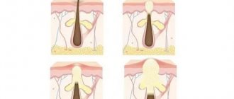
Complete fistulas have an external and internal opening, incomplete fistulas have only an internal opening.
Those opening in the skin are called external fistulas, while fistulas connecting organs are called internal .
Depending on the number of organs involved in the process, internal fistulas can be combined or isolated . Isolated fistulas are named after the organ that gave rise to them: pancreatic, biliary, intestinal, vaginal, ureteric, and so on.
When there is an anastomosis of two or more organs - a combined type of fistula - a “combined” name is used, so with a fistula between the rectum and the vagina - rectovaginal fistula, with a fistula from the gallbladder to the stomach - cholecystogastric fistula, between the pancreas and the stomach wall - pancreatogastric or pancreas-gastric fistula.
According to the number of tracts, fistulas are divided into single-channel or simple and multi-channel or complex , as well as branched or indirect and unbranched or direct .
According to the condition of the tissues and course - infected or complicated fistulas, as a rule, purulent and “clean” uninfected or uncomplicated fistulas with the release, for example, of bile or pancreatic juice.
| Depending on the number of holes | Complete fistulas | They have two openings - they connect two organs to each other or one organ to the surface of the skin. |
| Incomplete fistulas | They only have one hole. | |
| Depending on the location of the second hole (for complete fistulas) | Internal fistulas | Connects internal organs. |
| External fistulas | Connect internal organs to the surface of the skin. | |
| Depending on the number of organs involved | Isolated fistulas | They are named after only one organ that gave rise to the fistula. |
| Combined fistulas | Two or more organs are involved. | |
| Depending on the course of the fistula | Single channel (simple) | One fistula tract. |
| Multichannel (complex) | Several fistula tracts. | |
| Branched (indirect) | There is a branching of the fistula. | |
| Unbranched (straight) | No ramifications. | |
| Depending on the presence of complications | Uncomplicated (uninfected) | There are no signs of infection. |
| Complicated (infected) | There are signs of an infectious process in the fistula. | |
| Depending on the current | Primary | The fistula was discovered for the first time. |
| Recurrent | The process either subsides or reactivates: inflammation develops, new fistula tracts appear or old ones open. |
Fistulas are divided into primary and chronically occurring - recurrent , when the process either subsides or becomes inflamed again with the formation of new passages and, sometimes, the closure of old ones.
A rectal fistula is classified in relation to the anal sphincter, and fistulas that are localized above the anus and go around it like a horseshoe, with an opening into the rectum, are also graded according to 4 degrees of complexity.
Complete pancreatic fistulas can be small with the release of up to half a glass of juice, medium - up to 700 milliliters and large.
There are as many classification options as there are types of fistulas in human nature.
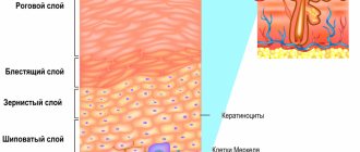
What is staphylococcus
It is a nonmotile, spherical, Gram-positive microorganism that forms numerous colonies. Staphylococci are anaerobic - they can exist and reproduce without oxygen, in a closed environment. There are more than twenty strains of bacteria. Some of them are quite harmless, while others cause powerful pathological reactions in the human body. Parts of the body and organs most vulnerable to staphylococci:
- leather;
- mucous membranes;
- connective tissue;
- subcutaneous tissue;
- nerve tissue;
- heart muscle.
By infecting them, microbes produce toxic substances and provoke inflammation, which in severe cases leads to sepsis, irreversible changes in structures or permanent dysfunction.
Causes of fistulas
Most fistulas are formed as a result of a complicated course of the inflammatory process : in acute pancreatitis, inflammation of the tissue surrounding the rectum - paraproctitis, ulcerative necrotizing colitis - Crohn's disease.
Destructive processes in internal organs can also give rise to the formation of a fistula tract, as occurs when pressure sores of the gallbladder wall are caused by a large stone, when compression of the tissues leads to their thinning and subsequent rupture. In three out of four women with cholelithiasis, bedsores are the leading cause of the formation of a biliary fistula.
A similar mechanism of local tissue destruction is activated when a gastric ulcer perforates or penetrates into the pancreas, resulting in the opening of the gastro-pancreatic fistulous tract.
Some researchers believe that the cause of fistula formation is ischemic changes in the rectal wall with frequent use of rectal suppositories with NSAIDs.
Surgical , birth and spontaneous are one of the leading causes of fistula formation. Surgical injuries can be accidental - not noticed during inspection and almost microscopic dissections of tissue in the operation area and delayed, when the body rejects the materials used in surgery: suture silk or stapler staples.
In addition to accidental surgical trauma, fistulas can be caused by surgical assistance during the opening of purulent paraproctitis. We are not talking about inadequate surgery, it’s just such an unfavorable place: poor blood supply to fiber - the main site of action, with the impossibility of stopping the transit through the intestine of feces, which are replete with intestinal microflora. There is no sterility and pathogenic microorganisms are always present - there are no conditions for the surgical wound to heal.
Injuries during childbirth , especially ruptures due to inadequate obstetric care, as well as injuries undetected during postpartum examination, are the most common cause of vaginal fistulas.
Changes in soft tissues after long-term radiation therapy , especially with combined irradiation of cervical or uterine cancer, and rectal cancer, can also lead to the formation of fistulas. Several factors predispose to the formation of a tract: impaired tissue nutrition as a result of post-radiation fibrosis in the presence of abundant intestinal and vaginal microflora. Post-radiation fistulas are quite difficult to differentiate from true malignant fistulas that form due to necrosis of a progressive or recurrent malignant tumor.
Pancreatic cysts, filled with caustic pancreatic secretions, are able to spontaneously find a way out for their contents, melting the tissue with enzymes and forming fistulous tracts from the abdominal cavity to the chest - between the pancreas and the pleura or bronchi.
A similar result, only with access to the skin, is possible with puncture drainage of cystic cavities of the pancreas, performed for therapeutic purposes in a weakened patient, or with drainage of a common bile duct blocked by a stone or tumor.
Each fistula has its own specific cause and combination of unfavorable conditions for healing.
Symptoms of appearance
The process of fistula formation is difficult to track; it can take several days, as happens with acute pancreatitis, or several months, as with post-radiation tissue changes.
Manifestations at the initial stage of fistula formation are due to its root cause, as a rule, a local inflammatory process resulting in purulent melting of tissue with pain and infiltration, often intoxication and fever.
Without an exacerbation of the inflammatory reaction, the fistula can be felt as a cord. The size of the compaction around the fistula tract is due to inflammatory infiltration and branching of the fistula tracts themselves, cicatricial changes in the surrounding tissues, previously involved in the inflammatory conglomerate.
A formed fistula has an entrance and, sometimes, an exit, the tissues around it are compacted, and discharge can be squeezed out of the hole: pus, bile, pancreatic juice, and so on. With a fistula, feces may leak from the intestine into the vagina from the genital organs; when there is an anastomosis of the intestine with the bladder, urine leaks from the anus. The discharge from the intestinal fistula has a fecal smell, and the purulent secretion from the vagina also has a specific smell. The smell of a discharged fistula, leading from the zone of disintegration of a malignant tumor, seems especially heavy to others.
Inflammation causes pain ranging from slight discomfort to unbearable pain. Tumor fistulas do not hurt because they form inside a disintegrating tumor.
When the infection activates with the formation of streaks of purulent contents, a general reaction occurs: intoxication, high temperature, sweating and pallor, palpitations and rapid breathing.
Blood in stool
The appearance of blood in the stool is an alarming symptom that is the first sign of many dangerous pathologies. Even if a person feels well, but observes regular bloody discharge from the anus, you need to visit a proctologist to find out the reasons for the deviation.
Causes of blood in stool
Bloody impurities in stool are a symptom of diseases in various parts of the gastrointestinal tract.
Blood appears in feces:
- with varicose veins of the esophagus or anal canal;
- hemorrhagic gastritis;
- stomach ulcer, duodenal ulcer;
- polyposis;
- cryptite;
- diverticulosis of the large intestine;
- nonspecific ulcerative colitis;
- proctitis;
- Crohn's disease;
- mesenteric thrombosis;
- vasculitis;
- hemorrhagic diathesis;
- liver cirrhosis;
- hemangiomas;
- terminal ileitis of various etiologies;
- infectious lesions.
Attention: bloody discharge from the anus is one of the early symptoms of intestinal and stomach cancer.
Bloody impurities in the stool can appear as a result of mechanical damage to the anus. For example, during anal sex, improper use of enemas. Sometimes bleeding occurs during a course of taking certain pharmaceutical drugs (anticoagulants, NSAIDs and others). In children, blood in the stool is observed with an allergy to cow's milk proteins or lactase deficiency.
Additional symptoms
Gastrointestinal bleeding can be acute or chronic. In the first case, large-scale blood loss develops rapidly (within a few minutes). The patient has:
- insatiable thirst;
- nausea;
- dry mouth;
- vomiting, the color and consistency of which resembles coffee grounds;
- a sharp drop in blood pressure;
- weakness;
- acute abdominal pain;
- muscle tremors;
- loss of consciousness;
- urinary retention.
Attention: this patient’s condition requires urgent medical attention.
Chronic bleeding can be recognized by the following accompanying signs:
- general malaise;
- brittle nails;
- blanching of the skin and visible mucous membranes;
- dizziness;
- increased fatigue;
- cardiopalmus;
- noise in ears;
- low blood pressure;
- hair loss;
- fainting;
- heartache.
As the disease develops, its characteristic clinical picture appears. At the initial stages, only a proctologist can determine the cause of the deviation.
Diagnosis of blood in stool
A visit to the clinic begins with a patient interview and palpation. The nature of bloody discharge has a high diagnostic value:
- bright scarlet streams or droplets not mixed with feces, smears on toilet paper - a sign of hemorrhoidal bleeding, anal fissure;
- red streaks are evidence of problems with the sigmoid colon;
- dark brown bloody inclusions, evenly connected with feces, are characteristic of diseases of the initial part of the large intestine;
- black, pasty stools indicate severe bleeding in the stomach, small intestine, or esophagus.
Based on the information received, the doctor establishes a presumptive diagnosis. To confirm (or refute) the patient is sent for additional examinations.
To determine the cause of bleeding and the degree of development of the disease, different methods are used:
- general blood analysis;
- endoscopic colonoscopy with collection of histological material;
- X-ray with contrast agent;
- examination of feces for helminth eggs, occult blood;
- Ultrasound;
- sigmoidoscopy.
The set of diagnostic procedures is selected individually. After receiving the examination results, the doctor prescribes effective therapy for the patient.
Treatment
Therapy for gastrointestinal pathologies, which are characterized by the appearance of blood in the stool, is carried out in two directions: eliminating the cause of the deviation, stopping the bleeding.
In drug treatment, drugs of different pharmacological groups are used, depending on the diagnosis:
- laxatives;
- venotonics;
- antibacterial;
- cytostatics;
- hormonal;
- glucocorticosteroids;
- antiviral and others.
Attention: the use of external or systemic medications without a doctor’s prescription leads to serious complications.
To stop intestinal bleeding, drugs that stimulate thrombus formation are used. When it is not possible to cope with the problem using conservative methods, the patient undergoes surgery.
Since spotting is accompanied by anemia, the patient is advised to take iron supplements to correct hemoglobin levels. In severe cases, the patient is given a transfusion of blood plasma or its components.
During the rehabilitation period, a person is recommended a special gentle diet with the exclusion of foods that injure the intestinal mucosa.
Prevention of blood in stool
There is no specific prevention of intestinal bleeding, but the risks of developing gastrointestinal pathologies can be reduced.
To do this you need:
- eat small portions, 4–6 times a day;
- add fiber to your diet, which is necessary for the normal functioning of the digestive tract;
- avoid stress;
- minimize the consumption of semi-finished products, fast food, fatty, fried foods;
- drink 1.5–2 liters of fluid per day (if there are no kidney diseases);
- avoid hypothermia;
- give up alcohol and smoking;
- do exercises regularly.
To notice abnormalities in the gastrointestinal tract in the early stages, it is necessary to undergo annual preventive examinations. This is especially important for people with a hereditary predisposition to diseases of the digestive system.
The causes of blood in the stool are varied, and it is not possible to determine them on your own. The answer to the question of why a violation occurred can only be given by a qualified specialist. Most pathologies accompanied by bloody discharge are benign and respond well to treatment in the early stages of development.
Author
Zagirov Fizuli Abumuslimovich
phlebologist, surgeon, proctologist
18 years of experience
+7
Diagnostic methods
Diagnosis of a simple unbranched single-channel fistula is not difficult - it is enough to feel a cord in the local seal, from which contents can leak when pressed.
All external exits of the fistula are examined with a button probe, this is how the localization of the passages is determined. The probe is inserted from the side of the skin, carefully pushing it all the way; if a rectal fistula is being examined, then the passage of the probe inside the rectum is determined with the index finger.
Next, a test is carried out with a dye - methylene blue, which is injected into the outer hole with a syringe. In case of rectal fistula, a cotton swab is inserted into the intestine before the test, and the exact location of the internal opening is determined by the dye marks on it.
For any fistula, the paint output can be recorded during endoscopy: anoscopy, rectoscopy, colonoscopy, cystoscopy, colposcopy, and so on. Endoscopic examination is one of the leading ones and is performed repeatedly in the process of diagnosis and treatment.
In some cases, fistulography is performed - an x-ray of the anatomical area before and after the injection of a contrast agent into the fistula. The procedure is not required only for simple and short rectal fistulas without exacerbation of inflammation.
Imaging methods - CT and MRI also make it possible to clarify the localization of tracts and leaks, branching and the root cause of the disease.
When the rectum is involved, ultrasonography (ultrasound) with a special rectal sensor is informative, when a computer program allows you to see the pathology in a three-dimensional image. When planning the operation, the function of the anal sphincter is additionally determined.
| Examination stage | Techniques | What are they used for? |
| Techniques used in the doctor's office during the initial examination |
|
|
| Lab tests |
|
|
| Instrumental studies |
|
|
Treatment
Recurrent furunculosis is not always easy to treat. Single boils can be opened by a surgeon, followed by local treatment. Antibiotics are prescribed if furunculosis is severe, does not respond to local treatment, or develops against a background of suppressed immunity or concomitant diseases.
In case of relapses, it may be necessary to treat all carriers of the causative agent of furunculosis (staphylococcus) at the patient’s home. If harmful bacteria colonize the patient's skin, the doctor can try to destroy them using antiseptics for external use. Effective treatment requires control of concomitant diseases, such as diabetes.
For effective treatment of small boils it is also recommended:
- Apply warm compresses to boils for about 10 minutes at a time. This promotes their opening and drainage.
- Never squeeze boils; this can spread infections in the soft tissues.
- Prevent pollution. After treating boils, you need to wash your hands. The fabric used for the compress should be washed thoroughly in hot water.
Methods for treating fistulas
Fistulas rarely close on their own; this can only be hoped for by creating favorable conditions, for example, limiting and partially controlling the movement of feces through the rectum using cleansing enemas. In the vast majority of cases, conservative therapy is ineffective; the only radical treatment is surgical, that is, excision of the pathological area, including reconstruction of the missing tissue.
Technically simple surgical intervention, including endoscopic, and a hundred surgical modifications cannot cure about half of the patients who suffer from relapses. It is especially difficult to achieve success with intestinal and urinary fistulas, since they are always contaminated with microflora. In some cases, it is necessary to resort to the formation of an intestinal stoma, temporarily stopping the movement of feces through the pathologically changed area of the intestine for several months.
In isolated cases, they resort to “old-fashioned methods” of treatment with scraping the mucous membrane of the tract, burning it with chemical reagents and enzymes, achieving sticking of the walls. Better results - in approximately 50% - are achieved by introducing fibrin glue into the fistula tract, which glues the walls together.
Tampons made of biomaterials act similarly to glue, sealing the internal opening; emptying of the passage can cause the walls to stick together and close the fistula.
Until now, the role of antibiotics in the treatment of fistulas caused by inflammation has not been determined, since drugs are not able to penetrate into the infiltrate due to massive scar changes. However, with fistula tracts due to Crohn's disease, specific drug therapy is mandatory and not unsuccessful.
Surgery for fistulas using the LIFT method
Why do boils recur?
Many scientists call the tendency of family members to furunculosis to be the most significant risk factor for both “simple” boils and the recurrent course of the disease. In recurring furunculosis, colonization of the nose or other parts of the body by staphylococcus plays an important role (this can be the inguinal folds, ears, folds under the mammary glands).
Risk factors for recurrent furunculosis include:
- anemia;
- use of antibiotics the day before;
- diabetes;
- hospitalization;
- multiple lesions on the skin;
- concomitant skin diseases (wounds, atopic dermatitis);
- poor personal hygiene skills;
- obesity;
- diseases of the blood system and hematopoietic organs;
- suppressed immunity (but scientists believe this is rarely the main cause).
In many cases of furunculosis, doctors cannot detect any of these risk factors; anyone can develop the disease. Scientists emphasize that many risk factors for relapse are modifiable, that is, they can be influenced in order to prevent the disease.
Prevention of fistulas
Not all diseases can be prevented, especially fistulas, which complicate the course of purulent paraproctitis. However, it is possible to adequately treat the diseases that lead to paraproctitis - hemorrhoids and fissures, and this is precisely what will prevent the formation of fistulas.
Complicated childbirth cannot be prevented, but high-quality and timely obstetric care, attentive attention to the woman and a thorough postpartum examination are available preventive measures.
The high incidence of post-radiation tissue damage and fibrosis progressing over time forced oncologists to abandon high doses of radiation therapy and even change approaches to the treatment of malignant tumors of the genital area.
Particular importance is attached to the correct choice of method of surgical treatment of diseases of the hollow organs and adequate management of the postoperative period.
In our clinic, a complicated course of the disease is very rare, because we not only know about prevention methods, but also actively use them.
| Price : | |
| Primary consultation with a surgeon | 5,100 rub. |
| Repeated consultation with a surgeon | 4,600 rub. |
| Consultation with a surgeon Ph.D. | 6,900 rub. |
| Primary consultation with an oncologist | 5,100 rub. |
| Repeated consultation with an oncologist | 4,600 rub. |
| Consultation with oncologist Ph.D. | 6,900 rub. |
| Consultation with an oncologist, MD. | 10,500 rub. |
| Fistulography | RUB 41,800 |
Book a consultation 24 hours a day
+7+7+78
Bibliography:
- Shelygin Yu.A., Blagodarny L.A. /Handbook of coloproctology// -M.: Litterra; 2012.
- Becker A., Koltun L., Sayfan J. /Simple clinical examination predicts complexity of perianal fistula// Colorectal Dis.; 2006; 8.
- Gaertner WB, Hagerman GF, Finne CO, et al. /Fistula-associated anal adenocarcinoma: good results with aggressive therapy// Dis Colon Rectum; 2008; 51.
- Genadry RR, Creanga AA, Roenneburg ML, Wheeless CR /Complex obstetric fistulas // Int. J. Gynaecol. Obstet.; 2007; Vol. 99; Suppl. 1.
- Ommer A., Herold A., Berg E. / S3-Leitlinie: Rektovaginale Fisteln (ohne M. Crohn) // Coloproctology; 2012; Vol. 34.
- Sahni VA, Ahmad R., Burling D. / Which method is best for imaging of perianal fistula?// Abdom Imaging.; 2008; 33.
- Zoulek E., Karp DR, Davila GW/ Rectovaginal fistula as a complication to a Bartholin gland excision // Obstet. Gynecol.; 2011; Vol. 118; N 2.