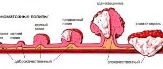
Vitiligo (leukoderma) is a peculiar skin disease characterized by depigmentation, manifested in the form of light spots throughout the body, including the face. They appear due to the lack of melanin production. The disease can develop in people of any age, but the largest percentage affects children and adolescents. Vitiligo in children occurs with certain characteristics.
The disease itself is not contagious or dangerous, but it does bring some discomfort and cosmetic defects. This pathology is characterized by gradual development and a long course.
As the child gets older, the spots increase in size and take on different shapes. If foci of vitiligo are localized in open areas of the body, then under the influence of sunlight, dryness, thickening of the skin in the area of depigmentation, as well as peeling and redness may appear. It is worth noting that sun tanning causes spots to become more noticeable.
Treatment of vitiligo in children with Melagenin Plus lotion
Types of childhood vitiligo
Depending on where the foci of depigmentation are distributed, the following types of disease are distinguished:
Type A (Generalized appearance)
This type is characterized by a long course of the disease. Has the following forms:
- Acrofascial. It is characterized by damage to the distal parts of the limbs.
- Universal. Quite rare in children. Characterized by damage to a large area of skin (more than 80%).
- Mixed. Includes several subspecies; spots are localized over the entire surface of the body, including the head.
- Vulgar. Develops in two or more areas of the body.
Type B (Localized view)
The following forms are distinguished:
- Focal. Vitiligo spots are located only on one side of the body. This type of disease does not progress or spread.
- Zosteriform. It is characterized by a grouping of small lesions in one area of the body.
- Segmental. Depigmentation spreads along the location of large nerves and plexuses.
- Mucous. The spots are localized only on the mucous membranes, as well as on the genitals.
Stages of childhood vitiligo
The disease in children has the same stages of development as in adults:
- Initial. First, one small spot appears, which may not increase in size for a long time and is a single spot.
- Progressive. It is characterized by the appearance of a large number of new spots over several weeks and an increase in the size of old ones.
- Repigmentation. This stage is diagnosed if no new foci of the disease have appeared for several months, and existing spots darken, do not grow, and in some places completely disappear.
Parents should know that vitiligo is prone to recurrence. If you manage to completely get rid of the signs of the disease, it may return after some time.
Prevention of occurrence
White marks on the skin of babies are a common problem that cannot always be prevented, but by following the recommendations of doctors, the risk is reduced. To do this you need:
- Control UV radiation - do not sunbathe with your baby in the hottest sun. If the child is already an adult, convey to him information about the possible consequences.
- Walk outside more. Replace daily gatherings at home with a trip to the park.
- Teach your little one to toughen up and do gymnastics from childhood. Explain why this is so important.
- Monitor your son or daughter’s diet. Remove sausages, soda and other dangerous foods from it.
- Pay attention to your child's well-being. If signs of illness appear, go to the clinic or call a doctor at home.
- Maintain hygiene. After a walk, make sure your hands are washed thoroughly.
In a family where a small child lives, a calm and friendly atmosphere is important. Children growing up in stressful environments often develop skin diseases and suffer from neuropsychiatric disorders.
The causes of white spots often remain unknown. If nothing bothers the child, doctors recommend taking a wait-and-see approach. For most toddlers, the problem disappears as unexpectedly as it appeared. This may mean that the body has dealt with the infection, stress, or other trigger on its own. If the cause is not established, then the decision on treatment is made individually.
And in this video, Dr. Komarovsky will tell you what skin problems occur in childhood.
Main symptoms
In general, symptoms of vitiligo in children begin to appear by the age of 9-10 years. At the same time, the clinical manifestations of the disease in infants and older children may be different and develop depending on the general condition of the child. Initially they are small in size, round in shape and pinkish in color. As the child grows, the spots also increase in size, take the form of blots and become milky white.
As a rule, depigmentation does not cause pain. However, too much sun exposure can lead to dryness, irritation and sometimes itching. If a child scratches the spots, it can cause infection of the wounds, as well as their inflammation.
If leukoderma is localized on the scalp, then in these places the hair becomes discolored and turns gray. In this case, the affected areas are less responsive to cold or heat. The disease does not affect the internal organs and systems of the body.
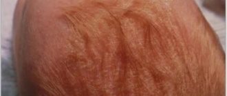
Dry eczema
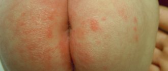
Sometimes white, dry, flaky patches on the skin of the legs can indicate dry eczema. With this disease, dryness and tightness of the skin first occurs, then peeling and unbearable itching occur, and cracks may occur. In addition to the legs, the hands, the space between the fingers, and even the face are often affected.
The causes of dry eczema can be bacterial and fungal infections, allergens, synthetic clothing, poor hygiene and other factors.
As you can see, the appearance of dry patches on the skin can be due to a variety of reasons.
The characteristics of the immune system and the tendency to allergic reactions play an important role. But in any case, when dry spots appear, it is important to consult a dermatologist and determine the cause of this skin condition. July 5, 2020
Author of the article: dermatologist Mak Vladimir Fedorovich
Causes of leukoderma in children
It is known that there is a genetic predisposition to vitiligo. The following factors may also influence the development of the pathological process:
- Improper functioning of the immune system. The child's body perceives melanocytes as foreign cells and begins to produce antigens for them.
- Chronic pathologies.
- Aggravated infectious processes.
- Metabolic disorder.
- Lack of vitamins and microelements.
- Chemotherapy.
- The influence of medications.
- Intoxication of the body.
- Chemical burns.
- Autoimmune diseases.
- Helminthiasis.
Sometimes the appearance of leukoderma in children is associated with unstable hormonal levels or psychological trauma.
Treatment methods
Basically, the treatment of childhood vitiligo is aimed at restoring metabolism in the child’s body, since most often it is metabolic disorders that become the cause that affects the insufficient production of skin pigment. For treatment to bring positive results, it is advisable to use an integrated approach.
Symptomatic therapy
Its main goal is the maximum restoration of the lost pigment formation function. For this use:
- Local application of corticosteroids.
- Photosensitizing agents that restore normal pigmentation in affected areas of the body.
- In the case of a generalized type of disease, photochemotherapy is used.
- Herbal medicines.
- Ultraviolet treatment (its plus is safe for children over 3 years old)
To make the cosmetic defect less noticeable, skin whitening procedures are performed to reduce the contrast between healthy and depigmented skin.
Drug treatment
The following groups of medications are used for drug therapy:
- Glucocorticoids.
- Antioxidants.
- Immunomodulatory agents.
- Photosensitizing hormonal drugs.
- Multivitamin complex.
Surgery
The main methods of this treatment:
- Autoplasty (affected skin is replaced with healthy skin).
- Transplantation of own or donor melanocytes.
- Transplantation of cultured epidermis.
- Application of tissue genetic engineering.
Such actions are carried out only in extreme cases, since there is a high probability of rejection or suppuration of the transplanted tissue.
It is possible to cure leukoderma, but the final result will primarily be influenced by timely diagnosis of the disease and a well-chosen course of therapy.
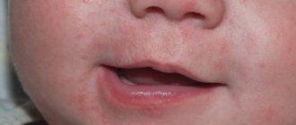
There are various causes of hypopigmented spots on a child's face, ranging from conditions that resolve on their own within a few weeks to conditions that can last a lifetime.
Causes of white spots on a child's face
White spots can be caused by the following skin conditions:
Milia
Milia are also called milk spots. These tiny white bumps usually appear on the face and rarely on the upper torso or limbs. Although milia can be seen at any age, it is common in newborns.
Causes and diagnosis of milia
Milia occurs due to entrapment of keratin (skin scales) beneath the surface of the skin. If there is no improvement after a couple of weeks, you should consult a doctor. Diagnosis is made by visual examination and no tests are required.
Prevention and treatment of milia
There is no cure for milia and it often goes away within a few weeks or months. There is no need to treat milia in children.
How can you reduce milia at home?
- Wash your child's face daily with mild soap and water.
- Pat your face dry after washing.
- Be careful not to scratch the milia to prevent skin damage and infection.
- Do not apply lotions or oils to your baby's face.
Note: Some may confuse baby pimples or Epstein pearls with milia. However, acne can cause red bumps and pustules on the face. Epstein's pearls are small, white-yellow cysts that often resemble milia, but they appear on the roof of the mouth and gums.
Pityriasis alba (lichen alba)
Pityriasis alba is a self-limiting skin condition that causes dry, thin, scaly and pale patches on the face. It is common in children and adolescents and is considered a type of eczema or dermatitis.
The name of this condition comes from the characteristic appearance of the skin.
Causes of white ptyriasis
The causes of white ptyriasis have not yet been clarified. However, atopic dermatitis and dry skin often coexist with it. Exposure to the sun can make it more visible by tanning the surrounding skin.
Suspected causes such as under- or over-bathing, low serum copper levels, ultraviolet radiation, or Malassezia yeast have not yet been proven to cause hypopigmentation.
Symptoms and signs of ptyriasis alba
One or more white spots, varying from 0.5 to 5 cm in diameter, are a characteristic sign of pityriasis alba. It may also cause mild itching in some children.
Diagnosis of pityriasis versicolor
The condition can be diagnosed through a physical examination by a doctor. A skin biopsy may show mild spongiotic (fluid accumulation between cells) dermatitis with decreased melanin pigment. A skin scraping may also be taken to rule out a yeast infection.
Treatment of white lichen
Treatment is not recommended in asymptomatic cases. Moisturizers are helpful for dry skin, and mild hydrocortisone (topical steroid) creams can help reduce itching and redness in the area.
Tacrolimus ointment, pimecrolimus cream, and calcineurin inhibitors have been shown to be effective in the treatment of pityriasis alba.
The appearance of the skin may gradually return to normal over several months or two to three years.
Prevention of white deprivation
Limited sun exposure may help reduce your risk of developing this disease.
Vitiligo
Vitiligo is a depigmentation of the skin due to the loss of melanocytes, which are the cells that produce the skin pigment called melanin. Vitiligo can affect both sun-exposed and unprotected areas of the body. Also, with the disease, depigmentation of the lips and gray hair are often observed.
Causes of vitiligo
The exact causes of vitiligo are unknown. The disease may be associated with dysfunction or loss of melanocytes (the cells that produce the pigment melanin), which give the skin its color. This may be due to genetic factors or an autoimmune condition in which the immune system attacks the melanocytes.
Vitiligo can occur at any age, but the onset of the disease is typical in children and adolescents.
Diagnosis of vitiligo
Vitiligo can be diagnosed through a physical examination. Rarely, a skin biopsy is performed to help confirm the diagnosis by the absence of melanocytes (pigment cells) on the skin. Thyroid disease and diabetes are checked as these may increase the risk of vitiligo in many people.
Treatment of vitiligo
Mild vitiligo may not require treatment: the spots will disappear over time. However, there are several treatments available to make your skin tone even.
Corticosteroid creams, photochemotherapy (PUVA), narrow band ultraviolet B (UVB) therapy, and depigmentation are a few treatment options. Using sunscreen can reduce tanning of the skin around vitiligo patches, and concealers can hide white spots on the skin.
Although vitiligo can be distressing to many people due to its appearance, it is not a medically dangerous condition. Vitiligo is not an infection, skin cancer, or a contagious disease.
Shingles
Shingles is a fungal skin infection that causes lighter or darker patches on the skin. The disease occurs at any age, but is most often seen in adolescents and young adults.
Causes of herpes zoster
Shingles is caused by a yeast that is usually present on the skin. Their excessive growth under the influence of environmental factors can lead to the appearance of spots on the skin. Malnutrition and excessive sweating (hyperhidrosis) can also lead to yeast growth.
Note: Shingles cannot be passed from one person to another (not contagious) since most people have Malassezia yeast on their skin. This condition is not caused by poor hygiene.
A weak immune system, oily and moist skin, or hot and humid climates may increase the risk of contracting shingles. Children taking corticosteroid medications may also be susceptible to this condition.
Signs and symptoms of shingles
Although white patches can appear on the face, they are often found on the chest, back, and forearms. The spots may be pink or light brown and have scales. Skin changes are usually limited to the outer layer of the skin and often do not cause any pain or itching.
Diagnosis of herpes zoster
A physical examination is sufficient to diagnose shingles. In rare cases, doctors may collect a skin scraping to confirm the diagnosis.
Treatment of herpes zoster
Shampoo containing selenium sulfide is the main treatment method. If the condition does not go away, then you can use antifungal or anti-dandruff shampoos. The skin may improve over a short period of time in many children. However, it may take several months to achieve an even skin tone. To prevent relapse of the disease, monthly use of shampoo is recommended.
Idiopathic guttate hypomelanosis
Idiopathic guttate hypomelanosis (IGH) is a skin condition with small, white, oval patches.
IGH is more common in older, fair-skinned people than in children and often goes undetected.
Causes of idiopathic guttate hypomelanosis
The exact cause of hypomelanosis is unknown. Although this has not yet been proven, it is believed that it may be related to sun exposure.
Diagnosis of idiopathic guttate hypomelanosis
A physical examination is sufficient to determine the condition. Rarely are biopsy samples taken. There is usually a decrease in the number of melanocytes in the affected areas, although they are not completely absent as in vitiligo.
Treatment of idiopathic guttate hypomelanosis
IGH spots are benign and do not require medical treatment. Most procedures are aimed at improving the cosmetic appearance of the skin.
Home remedies may include regular use of sunscreen and physical barriers to prevent sun exposure, as it can promote or accelerate hypomelanosis.
References:
- Milia; Healthychildren; The American Academy of Pediatrics
- Milia; MedlinePlus; The United States National Library of Medicine
- Pityriasis alba; DermNet NZ; The New Zealand Dermatological Society
- Donald N. Givler, et al.; Pityriasis Alba; The United States National Library of Medicine
- Pityriasis alba; C. S. Mott Children's Hospital; The University of Michigan 6. Pityriasis alba; The Australasian College of Dermatologists (ACD)
- Vitiligo; The mission of the National Institute of Arthritis and Musculoskeletal and Skin Diseases
- Vitiligo; National Health Service
- Diagnosing Vitiligo; NYU Langone Medical Center
- Vitiligo: Diagnosis and Treatment; The American Academy of Pediatrics
- Tinea Versicolor; Harvard Medical School
- Pityriasis Versicolor; National Health Service
- Falon Brown and Jonathan S. Crane; Idiopathic guttate hypomelanosis; The United States National Library of Medicine
- Idiopathic guttate hypomelanosis; DermNet NZ; The New Zealand Dermatological Society