What is a wen?
Wen (lipoma or milium) is a benign formation that does not pose any threat to human health. This is just an unpleasant cosmetic flaw. Wen differs from other formations in its mobility and softness.
Last time we discussed another benign neoplasm - fibroma. Read more about fibroid removal here.
They do not cause any discomfort or pain, as they represent overgrown adipose tissue. Wen can appear in any part of the body. As a rule, on the face they are observed mainly in the upper part.
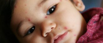
Very often, wen appears on the face of newborn children. This problem bothers adults, causing many problems. Parents should know that this phenomenon is not considered a pathology and does not pose a danger to the baby’s health.
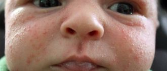
Externally, wen resemble small closed white pimples. They appear in groups of several formations on the forehead, near the eyes, as well as on the nose and chin. Typically, wen disappears on its own a few weeks after the baby is born.
Description
Wen is a benign tumor formation formed from connective tissue.
Lipoma in a child develops in the subcutaneous layer of fibrous structures. If their growth is noted, bone tissue may be damaged.
Wen itching
When a lump itches and grows, it is important to immediately consult a specialist and determine the causes, since a benign tumor can develop into a malignant one.
In a baby, they may appear around the eyes, in the area of the chin, on the forehead and cheeks.
Externally, the neoplasms resemble white pimples with no purulent contents inside the cavity. Sometimes lipomas can appear as acne.
It is also worth noting that the wen in a child after birth disappears on its own after a month and a half. If this does not happen, then you need to consult a pediatrician or dermatologist.
Causes of wen on the face of a newborn
Most parents perceive wen on the face of a newborn baby as a fairly serious danger to his life. Therefore, they begin to actively fight them, not realizing that they are only aggravating the situation and preventing the child’s skin from cleansing.
The appearance of wen on the face of newborns has not yet been fully studied, so identifying the true cause of this phenomenon can be very difficult.
Today we have only a few assumptions:
- The main cause of wen in young children is associated with immaturity of the skin tissue, which causes blockage of the sebaceous glands. Such formations do not require any treatment and disappear on their own after a month from the date of birth of the child.
- Active release of hormones in a pregnant woman immediately before childbirth.
- Artificial feeding.
- Improper metabolism.
- Genetic predisposition (if one of the parents has similar rashes).
- Improper skin care for a newborn, use of linen and clothing made from non-natural materials.
Types of wen on the face
There are diseases that pose a danger to the health of children, which in appearance are very similar to wen:
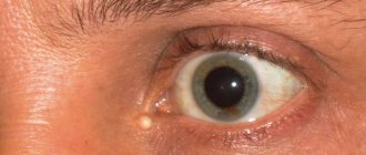
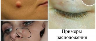
- Milia are small white formations that protrude above the skin. Another name for them is whiteheads that do not have a duct. Milia are filled with sebum and can appear on any part of the facial skin, either singly or in groups. Typical locations are cheekbones, forehead, nasolabial folds and wings of the nose. These formations are held quite firmly on the skin, so they are practically motionless. In case of multiple milia rash, the skin looks bumpy and uneven.
- Xanthelasmas . Their main difference from milia is their larger sizes and shades. As a rule, they have yellow tints. In addition, xanthelasmas have the ability to actively grow, increase in size and connect with each other. This type of wen is a mobile formation and requires fixation during removal. Location: upper and lower eyelids.
Usually, wen on the face does not have any clinical picture. If you press on it, there is a sensation under the skin of a foreign round movable formation.
Symptoms of a wen on a child’s face
In most cases, wen on the face develops without any symptoms. A characteristic feature of wen is that it appears on areas of the body with minimal fat. On the face they appear in the form of a protruding tubercle.
Lipomas on the face of newborn children are a completely natural phenomenon. They are rashes of small diameter. Their location is the forehead, nose and chin.
Xanthelasmas can be seen through thin skin in the form of yellow plaques of various shapes and sizes with blurred contours. Over time, their diameter may increase, but usually this happens for a very long time.
Xanthelasmas have a soft consistency and begin to move upon palpation. Unlike a subcutaneous pimple, it is impossible to squeeze out a wen. Due to the close location of the wen to the nerve endings, pain may occur when pressed.
Wen are often confused with other types of formations (atheroma or cyst) that require surgical intervention due to rapid growth.
You might be interested! Lipoma on the human body: what is it and why is it dangerous?
Results and discussion
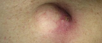
All patients with fatty tumors underwent surgery. In children with lipomas, in almost 85% of cases, classical excision of the tumor with capsule was performed. In recent years, the wound has been sutured with an intradermal furrier's suture. The advantage of this tactic is a fairly high radicalism and the absence of complications and tumor relapses in our patients. Minimally invasive treatment methods using video surgical techniques, as well as liposuction of lipomas, were not used. Despite the best cosmetic result, after using these techniques there is a high probability of tumor recurrence.
It should be noted that when the tumor was huge, removal was carried out in several stages. Here is one of our observations. Child G., 1 year 1 month. (case history No. 2720), was admitted to the pediatric surgery clinic on March 14, 2007 due to a huge tumor-like formation in the back. Ill since birth, when a back lipoma was diagnosed in the maternity hospital. Upon admission, the general condition was satisfactory. From the chest organs without pathology. In the back area, a tumor-like formation with a pasty consistency, painless, and prone to growth was palpated. On March 16, 2007, an operation was performed (surgeon - V.K. Litovka, candidate of medical sciences). Two bordering incisions in the transverse direction, in the cervico-suprascapular region, up to 20 cm long, cut the skin, fatty tissue, and superficial fascia around the tumor-like formation. Hemostasis. The tumor is represented by compacted fatty tissue, penetrated by small convoluted vessels, and in some places it grows into muscles. The thickness of the tumor-like mass is up to 5 cm. After excision of the skin flap with the tumor, several lipomatous nodes with a diameter of up to 3 cm are additionally excised, which grow into the muscles, up to the costal fascia. Hemostasis. The wound is sutured tightly in layers up to two rubber seals placed at the corners deep into the wound. Stitches on the skin. Aseptic dressing.
Macroscopic specimen: tumor-like formation, represented by compacted fatty tissue, whitish-yellow in color. The overall dimensions of the tumor are 19 ´ 5.5 ´ 4 cm. Pathohistological conclusion (No. 2067-98): fibrolipoma with invasion into muscle tissue. The postoperative period is smooth. The wound healed by primary intention. On March 22, 2007, the child was discharged home. Three months later, the girl was operated on again, and then another 6 months later (Fig. 3).
Subsequently, the patient was sent for consultation to OKHMADET and the National Cancer Institute (Kiev) to decide on further treatment tactics, since the tumor is prone to growth.
It is very difficult to diagnose mediastinal liposarcoma before surgery. The tumor manifests itself as intrathoracic compartment syndrome, so most often children are hospitalized for suspected pneumonia complicated by pleurisy. To confirm this, we present our observation. Patient E., 7 years old (case history No. 4471), was admitted on May 20, 1994 with complaints of cough, shortness of breath, and fever. She has been sick for about three weeks. He was treated as an outpatient for bronchopneumonia on the right. There was no improvement. X-ray suspected pleurisy (Fig. 4). The patient was referred to our clinic. During pleural puncture, up to 1.5 liters of serous-hemorrhagic effusion was obtained. Non-Hodgkin's lymphoma (mesothelioma?) was suspected. A course of polychemotherapy (PCT) was carried out: vincristine, cyclophosphamide, Vepezid, carboplatin.
On June 1, 1994, surgical intervention was performed (surgeon - V.K. Litovka, candidate of medical sciences). Thoracotomy in the VI intercostal space on the right using an anterolateral approach 20 cm long. Hemostasis. The costal pleura was dissected - up to 100 ml of hemorrhagic fluid with blood clots, fibrin and pieces of disintegrating tumor were released. The audit established that the entire pleural cavity is filled with a tumor-like formation of a cherry-gray color, which compresses and grows into the lung, vena cava and pericardium. The integrity of the tumor capsule is broken in places (tumor with disintegration) with symptoms of capillary bleeding. The abundance of disintegrating tumor tissue (approximately 1/3 of the tumor mass) was removed. The tumor was declared inoperable. A biopsy of the tumor was performed, closer to the pericardium, where it was without signs of decay, with stitching with catgut. There is no bleeding. The wound is sutured tightly. Through an additional incision, a drainage tube was inserted into the pleural cavity in the 7th intercostal space along the posterior axillary line. Bulau system. Toilet. Aseptic dressing.
Macropreparation: pieces of tumor, whitish consistency, the color of fish meat, in places pink-gray with areas of hemorrhage and necrosis. Diagnosis after surgery: angiofibrosarcoma of the mediastinum with invasion of the lung, vena cava, and pericardium. Tumor pleurisy. Pathohistological examination (No. 4141-55): round cell liposarcoma. The wound healed by primary intention. On June 15, 1994, the boy was discharged home. Two more courses of PCT were carried out. Due to tumor intoxication and dissemination of the tumor process, the child died 4 months later.
In another case of ours, the tumor also turned out to be malignant, but recovery was achieved. We present this case. Child M., 4 years old (case history No. 6638), was admitted to the surgical department of the clinic on July 23, 1998, with parents complaining of an increase in the boy’s abdominal volume and periodic pain. She has been sick for about 1.5 months. We went to the clinic. The operation was temporarily abstained. During this time, the stomach increased in size. Upon palpation, the tumor began to occupy almost the entire abdominal cavity. The general condition upon admission was severe. The skin is pale. Examined radiographically and sonographically. A diagnosis of lymphosarcoma of the abdominal cavity was made. A preoperative course of PCT with vincristine, cyclophosphamide and doxorubicin was carried out.
On August 7, 1998, an operation was performed (surgeon - Prof. I.P. Zhurilo). Median laparotomy 12 cm long. Hemostasis. Upon opening of the peritoneum, a tumor measuring 12 × 10 × 8 cm was discovered, adjacent to the anterior abdominal wall. A strand of the omentum and the tip of the appendix are fixed to the tumor capsule. The omentum was resected within healthy tissues, and the appendix was separated from the tumor. Thus, the upper pole of the tumor is released and dislocated into the wound. During the audit, it was established that the base of the tumor originates from the pelvic cavity and is intimately attached to the bottom of the bladder. Using sharp and blunt methods, the tumor was isolated, the feeding vessels were sutured, and removed. Seromuscular sutures were placed at the bottom of the bladder, including where the tumor was intimately fused to the wall. There is no bleeding. The liver is macroscopically unchanged, the lymph nodes of the intestinal mesentery are slightly enlarged. One of them was taken for biopsy. Layer-by-layer suturing of the wound tightly. Stitches on the skin. Aseptic sticker.
Macroscopic specimens: tumor measuring 12 ´ 10 ´ 8 cm, dense consistency, variegated appearance. The lymph node measures 2 ´ 1.5 ´ 1 cm, yellow. Diagnosis after surgery: rhabdomyosarcoma of the bladder (?), teratoblastoma of the pelvis (?). Pathohistological examination (No. 6558-93): liposarcoma of mixed structure, including large cell, myxoid and polymorphic cell variants, with extensive necrosis, hemorrhage and vascular thrombosis. The postoperative course is smooth. The sutures were removed and the wound healed by primary intention. A course of PCT (vincristine, adriblastine, cyclophosphamide, cisplatin) and radiation therapy (40 Gy) was administered. PCT courses were conducted throughout the year. Currently, there is no evidence of tumor recurrence or metastasis.
Among the six patients with liposarcoma, 3 (50%) died. In the remaining patients, after complex (combined) therapy, there was no evidence of tumor recurrence and metastases. The children are healthy.
Diagnostics
Apart from external manifestations, there are no other symptoms of the appearance of wen in children. They are diagnosed by a surgeon or dermatologist through external examination of the skin and palpation. The doctor will also find out from the parents all the necessary information: the time of appearance and the intensity of growth of the wen. After receiving the data, the doctor makes a diagnosis.
If a serious disease (malignant neoplasm) is suspected, the doctor may prescribe a histological examination. Sometimes, in order to avoid the spread of wen to other parts of the newborn’s body, additional procedures (MRI, computed tomography) are performed.
What does it look like
When a wen forms on the outside of the skin, diagnosis does not cause difficulties.
The main signs of lipoma in childhood are:
- pronounced mobility;
- soft and elastic consistency;
- lack of fusion with the skin;
- size 1.5-10 centimeters;
- lobular structure in large neoplasms;
- painlessness;
- oval or round shape for small wen.
Diagnosis of lipoma
It is not possible to determine a wen, which is located deep in the tissues or on internal organs, by external examination.
The internal structure of the formation consists of mature fat cells. They are, as a rule, united into lobular structures, and these, in turn, are covered with dense capsules.
Lipomas can form not only where there is adipose tissue; in the case when the growth of a wen is observed in a place where there is no fat, then this condition indicates the development of ectopia. Thus, in most cases, formations that are congenital in nature begin to form.
How to get rid of wen?
We have already covered in detail how to get rid of wen on the face, here we will talk about the features of getting rid of them for children.
Many parents, having noticed wen on their baby’s face, think that they can simply be squeezed out. This cannot be done, if only because, in this way, an infection can be introduced into the child’s body. As a result, a serious inflammatory process may begin, which will be very difficult to cure. There are other methods of getting rid of wen in newborns.
Treatment methods:
- Conservative treatment. This method involves the use of special medications that are administered inside the formation. Thanks to their absorbable action, they help to get rid of the wen easily and quickly. The only drawback of the conservative method is that one session will not help solve this problem. It is necessary to carry out a full course of treatment, which is very difficult for a newborn baby.
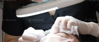
- Surgical method. There are cases when a wen can be dangerous to the child's health and must be removed through surgery. This method involves the use of local or general anesthesia, depending on the volume of formations and the individual characteristics of the child’s body. During the operation, the doctor makes an incision and squeezes out the contents of the wen. After this, he cleans the lipoma capsule itself.
- Laser method. This is the most effective and gentle method, since the laser beam has a positive effect on the regeneration process. It has coagulating and antibacterial characteristics, thanks to which wen removal is achieved without blood loss, scarring and side effects. The laser method is most suitable for eliminating formations on the face, since after a week not a trace remains of the wounds. After such an operation, the specimen is sent for a special histological examination.
- Puncture-aspiration method. This procedure is reminiscent of cosmetic surgery using a needle. The surgeon injects it into the tumor and sucks out the contents of the wen, preserving the membrane. The disadvantage of the puncture-aspiration method is the possibility of reappearance of the wen.
Surgical treatment of lipoma
The use of surgical methods is justified if the lipoma is large or growing rapidly.
The risk increases if the tumor is in the neck, as it can block important blood vessels.
The situation is the same with wen located in the ears: if the wen begins to grow, it can block part of the auditory canal and reduce hearing acuity.
In some cases, changes may be irreversible, so parents should not refuse the proposed surgical treatment if such indications exist.
There are several methods of radical therapy that will help remove fatty tissue in childhood.
- Laser excision.
Using a special laser knife, the lipoma and its capsule are excised from the connective tissue. The operation can be performed under general anesthesia or local anesthesia. For children under 10 years of age, use of the method is recommended only if there are serious indications. Some clinics do not perform this operation on adolescents under fourteen years of age.
- Electrothermocoagulation.
This is the cauterization of the tumor with electric current. This treatment method is considered effective only for small lipomas, since it is not always possible to control the complete removal of the tumor. If the pathological tissue is not completely excised, the chance of recurrence will be about 38-40%.
- Aspiration method.
A special needle with a thin tube is inserted into the formation, through which the doctor pumps out the contents of the capsule. The method is also not effective enough, since there remains a risk of refilling the capsule.
Important! Surgical treatment of lipoma in childhood is used only in the absence of effect from conservative therapy or in the presence of serious indications. In all other cases, experts advise fighting milia (if necessary) using medications.
Prevention
There are no special recommendations for the prevention of wen in children, since their occurrence has not yet been fully studied.
It is only known that in order to avoid their occurrence it is necessary:
- carefully choose baby care products;
- make changes to the baby’s diet: eliminate sweets and fatty foods, add more vitamins and healthy quality foods;
- Maintain child hygiene and monitor the condition of facial skin;
- An equally important condition is the timely passage of medical examinations, as well as treatment and removal of formations in order to eliminate consequences dangerous to the child’s health;
- never self-medicate.
If wen does not need to be treated, it is good to wipe the child’s face with decoctions of string and chamomile. It is also necessary to carefully ensure that the baby does not scratch his own face and cause an infection. Therefore, cut his nails more often and wear special gloves.
If an infection occurs, contact your pediatrician, who will prescribe you the most effective antibacterial agent.
Skin care after wen removal
For several days after removal, the problem area must be treated with a cotton swab soaked in iodine or any alcohol tincture (you can use calendula or St. John's wort).
This is necessary to disinfect the skin and prevent re-infection. For this purpose, medicinal antiseptic solutions, for example, Miramistin or Chlorhexidine, work well.
After removing the wen, it is important to maintain facial skin hygiene. While the baby is small, instead of washing, you need to wipe the skin with a cotton swab dipped in boiled water. The procedure must be repeated 2 times a day (further - as necessary, for example, if the child spits up or gets dirty).