Erysipelas, or simply erysipelas, is an infectious-allergic disease that affects the skin and subcutaneous tissue. The disease is quite common and prone to relapse. It ranks fourth among all infectious diseases and is also becoming more common over time. Thus, over the past twenty years, the number of relapses of this disease has increased by 25%. Moreover, a severe form of the disease is becoming more common - now it is about 80% of cases, although 50-60 years ago only 30 percent of patients were diagnosed with a severe form.
Causes of the disease
Streptococcus lives in the body of almost every person, and many people are its carriers. But the development of erysipelas and other streptococcal diseases does not occur if provoking factors are absent.
The occurrence of pathology is possible as a result of:
- damage to the dermis due to purulent, viral infection;
- circulatory disorders due to post-traumatic scars after surgery;
- decreased immunity;
- taking a number of drugs - cytostatics, steroids;
- the presence of pathology in metabolic processes;
- diseases of the immune system;
- AIDS;
- presence of bad habits.
Erysipelas is transmitted by airborne droplets or through direct contact with patients. It quickly begins to develop against the background of diabetes mellitus, sudden changes in temperature, poor nutrition, viral infectious diseases, and chronic ailments in the body.
Another well-known skin disease, streptoderma, is also caused by streptococci. Here you will find information about the treatment of streptoderma in adults.
Provoking factors
Inflammation in the hand can be caused by:
- surgery to remove mammary glands in women;
- excessive insolation;
- overheating or hypothermia;
- infection of abrasions, scratches, bruises, cuts with sharp objects.
In addition, the risk group includes people with pathologies such as:
- diabetes;
- alcoholism;
- obesity;
- varicose veins;
- lymphostasis;
- tonsillitis;
- caries;
- periodontitis;
- fungal infection of the feet;
- thrombophlebitis.
Symptoms of the disease
In the first 10 days after infection, the disease occurs without symptoms. After this, the patient begins to be bothered by the following symptoms:
- moderate headache;
- muscle pain;
- feeling of tiredness, fatigue;
- temperature increase;
- nausea;
- vomit;
- diarrhea.
Some patients also experience decreased appetite, often leading to anorexia. Voluminous red spots appear on the skin.
Characteristic symptoms of erysipelas on the hand
Microorganisms, penetrating into the pores of the skin on the hand, first remain for some time in the incubation period of up to 2-3 days . Infection may not occur if you immediately treat the area with an antiseptic in the event of an unexpected cut or if your immune system is fairly stable.
Otherwise, the primary and characteristic signs of the development of erysipelas are as follows:
- a sharp increase in body temperature;
- nausea;
- dizziness;
- increased fatigue;
- body aches;
- chills;
- loss of appetite;
- redness appears on the hand in the form of a pink-red spot or a roller with uneven edges with tongues of flame;
- further – peeling, burning sensation, swelling at the site of the lesion;
- in some cases, hemorrhages or blisters with serous or bloody fluid appear within the lesion.
Symptoms of erysipelas of the lower extremities
- quite noticeable headache
- high temperature, which is very difficult to bring down
- chills
- vomiting like food poisoning
- burning sensation on the skin
- severe redness on the skin
- The area of skin affected by erysipelas differs from healthy skin - it is either convex or separated by a small ridge.
If you notice such symptoms, you should immediately consult a doctor so as not to start the disease, because erysipelas can become more complicated.
This is what erysipelas looks like
If erysipelas is a consequence of varicose veins, then such a complication may be thrombophlebitis, in which inflammation spreads to the wall of the veins, and then a blood clot forms in the lumen of the vein. Another complication of erysipelas with varicose veins is tissue necrosis, leading to the appearance of trophic ulcers that are difficult to treat.
initial stage
The appearance of a pink spot when streptococcus penetrates the skin occurs within a few hours. The place begins to burn, burn, turn red, resembling tongues of flame. The skin becomes swollen and hot to the touch. The infection quickly spreads further.
Inflammation begins with:
- pain, aches in joints and muscles;
- increased temperature, fever;
- deterioration of general health;
- the appearance of nausea and vomiting;
- headache, dizziness;
- increased heart rate.
At first, erysipelas has an acute course and the symptoms are not specific. After 1-2 days , severe intoxication of the body occurs, some even experience hallucinations and delusions. Toxic damage to the kidneys and heart may occur. The patient feels nauseous, shivering, and sleepy. At the initial stage, erysipelas is similar to the flu.
Features of erysipelas on the hand
The peculiarity of erysipelas on the hand is that when it gets under the skin, the microorganism quickly penetrates into the deep layers of the dermis, leading to a sharp increase in temperature, chills, and even loss of consciousness. This unpleasant disease, which can greatly worsen the quality of life, interferes with ordinary household activities.
If the disease is not treated, the outcome of such a disease is extremely unfavorable. A minor lesion on the skin can lead to disruption of tissue trophism and gangrene.
Features of erysipelas on the leg
Erysipelas on the leg is a consequence of the penetration of hemolytic streptococcus through wounds with further proliferation of pathogenic flora in the vessels of the leg. This is a common occurrence when erysipelas develops specifically on the lower leg. Manifested by swelling and redness. At first it seems that the cause is an allergy or an insect bite.
The affected area begins to itch and swell very much in the first hours, hyperemia quickly spreads with pain to the adjacent tissues.
Erysipelas on the leg can lead to:
- lymph flow dysfunction;
- elephantiasis;
- development of gangrene.
A characteristic feature of erysipelas on the leg is the severe course of the disease.
You might be interested! Rash in children: what happens and where does it most often appear?
If you don’t know what kind of red spots on your legs are itchy, you can look at the article on the website.
Features of erysipelas on the body
Erysipelas can appear on the face and almost any part of the body. Sometimes in the form of a red hot spot it is localized on the torso after surgery or suturing. In children, the infection can penetrate into the umbilical wound or in the perineum due to diaper rash, scratching or abrasion of the skin.
The best ointment for erysipelas. External remedies for the treatment of erysipelas
Treatment with ointments is effective in the case of local therapy, when there is a need to destroy external lesions and reduce pain. In the case of a bullous form of the lesion, the surgeon usually squeezes out the contents of the resulting blisters. Subsequently, a bandage consisting of tissue in furacilin or rivanol is applied to the affected areas. For any type of pathology, ointments prepared at home can be widely used.
Streptocide
The composition is used in the form of powder, tablets, ointment. It has antimicrobial properties against streptococci and also fights inflammation. This is ointment 10%, liniment 5%. The product is used according to the instructions and in accordance with the recommendations of the treating specialist. Application - in the form of a compress, which involves first spreading the medicine on the bandage and then applying it to the leg.
Vishnevsky ointment
If the patient does not have any complications, it is permissible to use Vishnevsky ointment. The effectiveness of this medication can be explained by the fact that its composition is full of substances that help increase the level of exudation and speed up the opening of the bubbles. Anti-erysipelas balm is applied to a gauze bandage, which is subsequently used to wrap damaged skin areas. This lotion must be changed every 12 hours.
Castoreum
This healing composition has powerful and pronounced bactericidal properties, and also effectively heals wounds and increases overall immune defense. Therefore, it is universally effective in the case of erysipelas on the leg. It is recommended to use this drug in powder form.
Tetracycline
A broad-spectrum antibiotic that helps with erysipelas. In this situation, it should be used in ointment form. The product is inexpensive, its use is simple: you need to take a thin layer of the composition and apply it to the affected area, repeating three times a day. If there are special instructions from the doctor, you need to follow them directly.
Erythromycin
The medicine is antibacterial. It was first used to treat this disease. The advantages are high efficiency, affordable cost and the absence of contraindications except for individual intolerance. There is a possibility that redness and swelling will develop in the application area.
Naftalan
The ointment has a specific odor due to the presence of naphthenic hydrocarbons in the composition. Before starting therapy, it is necessary to treat the skin using an aqueous solution of furatsilin, and then apply the medicine, warming it to 38 degrees in the palms. Then the affected area is covered with a bandage for 20 minutes. The disadvantage is a large number of contraindications.
Sintomycin
Due to its versatility, the product is used for various skin diseases, including erysipelas on the leg. It is affordable, easy to use and has a pronounced effect after several applications.
Ichthyol ointment
The drug provides assistance at any stage of the lesion, but most often begins to be used at the very beginning of the disease. The medication promotes instant healing of wounds, eliminates redness and effectively combats inflammation.
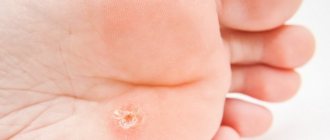
Forms and photos of erysipelas
Erysipelas can occur in erythematous, erythematous-bullous, erythematous-hemorrhagic, hemorrhagic or recurrent form
Erythematous form
The erythematous form is similar in nature to erythema, when the affected area swells, hurts, and acquires a bright color with clear boundaries in the form of a red spot rising above the skin. The spot looks like flames. Further, the erythema begins to peel off, itch, hurt on palpation, swell, and thicken.
The spot forms an infiltrated ridge that rises above the surface of the skin. Intoxication of the body develops. The duration of this form can vary and can last up to 1 week. In this case, there is an increase in lymph nodes, thickening and pain.
You can see what the erythematous form of erysipelas looks like in the photo:
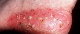
Erythematous-bullous form
The erythematous-bullous form appears in the upper layer of the skin in the form of bubbles with transparent contents, which subsequently burst. Brown areas covered with crusts form. Further, the crusts are rejected, erosion appears in places with further transformation into an ulcer with the subsequent formation of crusts. Further, the crusts fall off, but scars remain.
You can see what the erythematous-bullous form looks like in the photo:
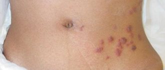
Erythematous-hemorrhagic form
The erythematous-hemorrhagic form is a fairly common occurrence with the appearance of a red spot in the form of hemorrhage. The duration of the course is 2 weeks with an increase in temperature and the formation of necrosis at the site of the lesion. This form occurs similarly to the erythematous form. There may be hemorrhages in the affected areas.
Photo:
Bullous-hemorrhagic form
The bullous-hemorrhagic form is similar to the erythematous-bullous form. The difference is the appearance in places of erythema of vesicles filled with bloody fluid. This is a more severe form of the disease. When the vesicles are opened, bloody contents flow out, the areas are erosive, leaving behind areas of necrosis and scars.
The photo clearly shows the bullous-hemorrhagic form of erysipelas:
Recurrent erysipelas
Recurrent erysipelas , when more than 2 weeks have passed since the initial infection, and the inflammation is recurrent. But it proceeds quite easily. The temperature is low, the chills are not strong. The development of a relapse is possible after undergoing primary erysipelas, if the treatment was not entirely adequate or there are concomitant foci of infection.
Photo:
Levomekol for erysipelas. General idea of the disease
Erysipelas (erysipelas) is an infectious and allergic disease that affects the skin, mucous membranes and lymphatic system. The cause of the disease is group A beta-hemolytic streptococcus. The name of the disease comes from the French word “rouge”, meaning “red”, because with erysipelas, a characteristic sign is the formation of red spots on the patient’s skin.
Erysipelas is one of the most common diseases caused by infections, right after respiratory and intestinal diseases. In addition, the patient, having recovered from erysipelas, runs the risk of encountering the manifestation of this disease again in the coming years.
In addition, medical scientists note with alarm that today most cases of erysipelas occur in a severe form, while the percentage of mild forms has decreased significantly. Up to a third of all cases of the disease are associated with impaired blood and lymph circulation. Antibiotics for erysipelas of the leg must be combined with drugs that normalize the circulation of fluids. There is also the possibility of developing severe complications of the disease, leading to death.
Erysipelas can affect a patient of any age and gender category, but most cases of the disease are observed among women over 50 years of age. There are also frequent cases of infection with streptococcus in infants, which subsequently also causes erysipelas. There are also statistical data suggesting a predisposition to erysipelas in people with blood group III.
Most often, the disease is transmitted through direct contact with an infected person through skin lesions - wounds, abrasions, etc. Also, if the pathogen is carried, the disease may enter the active phase after a pathological decrease in immunity.
The most common “targets” of the disease are the arms and legs, less often the face and head. The disease has several stages of development, each with its own distinctive signs, the main one of which is the appearance of an area of redness protruding above the surface of healthy skin as a dense convex ridge.
Complications
With proper treatment, erysipelas on the arm can go away on its own after 2-3 weeks. Redness and swelling will subside and will soon disappear altogether. But pigmentation may remain. Relapses are possible.
New erysipelas can subsequently lead to:
- stagnation of lymph;
- lymph circulation insufficiency;
- pulmonary embolism;
- sepsis;
- dead skin;
- thrombophlebitis.
All this indicates untimely treatment and progression of the disease.
Complications, as a rule, result from untimely consultation with doctors, self-medication, or the addition of a secondary infection. The risk group includes people with diabetes, HIV-infected people, and those who have had meningitis or pneumonia.
Erysipelas with complications can lead to the formation of trophic ulcers on the arm, lymphostasis, abscess, suppuration and thickening of the skin, which will significantly complicate treatment and may even endanger the life of the patient himself.
Ointment for erysipelas on the leg. Erysipelas and the reasons for its appearance
Erysipelas is an infectious disease manifested by acute inflammation of the skin in a certain part of the body.
The culprit of the infection is group A streptococcus, which penetrates the skin through lesions of various types. Small cuts, abrasions, scratches, scratches, and an insect bite can become an open portal for him.
The bacterium itself can remain in the skin for a long time without revealing itself in any way. Often carriers of a gram-positive microbe do not even suspect that they are at risk of disease. But the inflammatory process begins to develop rapidly as soon as it is provoked by external factors:
- injuries;
- sudden change in temperature;
- Tan;
- stressful situations;
- breakdown.
In addition to these factors, erysipelas can develop as a consequence of other diseases:
- obesity;
- alcoholism;
- diabetes;
- varicose veins;
- trophic ulcers;
- thrombophlebitis;
- fungus on the feet;
- chronic somatic diseases that reduce the performance of the immune system.
If this is what caused the erysipelas on the leg, then treatment should begin with these pathologies.
Those most at risk for erysipelas are men of working age and women over 40 years of age. Especially if the type of employment involves heavy physical labor. Infants also suffer from erysipelas. But for them this is a special danger that can lead to death.
Erysipelas ranks 4th in the ranking of infectious infections of the body. The first places were distributed among acute respiratory diseases, intestinal infections and viral hepatitis.
Before starting treatment for erysipelas on the leg, it is necessary to correctly identify the disease itself based on its symptoms.
Treatment
When visiting the clinic, the doctor will first examine the skin, identify the nature, location, degree of damage and shape of the erysipelas. Most likely, the patient with obvious clinical signs will be sent to hospitalization in the infectious diseases department.
For the treatment of erysipelas the following is prescribed:
- antiallergic drugs (Suprastin, Diazolin, Tavegil);
- sulfonamides (Biseptol, Streptocide);
- nitrofurans (Furadonin, Furazolidone) to kill bacteria;
- corticosteroids (Prednisolone) to clear up the infection;
- biostimulants (Pentoxyl, Methyluracil) to stimulate the formation of new healthy immune cells and skin regeneration;
- vitamins (ascorbic acid, Ascorutin) to strengthen vascular walls damaged by bacteria, increase proteolytic enzymes (trypsin, lidase, tactivin).
In addition, treatment is carried out in a hospital with the appointment of:
- benzylpenicillin, as the main antibiotic for streptococcal infections;
- cephalosporins to suppress pathogenic flora in the event of an abscess or phlegmon. The course of treatment is up to 10 days.
Treatment also includes the following:
- Detoxification therapy is carried out in severe cases of the disease by administering intravenously hemodez or saline solution with glucose.
- It is possible to prescribe cardiovascular, antipyretic, diuretic drugs , as well as treat the affected area by applying applications from a dimexide solution, enteroseptol powders to kill bacteria in the affected areas and prevent the addition of another infection.
- Patients are advised to wash their wounds themselves with furatsilin and other solutions with antimicrobial effects to kill bacteria. An oxycyclosol aerosol will help, applying bandages with syntomycin ointment, Vishnevsky's liniment, to relieve inflammation and heal wounds.
You can’t warm up areas - it will only speed up the movement of streptococci through the blood and the spread of bacteria throughout the body. The main treatment is antibiotics, and in no case should you resort to homemade formulations and prescriptions without the knowledge of your doctor.
Those who are sick must strengthen their immune system, take vitamins, multivitamins, and antiallergic medications. Electrophoresis, laser therapy, ultraviolet irradiation, high-frequency magnetic therapy, and physical therapy are indicated for complete suppression of pathogenic microflora.
Erysipelas: CLINIC, DIAGNOSTICS, TREATMENT
• According to the nature of local manifestations: a) erythematous; b) erythematous-bullous; c) erythematous-hemorrhagic; d) bullous-hemorrhagic. • According to the degree of intoxication (severity): I - mild; II - moderate; III - heavy. • By flow rate: a) primary; b) recurrent (occurring after 2 years, different localization of the process) c) recurrent. If there are at least three relapses of erysipelas per year, the definition of “frequently recurrent erysipelas” is appropriate. • According to the prevalence of local manifestations: a) localized erysipelas; b) common (migratory) erysipelas; c) metastatic erysipelas with the appearance of distant foci of inflammation. • Complications of erysipelas: a) local b) general. • Consequences of erysipelas: a) persistent lymphostasis (lymphatic edema, lymphedema); b) secondary elephantiasis (fibredema). Primary, repeated erysipelas and the so-called late relapses of the disease (after 6 - 12 months and later) are an acute cyclic infectious process that occurs as a result of exogenous infection with group A b-hemolytic streptococcus. The source of infection in this case is both patients with a variety of streptococcal infections and and healthy streptococcus bacteria carriers. The main transmission mechanism is contact (microtraumas, abrasions, skin rash, etc.). The airborne transmission mechanism of streptococcus with primary damage to the nasopharynx and subsequent introduction of the microbe to the skin by hand, as well as by lymphogenous and hematogenous routes, is also of particular importance. Recurrent erysipelas, in which early and frequent relapses of the disease occur, is formed after a primary or recurrent erysipelas due to inadequate treatment, the presence of unfavorable background and concomitant diseases (varicose veins, mycoses, diabetes mellitus, chronic tonsillitis, sinusitis, etc.), the development of secondary immune deficiency, defects in nonspecific defense of the body. Foci of chronic endogenous infection form in the skin and regional lymph nodes. Along with the bacterial forms of group A streptococcus, when the process is chronic, the L-forms of the pathogen are also of great importance, persisting for a long time in the macrophages of the skin and organs of the mononuclear-phagocytic system. Reversion of L-forms of streptococcus into the original bacterial forms leads to another relapse of the disease. Erysipelas usually occurs against the background of pronounced sensitization to b-hemolytic streptococcus and is accompanied by the formation of fixed immune complexes in the dermis, including perivascularly. When infected with streptococcus, the disease develops only in individuals who have a congenital or acquired predisposition to it. Infectious-allergic and immunocomplex mechanisms of inflammation in erysipelas determine its serous or serous-hemorrhagic nature. The addition of purulent inflammation indicates a complicated course of the disease. Patients with erysipelas are less contagious. Women get erysipelas more often than men, especially the recurrent form of the disease. In more than 60% of cases, erysipelas occurs in people aged 40 years and older. Unlike other streptococcal infections, erysipelas is characterized by a distinct summer-autumn seasonality. In recent years, there has been an increase in the number of cases of hemorrhagic erysipelas, which is characterized by slow tissue repair at the site of inflammation, a tendency towards a protracted (chronic) course of the infectious process, and a high frequency of complications. Clinical picture of erysipelas
The incubation period ranges from several hours to 3 - 5 days. In patients with recurrent erysipelas, the development of another attack of the disease is often preceded by hypothermia and stress. In the vast majority of cases, the disease begins acutely. Initial period
The disease is characterized by the rapid development of intoxication symptoms, which in more than half of patients (usually when erysipelas is localized on the lower extremities) precede the onset of local manifestations of the disease by several hours to 1–2 days.
Headache, general weakness, chills, and muscle pain are noted. Nausea and vomiting occur in 25–30% of patients. Already in the first hours of illness, the temperature rises to 38 - 40°C. In areas of the skin in the area of future local manifestations, a number of patients experience paresthesia, a feeling of fullness or burning, and mild pain. Often pain also occurs in the area of enlarged regional lymph nodes. The height of the disease
occurs within a period of several hours to 1 - 2 days after the first manifestations of the disease.
General toxic manifestations and fever reach their maximum. Characteristic local manifestations of erysipelas occur. Most often, the inflammatory process is localized on the lower extremities (60 - 70%), less often on the face (20 - 30%) and upper extremities (4 - 7%), very rarely only on the torso, in the area of the mammary gland, perineum, external genitalia . With timely treatment and uncomplicated course of erysipelas, the duration of fever usually does not exceed 5 days. In 10 - 15% of patients, fever persists for longer than 7 days, which is usually observed with a widespread process and insufficient adequate etiotropic therapy. The longest febrile period is observed with bullous hemorrhagic erysipelas. More than 70% of patients with erysipelas develop regional lymphadenitis, which develops in all forms of the disease. The period of convalescence.
Normalization of temperature and disappearance of symptoms of intoxication are observed with erysipelas earlier than the disappearance of local manifestations.
Acute local manifestations of the disease persist for up to 5 - 8 days, in hemorrhagic forms - up to 12 - 18 days or more. Residual effects of erysipelas that persist for several weeks and months include pasty and pigmented skin, congestive hyperemia at the site of faded erythema, dense dry crusts at the site of bullae, and edematous syndrome. Unfavorable prognostic significance (the likelihood of an early relapse) are persistent enlarged and painful lymph nodes, skin infiltrates in the area of the extinguished source of inflammation, and low-grade fever. Prognostically unfavorable is also the long-term persistence of lymphatic edema (lymphostasis), which should be considered as an early stage of secondary elephantiasis. Hyperpigmentation of skin areas on the lower extremities in patients who have suffered from bullous hemorrhagic erysipelas can persist for life. Erythematous erysipelas
can be either an independent clinical form of erysipelas or the initial stage of development of other forms of erysipelas.
A small red or pink spot appears on the skin, which within a few hours turns into the characteristic erythema erysipelas. Erythema is a clearly demarcated area of hyperemic skin with uneven boundaries in the form of teeth and tongues. The skin in the area of erythema is infiltrated, tense, hot to the touch, moderately painful on palpation (more along the periphery of the erythema). In some cases, a “peripheral ridge” can be detected in the form of infiltrated and raised edges of erythema. Along with hyperemia and infiltration of the skin, its edema develops, spreading beyond the erythema. Erythematous-bullous
erysipelas develops in a period of several hours to 2 - 5 days against the background of erythema erysipelas.
The development of blisters is associated with increased exudation at the site of inflammation and detachment of the epidermis from the dermis by accumulated fluid. When the surfaces of the blisters are damaged or they spontaneously rupture, exudate leaks out, and erosion often occurs in large quantities at the site of the blisters. While maintaining the integrity of the blisters, they gradually shrink to form yellow or brown crusts. Erythematous-hemorrhagic
erysipelas develops against the background of erythematous erysipelas within 1 to 3 days from the onset of the disease, sometimes later.
Hemorrhages of various sizes appear - from small petechiae to extensive confluent hemorrhages, sometimes throughout the entire erythema. Bullous-hemorrhagic
erysipelas transforms from the erythematous-bullous or erythematous-hemorrhagic form and occurs as a result of deep damage to the capillaries and blood vessels of the reticular and papillary layers of the dermis.
Bullous elements are filled with hemorrhagic and fibro-hemorrhagic exudate, and extensive hemorrhages occur in the skin in the area of erythema. The resulting blisters come in different sizes and are dark in color with translucent yellow inclusions of fibrin. The blisters may also contain predominantly fibrinous exudate. The appearance of extensive flattened blisters, dense on palpation due to significant deposition of fibrin in them, is possible. In patients with active repair in the lesion, brown crusts quickly form at the site of the blisters. In other cases, the covers of the blisters rupture and are rejected along with clots of fibrinous-hemorrhagic contents, exposing the eroded surface. In most patients, its gradual epithelization occurs. With significant hemorrhages in the bottom of the bladder and the thickness of the skin, necrosis may develop, sometimes with the addition of secondary suppuration and the formation of ulcers. According to the specialized erysipelas department of the 2nd Clinical Infectious Diseases Hospital (Moscow), in patients hospitalized in the department in 1997, the erythematous or erythematous-bullous form was diagnosed in 5.2% of cases, the erythematous-hemorrhagic form - in 48.8% , bullous-hemorrhagic - in 46%. The severity criteria
for erysipelas are the severity of intoxication and the prevalence of the local process.
The mild (I) form of erysipelas includes cases with minor intoxication, low-grade fever, and a localized (usually erythematous) local process. The moderate (II) form of the disease is characterized by severe intoxication. There is general weakness, headache, chills, muscle pain, sometimes nausea, vomiting, fever up to 38 - 40 ° C, tachycardia, and in almost half of the patients - hypotension. The local process can be either localized or widespread (involves two anatomical areas) in nature. Severe (III) form of erysipelas includes cases of illness with severe intoxication: intense headache, repeated vomiting, hyperthermia (above 40°C), sometimes blackout, symptoms of meningism, convulsions. Significant tachycardia and often hypotension are observed; in elderly and senile people, with late treatment, the development of acute cardiovascular failure is possible. Widespread bullous-hemorrhagic erysipelas with extensive blisters should be considered severe in the absence of pronounced toxicosis and hyperthermia. With different localization of erysipelas, the clinical course of the disease and its prognosis have their own characteristics. Erysipelas of the lower extremities is the most common localization of the disease (60 - 70%). Characteristic are hemorrhagic forms of the disease with the development of extensive hemorrhages, large blisters with subsequent formation of erosions and other skin defects. For this localization of the process, the most typical lesions of the lymphatic system are lymphangitis, periadenitis, and a chronically relapsing course of the disease. The latter is largely facilitated by background concomitant conditions - chronic venous insufficiency, primary disorders of lymph circulation, mycoses, etc. Erysipelas (20 - 30%) is usually observed in primary and recurrent forms of the disease. With it, it is relatively rare that the disease often recurs. With timely treatment, facial erysipelas is easier than erysipelas of other localizations. It is often preceded by tonsillitis, acute respiratory diseases, exacerbations of chronic sinusitis, otitis media, and caries. Erysipelas of the upper extremities (5 - 7%), as a rule, occurs against the background of postoperative lymphostasis (elephantiasis) in women operated on for a breast tumor. Erysipelas of this localization in women has a tendency to recur. One of the main aspects of the problem of erysipelas as a streptococcal infection is the tendency of the disease to have a chronically relapsing course (in 25 - 35% of all cases). Relapses of erysipelas can be late
(occur a year or more after the previous outbreak of erysipelas with the same localization of the local inflammatory process), seasonal (occur annually for many years, most often in the summer-autumn period).
Late and seasonal relapses of the disease, usually the result of reinfection, do not differ in clinical course from typical primary erysipelas, although they occur against the background of persistent lymphostasis and other consequences of previous outbreaks of the disease. Early
and
frequent
relapses (3 relapses per year or more) are exacerbations of a chronic disease.
In more than 70% of patients, often recurrent erysipelas occurs against the background of various concomitant conditions, accompanied by disturbances in skin trophism, a decrease in its barrier functions, and local immunodeficiency. These include primary lymphostasis and elephantiasis of various etiologies, chronic venous insufficiency (postthrombophlebitic syndrome, varicose veins), fungal skin lesions, diaper rash, etc. Chronic ENT infection, diabetes mellitus, and obesity are of particular importance for the formation of recurrent erysipelas. The combination of two or three of the listed background diseases significantly increases the possibility of frequent relapses of the disease, and those suffering from them constitute a risk group. Complications
of erysipelas, mainly of a local nature, are observed in 5 - 8% of patients.
Local complications of erysipelas include abscesses, phlegmons, skin necrosis, pustulization of bullae, phlebitis, thrombophlebitis, lymphangitis, and periadenitis. Complications most often occur in patients with bullous hemorrhagic erysipelas. With thrombophlebitis, the subcutaneous and less often the deep veins of the leg are affected. Treatment of these complications must be carried out in purulent surgical departments. Common complications that develop in patients with erysipelas quite rarely include sepsis, toxic-infectious shock, acute cardiovascular failure, pulmonary embolism, etc. The consequences
of erysipelas include persistent lymphostasis (lymphedema) and secondary elephantiasis itself (fibredema), which are two stages of one process. According to modern concepts, persistent lymphostasis and elephantiasis in most cases develop in patients with erysipelas against the background of already existing functional insufficiency of lymph circulation of the skin (congenital, post-traumatic, etc.). Recurrent erysipelas that occurs against this background significantly enhances existing (sometimes subclinical) lymph circulation disorders, leading to the formation of consequences of the disease. Successful anti-recurrence treatment of erysipelas (including repeated courses of physical therapy) can lead to a significant reduction in lymphedema. In case of already formed secondary elephantiasis, only surgical treatment is effective.
Laboratory diagnostics
Due to the rare isolation of b-hemolytic streptococcus from the blood of patients and from the source of inflammation, conducting routine bacteriological studies is impractical. Elevated titers of antistreptolysin-O and other antistreptococcal antibodies, identification of bacterial and L-forms of streptococcus in the blood of patients have a certain diagnostic significance, which is especially important when predicting relapses in convalescents. Recently, polymerase chain reaction has begun to be used to diagnose streptococcal infections. In most patients with erysipelas, at the height of the disease, moderate neutrophilic leukocytosis with a shift to the left, aneosinophilia, and moderately increased ESR are usually observed. In patients with frequent relapses of the disease, leukopenia may occur. In severe cases of erysipelas and its purulent complications, hyperleukocytosis may be detected, sometimes with the development of a leukemoid reaction and toxic granularity of neutrophils. Changed hemogram parameters usually normalize during the period of convalescence. Changes in the T- and B-immune systems are most typical for the recurrent form of the disease. They reflect signs of secondary immune deficiency, usually occurring in the hypersuppressor mode. For patients with hemorrhagic erysipelas, pronounced disturbances of hemostasis and fibrinolysis are typical, manifested by an increase in the blood level of fibrinogen, PDP, RKMP, an increase or decrease in the amount of plasminogen, plasmin, antithrombin III, an increase in the level of platelet factor 4, and a decrease in their number. Moreover, the activity of various components of hemostasis and fibrinolysis varies significantly in individual patients.
Diagnostic criteria and differential diagnosis
Diagnostic criteria for erysipelas in typical cases are: • acute onset of the disease with severe symptoms of intoxication, increased body temperature to 38-39°C and above; • predominant localization of the local inflammatory process on the lower extremities and face; • development of typical local manifestations with characteristic erythema, possible local hemorrhagic syndrome; • development of regional lymphadenitis; • absence of severe pain in the area of inflammation at rest. Differential diagnosis for erysipelas should be carried out with more than 50 diseases related to the clinic of surgical, skin, infectious and internal diseases. First of all, it is necessary to exclude abscess, phlegmon, suppurating hematoma, thrombophlebitis (phlebitis), dermatitis, eczema, herpes zoster, erysipeloid, erythema nodosum.
Treatment
Treatment of patients with erysipelas should be carried out taking into account the form of the disease, primarily its frequency (primary, repeated, recurrent, often recurrent erysipelas), as well as the degree of intoxication, the nature of local lesions, the presence of complications and consequences. Currently, most patients with mild erysipelas and many patients with moderate forms of the disease are treated in a clinic. Indications for compulsory hospitalization in infectious diseases hospitals (departments) are: • severe erysipelas with severe intoxication or widespread skin lesions (especially in the bullous-hemorrhagic form of erysipelas); • frequent relapses of erysipelas, regardless of the degree of intoxication, the nature of the local process; • presence of severe common concomitant diseases; • old age or childhood. Antibacterial therapy occupies the most important place in the complex treatment of patients with erysipelas (as well as other streptococcal infections). When treating patients in a clinic and at home, it is advisable to prescribe antibiotics orally: erythromycin 0.3 g 4 times a day, oletethrin 0.25 g 4 - 5 times a day, doxycycline 0.1 g 2 times a day, spiramycin 3 million IU 2 times a day (course of treatment 7 - 10 days); azithromycin - on the 1st day 0.5 g, then for 4 days 0.25 g 1 time per day (or 0.5 g for 5 days); ciprofloxacin - 0.5 g 2 - 3 times a day (5 - 7 days); biseptol (sulfatone) - 0.96 g 2 - 3 times a day for 7 - 10 days; rifampicin - 0.3 - 0.45 g 2 times a day (7 - 10 days). In case of intolerance to antibiotics, furazolidone is indicated - 0.1 g 4 times a day (10 days); delagil 0.25 g 2 times a day (10 days). It is advisable to treat erysipelas in a hospital setting with benzylpenicillin in a daily dose of 6 - 12 million units, a course of 7 - 10 days. In severe cases of the disease, the development of complications (abscess, phlegmon, etc.), a combination of benzylpenicillin and gentamicin (240 mg once a day) and the prescription of cephalosporins are possible. For severe skin infiltration at the site of inflammation, non-steroidal anti-inflammatory drugs are indicated: chlotazol 0.1 - 0.2 g 3 times or butadione 0.15 g 3 times a day for 10 - 15 days. Patients with erysipelas need to be prescribed a complex of B vitamins, vitamin A, rutin, ascorbic acid, a course of treatment of 2 - 4 weeks. In case of severe erysipelas, parenteral detoxification therapy is carried out (hemodez, rheopolyglucin, 5% glucose solution, saline solution) with the addition of 5 - 10 ml of 5% ascorbic acid solution, 60 - 90 mg of prednisolone. Cardiovascular, diuretic, and antipyretic drugs are prescribed. Pathogenetic therapy of local hemorrhagic syndrome is effective with earlier treatment (in the first 3-4 days), when it prevents the development of extensive hemorrhages and bullae. The choice of drug is carried out taking into account the initial state of hemostasis and fibrinolysis (according to coagulogram data). In case of clearly expressed hypercoagulation phenomena, treatment with a direct-acting anticoagulant heparin (subcutaneous administration or by electrophoresis) and the antiplatelet agent trental at a dose of 0.2 g 3 times a day for 7 - 10 days is indicated. In the presence of pronounced activation of fibrinolysis in the early stages of the disease, it is advisable to treat with the fibrinolysis inhibitor Ambien at a dose of 0.25 g 3 times a day for 5 - 6 days. In the absence of pronounced hypercoagulation, it is also recommended to administer protease inhibitors - Contrical and Gordox - directly into the inflammation site by electrophoresis, for a course of treatment of 5 - 6 days.
Treatment of patients with recurrent erysipelas
Treatment of this form of the disease should be carried out in a hospital setting. It is mandatory to prescribe reserve antibiotics that were not used in the treatment of previous relapses. Cephalosporins (I or II generation) are prescribed intramuscularly at 0.5 - 1.0 g 3 - 4 times a day or lincomycin intramuscularly 0.6 g 3 times a day, rifampicin intramuscularly 0.25 g 3 times a day. The course of antibacterial therapy is 8 - 10 days. For particularly persistent relapses of erysipelas, two-course treatment is advisable. Antibiotics are consistently prescribed that have an optimal effect on bacterial and L-forms of streptococcus. The first course of antibiotic therapy is carried out with cephalosporins (7 - 8 days). After a 5-7 day break, a second course of treatment with lincomycin is carried out (6-7 days). For recurrent erysipelas, immunocorrective therapy (methyluracil, sodium nucleinate, prodigiosan, T-activin) is indicated.
Local therapy
Treatment of local manifestations of the disease is carried out only in its bullous forms with localization of the process on the extremities. The erythematous form of erysipelas does not require the use of local treatments, and many of them (ichthyol ointment, Vishnevsky balm, antibiotic ointments) are generally contraindicated. In the acute period of erysipelas, if there are intact blisters, they are carefully incised at one of the edges and after the exudate is released, bandages with a 0.1% solution of rivanol or a 0.02% solution of furatsilin are applied to the site of inflammation, changing them several times during the day. Tight bandaging is unacceptable. In the presence of extensive weeping erosions at the site of the opened blisters, local treatment begins with manganese baths for the extremities, followed by the application of the bandages listed above. For the treatment of local hemorrhagic syndrome with erythematous-hemorrhagic erysipelas, 5-10% dibunol liniment is prescribed in the form of applications in the area of inflammation 2 times a day for 5-7 days. Timely treatment of hemorrhagic syndrome significantly reduces the duration of the acute period of the disease, prevents the transformation of erythematous-hemorrhagic erysipelas into bullous-hemorrhagic, accelerates reparative processes, and prevents complications characteristic of hemorrhagic erysipelas.
Physiotherapy
Traditionally, in the acute period of erysipelas, ultraviolet radiation is prescribed to the area of inflammation in the area of regional lymph nodes. If skin infiltration, edematous syndrome, regional lymphadenitis persist during the convalescence period, ozokerite applications or dressings with heated naphthalan ointment (on the lower extremities), paraffin applications (on the face), lidase electrophoresis (especially in the initial stages of elephantiasis formation), calcium chloride, radon baths. Recent studies have shown the high effectiveness of low-intensity laser therapy for local inflammation, especially in hemorrhagic forms of erysipelas. Laser radiation is used in both the red and infrared ranges. The applied dose of laser radiation varies depending on the condition of the local hemorrhagic lesion and the presence of concomitant diseases.
Bicillin prevention of recurrent erysipelas
Bicillin prophylaxis is an integral part of complex dispensary treatment of patients suffering from a recurrent form of the disease. Prophylactic intramuscular administration of bicillin (5 - 1.5 million units) or retarpen (2.4 million units) prevents relapses of the disease associated with reinfection with streptococcus. While foci of endogenous infection persist, these drugs prevent the reversion of L-forms of streptococcus into the original bacterial forms, which helps prevent relapses. In case of frequent relapses (at least 3 in the last year) of erysipelas, continuous (year-round) bicillin prophylaxis is advisable for 2 - 3 years with an interval of drug administration of 3 - 4 weeks (in the first months the interval can be reduced to 2 weeks). In case of seasonal relapses, the drug is started to be administered a month before the start of the morbidity season in a given patient with an interval of 4 weeks for 3 to 4 months annually. If there are significant residual effects after erysipelas, the drug is administered at intervals of 4 weeks for 4 to 6 months. Clinical examination of patients with erysipelas should be carried out by doctors in the infectious diseases departments of polyclinics, with the involvement, if necessary, of doctors of other specialties.
Prevention
There are no specific specifics and prevention for erysipelas.
Development can be prevented if:
- do not neglect the rules of personal hygiene, wearing loose clothing and shoes made of natural fabrics;
- use soap containing lactic acid when showering to create a protective layer on the skin;
- immediately treat any damage or abrasions on the skin with antiseptics;
- Avoid ultraviolet irradiation, chapping, frostbite of extremities.
Erysipelas is a common ailment, and it can be treated fairly quickly with timely medication. An advanced disease will ultimately lead to a chronic relapsing course, scarring on the arm, swelling, and lymph stagnation.
Symptoms will recur from time to time, up to the appearance of stiffness in the joints, constant pain, limited mobility and disability. The appearance of a red, itchy and flaky spot on your hand cannot be ignored. It is possible that a streptococcal infection has occurred. The sooner, the better to seek advice from a dermatologist.
How to treat erysipelas on the leg at home. Treatment of erysipelas of the leg with medication
Erysipelas is usually treated with medication.
Immunomodulatory and/or desensitizing therapy is carried out simultaneously with antibiotics. Since harmful microorganisms produce toxins during their life, they can cause allergies in the patient. To prevent the development of allergic reactions during the treatment of erysipelas, patients are prescribed antihistamines. Often the pathology develops in the lower extremities. How to treat erysipelas on the leg? If the disease affects a limb, then the acute onset of the disease may occur only after a week. A person may suddenly develop symptoms of the disease such as muscle aches, migraines, high fever (up to 40°C), and general weakness. Often the diagnosis is made without analysis based on a combination of visual signs. Treatment of erysipelas of the leg is carried out with medication, both inpatient and outpatient.
Antibiotics for erysipelas
According to statistics, erysipelas ranks fourth in the prevalence of infectious diseases. How to treat erysipelas? Antibiotics have been and remain the priority in the fight against infection. The course is calculated by the doctor, depending on the form of the disease and the antibacterial drug. Immediately after starting to take antibiotics for erysipelas, the development of infection decreases and body temperature returns to normal. To treat erysipelas, 1st or 2nd generation antibacterial agents are used - cephalosporins (Cedex, Suprax, Vertsef) and penicillins (Retarpen, Benzylpenicillin, Ospen).
Ointment for erysipelas of the leg
When treating erysipelas on the leg, which is at an early stage, pastes for external use are not used. When the form of the disease becomes cystic, then Ichthyol ointment or Vishnevsky is prescribed. Naftalan gives excellent results at the recovery stage. Ichthyol ointment for erysipelas of the leg quickly helps get rid of itching, softens keratinization, and provides effective wound healing, provoking rapid skin regeneration.
The medicine has anti-inflammatory and antiseptic effects. For erysipelas, it is necessary to apply the product to the affected area, but not in its pure form, but in equal proportions with glycerin. The mixture is rubbed into a thin layer, then covered with gauze folded in 3-4 layers. The bandage is fixed with adhesive tape. It must be changed at least three times per day. The procedure is carried out until open wounds heal.
Vishnevsky ointment
How to treat erysipelas with Vishnevsky ointment? The topical preparation is also called balsamic liniment. The product contains three components: xeroform, birch tar and castor oil. Now the latter substance is often replaced with fish oil. Vishnevsky ointment has a pronounced anti-inflammatory and antiseptic effect. When treating skin pathologies, it helps restore the epidermis, accelerates the healing process, and has drying, antipruritic, and anesthetic properties.
In the absence of relapses, Vishnevsky ointment is prescribed for the treatment of erysipelas. The medicine promotes exudation and breakthrough of blisters. Apply the ointment to a gauze bandage in a thin layer, after which it should be applied to the affected area of the skin. The bandage is changed once every 12 hours. Since the drug can dilate blood vessels, doctors do not recommend using it for severe forms of erysipelas.
1.General information
Erysipelas, erysipelas - these and similar terms are used mainly in Slavic languages; in medical Latin and most Western European languages, the original ancient Greek name erysipelas was fixed, i.e. "red skin" In the old days there were also synonyms “holy fire”, “Saint Anthony’s fire” or, in Russian, “Antonov’s fire”, although some dictionaries define this term as one of the designations for gangrene.
Erysipelas is an acute skin infection (or recurrent exacerbation of a chronic one), the hallmarks of which are swelling and erythema - a bright red, “fiery” hue of the affected area. In general, erysipelas is one of the most common human infectious diseases; the localization of inflammation can be very different, however, due to the peculiarities of the anatomical structure, the disease often manifests itself on the skin of the face, primarily in the soft tissues of the external nose (vestibule, wings, nasal walls): a high concentration of sebaceous glands, capillaries and nerve endings facilitates the penetration and activation of the pathogen .
A must read! Help with treatment and hospitalization!