Erysipelas: what is it?
Erysipelas of the foot is an infectious disease caused by the activity of hemolytic streptococci. Inflammation and deformation usually affect a limited area of the skin. The course of the disease may be accompanied by intoxication. Most often, the disease occurs in men and women over the age of 45. However, it can also appear in children. For a child under the age of 1 year, erysipelas on the leg poses a mortal danger.
The disease is quite common. It ranks fourth after acute respiratory infections, hepatitis and gastrointestinal tract infections. There are several varieties of erysipelas. The list includes:
- Erythematous form. This type of disease is characterized by redness of the affected area. In this case, the erythema rises above the skin and has clear boundaries. It is uniform in color and has an irregular shape. Subsequently, inflammation may be added to the overall picture.
- Erythematous-hemorrhagic form. Almost copies the previous one. The only difference is that hemorrhages occur on the affected area of the skin.
- Erythematous-bullous form. At the first stage, the type of disease is no different from the erythematous form. However, after two to three days, the top layer on the affected area begins to peel off. Then bubbles appear, which are filled with a clear liquid. After they burst, brown crusts appear in their place. What is underneath is directly related to the treatment. If the patient was provided with timely, qualified assistance, after the scabs fall off, smooth pink skin will appear underneath them. If timely treatment is not carried out, there is a possibility of painful lesions. Subsequently, they can turn into trophic ulcers.
- Bullous-hemorrhagic form. Externally, this type of disease is similar to the previous form. The difference is that bubbles appear filled with liquid that contains blood.
Erysipelas can be systematized by symptoms. So, depending on the severity, the lesion can be mild, moderate or severe. There are different types of forms based on the number of times they appear. There are primary, recurrent and repeated erysipelas.
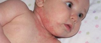
Erysipelas in children
The incubation period for children with erysipelas lasts from 2-3 hours to 3-5 days. The disease in most cases has an acute onset. Some cases are characterized by prodromal (preceding illness) phenomena: malaise, a feeling of heaviness in the affected limb, pain in the region of the regional lymph nodes, paresthesia. Paresthesia is understood as a feeling of numbness, tingling, or “pins and needles.”
The acute onset of the disease is accompanied by the following symptoms: headache, fever up to 38-40 °C, chills, nausea and vomiting, weakness. Severe forms of the disease are characterized by delirium and meningism.
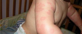
2-3 hours (or more) after the onset of symptoms of intoxication, erythema and severe swelling develop on the skin of the affected area, accompanied by sharp pain. The inflammatory process can develop on any part of the body, but most often it is concentrated on the skin of the face and legs, least often it is localized on the mucous membranes.
At the site of the lesion, the skin is hot, which can be checked by touch, painful and tense. The erythema quickly increases, erythematous spots merge with newly appearing ones, the skin becomes glossy, in some cases has a cyanotic tint (bluish).
The affected area protrudes above the level of healthy skin, delimited from it by an inflammatory ridge with scalloped (wavy) edges. There is enlargement and tenderness of regional lymph nodes. Often, against the background of erythema and edema, detachment of the epidermis occurs, which is why blisters (bullas) of oval or round shape and various sizes appear in the lesion, which are filled with serous hemorrhagic fluid.
General intoxication is as pronounced as local manifestations. In severe forms, bullous elements are observed, while in mild forms they are absent.
Classification. According to the nature of local manifestations, erysipelas can be erythematous, erythematous-bullous, erythematous-hemorrhagic, bullous-hemorrhagic.
Depending on the severity of intoxication, erysipelas in children is divided into mild, moderate and moderately severe. According to the frequency of the disease, erysipelas can be primary, repeated, or recurrent. According to the prevalence of the local process, widespread, localized, wandering, and metastatic erysipelas are distinguished.
Complications of erysipelas are local and general. Local ones include necrosis, abscess, and phlegmon. Common ones include pneumonia and sepsis.
Erythematous form of erysipelas is the most common, accounting for 50-60% of all cases. It is characterized by limited hyperemia of the skin with zigzag outlines in the form of teeth, tongues and arcs. Erythema can be minimal (barely noticeable) and maximum – bluish-purple. It is always accompanied by swelling, spreading beyond the erythema and involving the underlying adipose tissue. In some cases, swelling is the cause of compression of the blood vessels, then edema comes to the fore, and erythema moves to the background. In the affected area, the patient feels a burning sensation, pain and tension.
Regional lymphadenitis is rarely complicated by periadenitis and lymphangitis.
In the erythematous-bullous form, against the background of edema and hyperemia, bullous elements are formed, which contain clear liquid. These elements appear at different stages of the disease and can be either small vesicles or large blisters. Next, the blisters burst, their contents dry out, gray or gray-yellow crusts form in place of the blisters, and less often, erosions and ulcers with the development of granulations (juicy bright red tissue with a granular surface).
The erythematous-hemorrhagic form of erysipelas in children is characterized by the appearance of hemorrhages against the background of edema and hyperemia in the area of inflammation. In size they can range from petechiae to extensive ecchymoses. With this form of erysipelas, deep damage to blood vessels and lymphatic capillaries occurs with the development of complications in the form of necrosis and ulcers.
The bullous-hemorrhagic form is characterized by the appearance of blisters saturated with hemorrhagic contents. This form is considered the most severe and is extremely rare in children. The severity of the clinical form of erysipelas depends on the severity of general symptoms of intoxication and local inflammatory changes in the skin.
Flow. On average, the erythematous form of erysipelas lasts 7-10 days if treatment is carried out adequately and in a timely manner. At the site of erythema, after the disappearance of acute manifestations, peeling begins.
In bullous-hemorrhagic forms of erysipelas, brown or black crusts form in place of the blisters after they burst, and in some cases, erosions and ulcers.
After a child has had erysipelas, in some cases the pastiness and pigmentation of the skin, peeling persist for a long time, and sometimes elephantiasis develops.
Features of the disease in newborns and children of the 1st year of life. Erysipelas in newborns occurs in extremely rare cases. Most often, the process is concentrated in the navel area, during the first day it spreads along the anterior abdominal wall, goes down to the genitals, and spreads to the back and torso. In infants, skin hyperemia is less pronounced than in older children; the limiting ridge is not distinct.
Newborns are characterized by a widespread or wandering form of erysipelas. Intoxication can quickly increase, anxiety and hyperthermia appear. The child may refuse to breastfeed, convulsions, and septicopyemia are likely (a form of sepsis in which, along with general intoxication of the body, metastatic abscesses form in various tissues and organs).
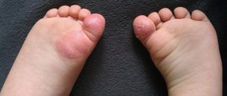
Erysipelas in babies under 12 months has a severe course. The inflammation is localized at the site of diaper rash or on the face. The process spreads quickly, sepsis and meningitis can develop.
What does a erysipelas on a leg look like?
Erysipelas on the leg is an affected area of bright red skin. Most often the limbs are affected. In rare cases, inflammation may occur in the genital area or torso. In the initial stage, shiny red spots appear on the leg. They spread quickly, turning into extensive outbreaks. Sometimes blisters form. The fluid in them may be clear or contain blood. In later stages of the disease, the affected area becomes covered with a crust. To better understand what a erysipelas on a leg looks like, it is recommended to study the photo.
Classification of erysipelas
There are several classifications of erysipelas on the legs:
- The first stage is classified depending on the severity of the disease. There are light, medium and heavy.
- The second stage of classification is determined by the scale of the affected areas. There are localized, limited and extensive forms.
- The third stage is determined depending on the nature of the manifestation. There is a primary, recurrent and repeated form of the disease.
Reasons for appearance
A streptococcal infection leads to the appearance of erysipelas on the leg. Typically, the pathogen enters the body through:
- diaper rash and cracks;
- skin damage resulting from injuries and bruises;
- insect bites or scratching;
- scratches or burns.
An old streptococcal infection can also provoke the disease. From the main focus, bacteria spread throughout the body, causing problems of varying severity. Having a healthy immune system prevents this from happening. However, if it is weakened for any reason, the risk of erysipelas on the leg increases. Frequently getting cold feet or getting too much tan can also lead to the problem. Stress or a sudden change in temperature are other factors that increase the likelihood of identifying a problem. There are a number of diseases against which erysipelas on the leg can develop. The list includes:
- alcoholism;
- overweight;
- varicose veins;
- fungal infections that are localized on the feet;
- trophic ulcers and thrombophlebitis;
- diabetes.
Most carriers of streptococcal infection do not even suspect it. Therefore, the appearance of erysipelas is often an unpleasant surprise.
Causes of erysipelas on the leg
The main cause of erysipelas is infection by streptococcus bacteria . Streptococci are gram-positive aerobic bacteria that live in the human body. Pathogenic microbes enter through open wounds caused by cuts, scrapes, cracks or burns. Sometimes carriers of streptococci do not even suspect its existence.
Of 100% of carriers, only 15% remain unaware, since their bacteria do not manifest themselves in any way throughout their lives. The remaining 85% of carriers suffer from various diseases that are caused by the proliferation of pathogens.
Erysipelas on the leg can occur at different ages. There is a tendency: in youth, it is mainly men who suffer from erysipelas, and in old age, erysipelas occurs more often in women.
Erysipelas is a deep type of pyoderma. Here you can read about pyoderma in children.
Causes of erysipelas:
- First of all, the disease occurs in people who, due to their profession or lifestyle, are constantly in unsanitary conditions.
- Erysipelas sometimes appears as a consequence of a sedentary lifestyle in older people. Trophic ulcers, bedsores and poor circulation are a favorable environment for the penetration and development of streptococcal bacteria.
- Erysipelas occurs in people with reduced immunity; this may be due to previous illnesses, severe stress and nervous exhaustion.
- Another reason for the appearance of erysipelas on the human body is the systematic exposure of the skin to UV rays, which leads to burns.
- Erysipelas often occurs in patients with diabetes, obesity and varicose veins. And also in people who suffer from alcoholism.
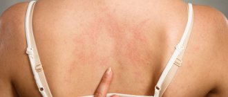
Symptoms of the disease
The symptoms of the disease are quite characteristic. However, they can confuse the doctor, leading to an erroneous diagnosis. The incubation period of the disease lasts from several hours to 3-4 days. Then the patient begins to experience the first symptoms of erysipelas on the leg. The person feels unwell and feels overwhelmed. All this arises against the background of general weakness. Then, quite suddenly, there is a rise in temperature. The person may experience headaches and chills. The temperature persists for the first few hours. The value of the indicator can reach up to 40 degrees. Additionally, there is pain in the muscles in the legs and lower back. A person complains of discomfort in the joints. In some cases, the patient may experience vomiting, diarrhea and nausea. Sometimes anorexia can develop against the background of the disease.
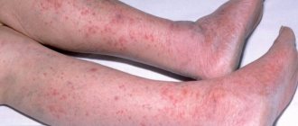
After a day, characteristic local signs are added to the general symptoms. The affected area begins to redden and swell. The person feels a burning sensation and pain. Tension is felt in the area where the erysipelas forms. Further symptoms directly depend on the type of disease.
The lesion has uneven jagged boundaries. Inflamed skin appears slightly shiny. It is dry and hot to the touch. Bright redness may be replaced by bluish, congestive edema. It occurs due to the fact that there is a local disturbance of blood microcirculation. Small hemorrhages often appear. They arise due to damage to the walls of blood vessels, as well as sweating of the formed elements.
On the second or third day of the disease, signs of lymphostasis with the development of dense lymphatic edema may be added to the general symptoms. It is at this moment that bubbles often appear within the lesion. They are filled with clear liquid. Sometimes it may contain blood.
After the blisters open, a dense brown crust appears at the site of the disease. Erysipelas gradually expands. However, if adequate treatment is carried out, the temperature returns to normal within 3-5 days. If the disease manifests itself in an erythematous form, its symptoms will disappear after 8-9 days. In hemorrhagic syndrome, they can persist for 12-16 days.
After the hyperemia and swelling of the skin decreases, its surface begins to itch and peel. During this period, some patients experience dark, congestive hyperemia and uneven hyperpigmentation. Subsequently, they will disappear on their own. If a person has suffered severe bullous hemorrhagic erysipelas, traces of the disease may persist for several years and even decades.
Forms of the disease
There are four forms of erysipelas on the legs.
All forms are similar, however, there are significant distinctive features:
- Erythematous . With this form, red erythema appears on the skin, which has clear boundaries with a uniform color. After some time, peeling occurs on the lesions. The erythematous form is also characteristic of erysipelas of the face.
- Erythematous-bullous. A form in which the erythema becomes covered with blisters containing clear liquid after a few days. If the patient received medical care on time, then after the blisters go away, healthy areas of skin can be seen underneath them. If treatment is not carried out, then a burst blister can lead to trophic ulcers and rotting wounds.
- Bullous-hemorrhagic. A form similar to erythematobullous, however, in this case the blisters are filled with blood.
- Erythematous-hemorrhagic. This form manifests itself in the form of subcutaneous hemorrhages in the area of erythema.
Treatment of erysipelas
If we are talking about the initial stage of the disease, treatment is carried out on an outpatient basis. In case of severe advanced forms, the patient is hospitalized. Usually the doctor prescribes antibiotics. The list includes:
- furazolidone;
- penicillin;
- erythromycin;
- oleandomycin;
- Biseptol.
They are usually prescribed in tablet form. If treatment is carried out inpatiently, the patient is prescribed courses of intravenous and intramuscular injections. In addition to antibiotics, other drugs are also used. So, to improve the general condition of the body, vitamins are prescribed. Inflammation is relieved using anti-inflammatory drugs. If the disease is severe and complicated by intoxication, detoxification drugs are additionally used. They can be used as a glucose solution or Reopoliglucin. Sometimes your doctor may prescribe them together.
To relieve pain and other manifestations of the disease, symptomatic treatment is carried out. In the process, vascular, antipyretic and diuretic drugs can be used. In some cases, agents may be prescribed that reduce the permeability of blood vessels.
To combat erysipelas, local medications are also used. Sometimes erythromycin ointment, furatsilin solution or enteroseptol are used. It may be prescribed in the form of powders or ointments.
If a patient is diagnosed with a bullous form of erysipelas, complex complex treatment is required. When the disease passes the acute stage, the surgeon removes the blisters. The opened bottom is covered with sterile dressings, which are soaked in a solution of furatsilin or rivanol. They must be changed at least once a day. In this case, the bandages should not be tight.
If the bullous-hemorrhagic form is diagnosed, applications are performed with dibunol ointment 5-10%. The procedure is repeated at least twice a day. It is carried out within a week.
In the acute period of the disease, ultrasound irradiation, exposure to electric current discharges, and laser therapy can also be used.
It is strictly forbidden to treat the affected area with ichthyol ointment or Vishnevsky balm. The above agents help to increase the flow of interstitial fluid. All this leads to the healing process slowing down.
Erysipelas
The incubation period is determined only in the case of post-traumatic erysipelas and ranges from several hours to five days. In the vast majority of cases (more than 90%), erysipelas has an acute onset (the time of onset of clinical symptoms is noted with an accuracy of hours), fever quickly develops, accompanied by symptoms of intoxication (chills, headache, weakness, body aches).
A severe course is characterized by the occurrence of vomiting of central origin, convulsions, and delirium. A few hours later (sometimes the next day), local symptoms appear: burning, itching, a feeling of fullness and moderate pain when palpated or pressed appear in a limited area of the skin or mucous membrane. Severe pain is characteristic of erysipelas of the scalp. There may be pain in the regional lymph nodes upon palpation and movement. Erythema and swelling appear in the area of the lesion.
The height of the period is characterized by the progression of intoxication, apathy, insomnia, nausea and vomiting, and symptoms from the central nervous system (loss of consciousness, delirium). The focal area is a dense, bright red spot with clearly defined, uneven boundaries (a symptom of “flames” or “geographic map”), with pronounced swelling. The color of erythema can range from cyanotic (with lymphostasis) to brownish (with trophic disturbances). There is a short-term (1-2 sec) disappearance of redness after pressure. In most cases, compaction, limited mobility and pain on palpation of regional lymph nodes are detected.
Fever and intoxication persist for about a week, after which the temperature returns to normal, regression of skin symptoms occurs somewhat later. Erythema leaves behind fine scaly peeling and sometimes pigmentation. Regional lymphadenitis and skin infiltration in some cases may persist for a long time, which is a sign of a probable early relapse. Persistent swelling is a symptom of developing lymphostasis. Erysipelas is most often localized on the lower extremities, then in frequency of development comes erysipelas of the face, upper extremities, and chest (erysipelas of the chest is most typical with the development of lymphostasis in the area of the postoperative scar).
Erythematous-hemorrhagic erysipelas is distinguished by the presence of local lesions in the area against the background of general erythema of hemorrhages: from small (petechiae) to extensive, confluent ones. Fever in this form of the disease usually lasts longer (up to two weeks) and regression of clinical manifestations occurs much more slowly. In addition, this form of erysipelas can be complicated by necrosis of local tissues.
In the erythematous-bullous form, vesicles (bulls), both small and quite large, with transparent contents of a serous nature, form in the area of erythema. Bubbles appear 2-3 days after the formation of erythema, open on their own, or they are opened with sterile scissors. Bullae with erysipelas usually do not leave scars. In the bullous-hemorrhagic form, the contents of the vesicles are serous-hemorrhagic in nature, and are often left behind after opening erosion and ulceration. This form is often complicated by phlegmon or necrosis; after recovery, scars and areas of pigmentation may remain.
Regardless of the form of the disease, erysipelas has features of its course in different age groups. In old age, primary and repeated inflammation is usually more severe, with an extended period of fever (up to a month) and exacerbation of existing chronic diseases. Inflammation of regional lymph nodes is usually not observed. The subsidence of clinical symptoms occurs slowly, and relapses are common: early (in the first six months) and late. The frequency of relapses also varies from rare episodes to frequent (3 or more times per year) exacerbations. Often recurrent erysipelas is considered chronic, while intoxication often becomes quite moderate, the erythema does not have clear boundaries and is paler, the lymph nodes are not changed.
Possible complications
If erysipelas is not treated, it can lead to serious complications. The disease can provoke:
- abscess;
- the appearance of an ulcer;
- sepsis;
- elephantiasis of the limbs;
- the beginning of necrotic processes;
- thrombophlebitis.
In severe cases, kidney disease and pathology of the cardiovascular system may occur. All of the above diseases pose a serious threat to health and can cause disability.
How to prevent it from appearing?
In order to prevent the occurrence of erysipelas on the leg, it is imperative to treat all skin diseases that appear in this area. The occurrence of a rash and other manifestations of a problem should be a reason for an immediate visit to a specialist.
If a person suffers from excessive sweating, it is necessary to use powder or talcum powder. High humidity is a favorable environment for the proliferation of microorganisms.
Avoid excessive exposure to ultraviolet radiation. The burns that the sun can leave on human skin can cause a number of serious diseases. At the same time, erysipelas is not the worst of them.
It is worth paying special attention to foot hygiene. When washing, you should use warm water, avoiding hypothermia. The use of soap products will prevent the accumulation of large numbers of microorganisms on the skin. When choosing a shower gel, you need to pay attention to the pH level.
Do not ignore the appearance of corns and cracked heels. A minor lesion can cause significant health problems in the future. If a person notices microcracks on the feet, it is necessary to immediately take measures to promote their rapid healing.