What do we know about moles?
What we have always heard about moles: “Does it bother you? Live in peace, don’t touch your mole!” This advice, which was very common in the past, convinced us that moles that were originally given by nature or that appear over the years should remain inviolable. If you touch it, you won’t be able to avoid trouble. This is partly true - indeed, removing moles can lead to the most dire consequences. But in fairness, it should also be noted that not every mole needs to be removed. The most harmless - flat moles
(pink to dark brown).
Usually they behave “quietly”, do not get injured and do not create inconvenience. The only thing you need to remember is to protect such moles (on open areas of the body) from sunlight. Convex moles
, especially if they are often touched by clothing, need to be treated more carefully. If they are on the back and in other places inaccessible to your view, you should entrust someone close to you with monitoring them. And, of course, if any changes occur—the mole becomes larger, turns from flat to convex, or changes its shape—you should immediately contact a specialist.
When can exposure to iodine cause harm?
The danger of using the drug is hidden in the consequences. There are reasons why you should not use this type of cauterization:
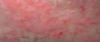
- The solution has a stimulating effect on blood circulation, provoking rapid growth of the nevus. Triggers the process of cell degeneration into a malignant tumor.
- There is a risk of burns and can cause serious complications.
- Infection and the onset of an inflammatory process are possible.
- Due to the coloring properties of the product (the brown color resembles the natural color of birthmarks), it becomes difficult to observe a benign nevus. You can skip the process of transformation into a malignant tumor.
Modern medicine has provided safe ways to remove unwanted formations on the body, without harm to health. Nevi, warts, and papillomas can be removed using laser, radio wave, cryotherapy, and surgery. The excision is painless and leaves no marks or scars. Re-removal is not required except in rare cases.
If there is the slightest change in a mole: itching, burning, color or shape, size have changed, you should not pick and treat the nevus yourself, especially cauterize it.
Before visiting a doctor, it is not advisable to smear the mole with iodine - this affects the natural color of the formation and complicates diagnosis.
Do not forget also about the contraindications for removing moles with iodine. These include:
- allergic reaction to the liquid or its components;
- during pregnancy, breastfeeding;
- children under three years of age;
- chronic diseases of the thyroid gland, kidneys, and liver;
- increased sensitivity to the product;
- pulmonary tuberculosis;
- if the formations are located in dangerous areas (faces, eyelids, mucous membranes, intimate areas).
No independence.
Some people believe that you can remove moles yourself by tying them, for example, with silk thread. This is a deep misconception. Attempts to bandage warts and papillomas with a thin silk thread often only provoke the growth of these formations. Today, there are celandine-based preparations on sale that are intended to eliminate warts. But it is better not to remove moles, papillomas, or warts yourself by any means. Celandine juice is similar in composition to iodine, so prolonged exposure to the drug causes skin burns. Cauterization with iodine is even more dangerous - it is more aggressive than celandine. Trying hard to get rid of papillomas with iodine, you can get scars on the skin. Only a dermatologist can properly remove a mole.
Advantages and disadvantages of mole removal with iodine
Iodine is a common drug that has a bactericidal, antifungal effect and disinfects the skin. Thanks to its composition, the product reduces the production of melanin, removing skin pigmentation. You can cauterize moles with iodine only after examination and identification of the benign nature of the formation. The method has a number of advantages:
- affordable price;
- availability of the drug (can be purchased at any pharmacy);
- has virtually no contraindications;
- efficiency;
- treatment can be carried out at home;
- side effects are minimal.
Like all medications, the drug has disadvantages in use, these include:
- It is not recommended to burn out large nevi; the tumor will remain in place;
- possible skin burn;
- allergic reaction to some components of the product.
Regular cauterization with iodine has a destructive effect on the formation, reducing it in size and drying it out.
The nevus becomes covered with a crust, which is not recommended to be peeled off. It should fall off on its own. Single papillomas, hanging and red warts can be treated. This is one of the budget, effective folk methods for removing growths at home. The action is aimed at destroying cells affected by the virus.
Removal methods.
Removal of moles and warts
- Cost: 2,000 rub.
More details
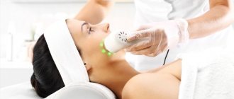
Mole removal can be carried out using a laser, radio wave coagulation or electrical coagulation. These manipulations are performed on an outpatient basis under local anesthesia. The choice of treatment method is determined by the doctor in each specific case. In my opinion, the most effective and painless method is radio wave coagulation. We use this technique in most cases, although in some cases we use excision with conventional surgical instruments followed by suturing. When using radio wave coagulation, tissue damage during material collection will be minimal, blood loss is reduced, and the rehabilitation period is shortened, while, for example, with the laser method, the healing process is extended over a fairly long period. Repeated formations do not appear after removal, but if precautions are not followed by the patient himself, infection may occur.
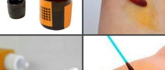
How to reduce a mole with iodine
Knowing the rules for using the product, you can avoid unpleasant consequences and complications. For the procedure to be effective and safe, the formation must be cauterized in the following way:
- Do not dilute iodine, since the product will not reduce nevi faster;
- to increase the rate of penetration into the epithelium, it is recommended to steam the body before the procedure by taking a hot shower or bath;
- Before the procedure, apply a thick cream or Vaseline to the skin around the growth. This will help avoid damage to healthy areas of the skin;
- Do not apply a cotton pad or gauze soaked in solution to the formation for a long time. This is fraught with inflammation of the affected area, itching, burning, and redness;
- To avoid scars and cicatrices, it is not recommended to pick or tear off the resulting crust. It should peel off on its own;
Instructions for removing formations with iodine:
- before smearing a mole with iodine, treat the skin with an antiseptic or soap;
- Gently dry the treated area with a towel;
- to protect the skin, apply cream or Vaseline around the growth;
- soak a cotton swab in the solution;
- Apply pointwise to the formation, avoiding contact with healthy areas of the epithelium.
Treatment is done twice a day for two weeks. During this time, warts and moles dry out and fall off. Continue to smear the area until the growth dries completely. It is impossible to cauterize the mole with iodine; if you pick it up or tear it off, it is better to treat it with an antiseptic and consult a dermatologist about its possible removal. Treatment of a damaged nevus should only be done with the permission of a doctor.
It is not recommended to use the method for people with a low pain threshold and sensitive skin. Itching, burning, and redness may occur.
To combat papillomavirus, blue iodine is an effective remedy (sold in pharmacies under the name “Iodinol”). The drug is taken orally and is prescribed only by a doctor. Effectively destroys cells infected with the virus, blocking their further spread throughout the body, improves the functioning of the gastrointestinal tract.
Unattended moles.
Some types of moles can degenerate into one of the most malignant tumors - melanoma (skin cancer). The initial site of formation of most melanomas is congenital pigment spots and warty moles. How does this dangerous development begin?
For example, after damage, a person only notices how the color of the mole begins to change, it enlarges, cracks, turns into an ulcer, and sometimes into a tumor. Therefore, without panicking, pay attention to the above symptoms, especially the progressive enlargement of the mole and change in its color. If all these symptoms occur, immediately rush to the dermatologist. The unfavorable prognosis of melanoma dictates the need for regular monitoring by a doctor. In addition to irritant factors (ultraviolet radiation, trauma), the development of melanoma can also be caused by genetic and endocrine factors. However, in the first place is, of course, excessive exposure to the sun. Ultraviolet radiation in large doses causes irreversible changes in skin cells, greatly increasing the risk of their degeneration. Light-skinned and fair-haired, red-haired people with blue and gray eyes are most susceptible to the mutagenic effects of sunlight. Those at risk also include those who have a lot of freckles, age spots and moles.
The effect of povidone-iodine on the eradication of highly oncogenic HPV types in women with cervical lesions
Relevance
Over the past decade, significant advances have been made in understanding the role of human papillomavirus (HPV) in the development of cervical cancer (CC) and other anogenital cancers. HPV is the main etiological agent of cervical dysplasia and carcinoma [1]. CC is the third most common gynecological cancer in developed countries. In Russia, the number of deaths from cervical cancer has increased by 3,500 women per year. Approximately half of patients with cervical cancer are women of reproductive age (under 50 years old) [2]. Recently, more than 200 women aged 20–39 years die annually for this reason. HPV is a common virus that is transmitted horizontally through heterosexual contact. Approximately 80% of all women are infected with HPV at some point in their lives, but in about 90% the human papillomavirus (PVI) infection resolves spontaneously within a few years. In the absence of independent elimination, cervical cancer may develop. Almost 100% of squamous intraepithelial lesions and CC, about 43% of vulvar tumors and 70% of vaginal tumors are associated with PVI, which annually causes 530 thousand new cases of CC and 21 thousand cases of vulvar and vaginal cancer worldwide [3]. In the absence of a screening strategy, there is an increase in the incidence of cervical cancer and vulvar cancer in young women [4]. HPV is a double-stranded DNA virus belonging to the genus Papilloma
in the family
Papovaviridae
[5]. It has been established that HPV is the most common sexually transmitted infection and is the main cause of cervical squamous intraepithelial lesions and invasive cervical cancer. There are more than 100 types that infect the epithelium of the reproductive tract. Transmission of HPV occurs primarily through skin-to-skin contact and probably through disruption of the epidermis, where the virus can infect basal squamous epithelial cells [6].
Types of HPV and their role in the development of cancer processes
To date, more than 100 HPV genotypes have been identified in the female reproductive tract. 13 HPV types (16, 18, 31, 33, 35, 39, 45, 51, 52, 56, 58, 59, and 68) are recognized as highly oncogenic (13HR) cancer-causing types. However, other types of HPV may also be associated with cancer. According to N. Munoz, two additional types of HPV - 73 and 82 - should be considered carcinogenic. G. Halec et al. demonstrated from a systematic review that HPV types 26, 53, 66, 67, 68, 70, 73 and 82 may be oncogenic [7]. One of the most important issues is that polymerase chain reaction (PCR)-based diagnostics used in epidemiological studies have limitations. Firstly, the PCR method detects not only highly oncogenic, but also other types of HPV. Second, some PCR tests using consensus primers have shown inconsistent results in identifying HPV types because the detection sensitivity of some HPV types is lower than that of others. Low-risk HPV types such as 6, 11, 40, 42, 43, 44, and 54 are associated with genital warts and low-risk anogenital lesions [8]. High-risk HPV types 16 and 18 together account for about 70% of cervical cancer cases, while low-risk HPV types 6 and 11 are responsible for 90% of genital warts [9]. The immune system plays an important role in controlling the development of cancer. The HPV genome encodes two oncoproteins - E6 and E7, capable of inactivating p53 proteins, and retinoblastoma (pRB) - regulators of cell proliferative activity. They are necessary for the emergence and maintenance of the malignant cell phenotype [10]. The adaptive immune response protects against HPV-induced diseases [11]. The progression of HPV-induced disease is associated with the lack of strong HPV-specific CD4+ and CD8+ T cell responses. Chemotherapy or radiation therapy influences immune regulatory activity and, in combination with vaccination, potentiates effective local HPV-specific T-cell immunity in mouse tumor models. Successful treatment of HPV-induced lesions can be achieved with low-dose cyclophosphamide, which modifies local immunity. Given the importance of the local microenvironment in the persistence of HPV-induced lesions and tumors, treatments that can locally shift the balance of immunoeffectors, such as cyclooxygenase-2 inhibitors, through the production of prostaglandin E2 and transforming growth factor β may be effective [12]. Vulvar carcinoma has two distinct carcinogenic pathways, one of which is associated with HPV type 16. It is the fourth most common invasive gynecological cancer, affecting mainly older women and is more common in Western countries [13]. Prognostic factors for the course of the disease include the presence or absence of metastases in the lymph nodes, as well as tumor size and recurrence. Relapse (hematogenous metastases) is observed in 40% of patients. The prognostic role of HPV in vulvar cancer is debated, with some studies showing better survival in women with HPV-positive vulvar tumors, others not [14]. An important issue remains the organization of screening programs. Such programs are being implemented effectively in New Zealand. National recommendations for a cervical cancer screening program recommend that all women aged 20 to 69 years who have ever had sexual intercourse be given a cervical smear within 3 years. If this is a primary screening smear or more than 5 years have passed since the previous test, it is recommended that the second smear be repeated one year after the first, and then after 3 years. After the introduction of this screening program, the incidence of cervical cancer decreased significantly, currently amounting to 5.4 per 100 thousand women [15]. In addition, a large study on the distribution of HPV genotypes in cervical samples was carried out in New Zealand. The material was collected from women with highly oncogenic HPV types (in 2009–2011) or suffering from invasive cervical cancer (in 2004–2010). The most common HPV types causing cervical neoplasia (cervical intraepithelial neoplasia - CIN 2 and 3) were HPV types 16 (51%), 52 (19%), 31 (17%), 33 (13%) and 18 (12 %). However, there was a trend toward increased rates of infection with HPV types 16 and 18 compared to other HPV genotypes in women aged 20–29 years. The most common HPV genotypes associated with invasive CC were HPV 16 (51%), 18 (21%), 31 (4%), 45 (3%) and 52 (3%) [16]. In 2008, New Zealand introduced the National Women's Immunization Program with the quadrivalent HPV vaccine (genotypes 6, 11, 16, 18). More recently, in 2013–2016, a study showed a decrease in the proportion of cervical intraepithelial neoplasia (CIN 2) associated with HPV 16/18 in a cohort of young New Zealand women, which may be associated with the implementation of the national HPV vaccination program. This study was significant because it was the first documented change in HPV genotype in women with cervical lesions after initiation of a vaccination program [17]. HPV type 16 is the most common genotype identified worldwide in patients with invasive CC, followed by HPV type 18. Eight HPV genotypes (16, 18, 31, 33, 35, 45, 52 and 58) lead to higher rates of CC progression [18]. The clinical guidelines of the Japanese Society of Obstetrics and Gynecology and the Japanese Association of Obstetricians and Gynecologists provide an algorithm for the management of patients with CIN, which includes HPV genotyping to determine the risk of progression to CIN 3 [19]. However, the relationship between HPV genotype and cancer prognosis remains controversial. Some studies have shown that HPV 18-positive tumors are associated with poor prognosis [20]. Other authors have stated that Chinese and British patients with tumors associated with HPV 16 and/or 18 have better survival, while in Taiwan favorable results were obtained for HPV 58- and 31-positive tumors [21]. On the contrary, other scientists have not found any associations between the HPV genotype and the prognosis of cervical cancer in Russian and Korean populations [22]. A cervical cytology study performed at three institutions in Japan demonstrated that HPV 18-positive tumors were associated with poor survival in women. Longer follow-up (102 months) confirmed that HPV 16-positive tumors correlated with better survival compared with HPV 18-positive tumors. These conflicting results may be due in part to geographic differences in HPV type prevalence [23].
Factors that increase susceptibility to HPV
Microbes are considered the main trigger factor for the development of malignant neoplasms. The Human Microbiome Project (HMP) has determined that 20% of all fatal malignancies are microbiologically induced. The ectocervix is colonized by microbes, whereas the endocervix and upper genital tract are considered virtually sterile in healthy women. Changes in the cervicovaginal microbiome and processes such as bacterial vaginosis, cervical inflammation and increased vaginal pH influence susceptibility to cervical HPV. Women in different ethnic groups have different vaginal microbiomes [24]. Most cervicovaginal infections and vaginal discomfort are caused by Gardnerella vaginalis
or
Atopobium vaginae
[25], as well as Candida albicans [26].
The cervical epithelium becomes vulnerable to infectious microbial agents such as HPV [27]. With the development of modern sequencing techniques, it is becoming clear that the microbiome influences susceptibility to cancer through the production of harmful metabolites and their impact on cellular function, since deregulated metabolism and inflammation are hallmarks of cancer [28]. The relationship of specific microbes (prokaryotic and eukaryotic) with HPV infections and cervical neoplasia remains one of the significant research issues. It is known that the microbiome of healthy women is dominated by Lactobacilli
.
This constant vaginal species produces lactic acid in the form of fermentation of products, which leads to a decrease in vaginal pH to 3.5–4.5, creating a chemical barrier to pathogens [29]. High levels of L. iners
are associated with the risk of developing HPV-associated cervical lesions.
An increase in body mass index correlates with an increase in the abundance of L. iners
, which makes obesity a significant factor in the development of CIN [30].
L. iners are more adaptable to pH changes and a variety of metabolic conditions than other lactobacilli. L. kitasatonis
and
L. crispatus
in the cervix in combination with highly oncogenic HPV types increase the risk of developing CIN 3. Floral diversity correlates with the severity of CIN.
The presence of Sneathia sanguinegens, Anaerococcus tetradius,
and
Peptostreptococcus anaerobius
in the vaginal microbiome of Caucasian, Asian, and Black women is often associated with the development of CIN.
P. micros
is an uncommon taxon commonly found in the oral cavity, found in the salivary microbiome and common in the presence of cervical lesions.
It has been suggested that P. micros can penetrate the cervicovaginal tract as a result of oral sex [31]. A direct connection of HPV with the dominance of three fungi of the genera Candida
,
Malassezia
and
Sporidiobolaceae was discovered.
Malassezia are lipophilic fungi that parasitize the upper layers of human skin and cause superficial fungal infections such as atopic dermatitis and psoriasis.
Malassezia
is often found on the foreskin and glans penis in men. Malassezia produce bioactive indolysines, including aryl hydrocarbon receptor (AhR) activators. AhR receptors mediate many skin functions, including promoting cell division, and it has been suggested that Malassezia may be involved in skin carcinogenesis. A recent study found that certain cervicovaginal tract microbiome structure correlates with both highly tumorigenic types of PVI and CIN severity in a population of women of reproductive age. Therefore, searching for features of the cervicogenital microbiome may be an important step in understanding the biology of cervical neoplasia and developing new therapeutic regimens targeting the microbiota [32].
Treatment of dysplastic changes in the cervix
Because CIN primarily affects women of reproductive age, there is a need for early identification and improved treatment strategies for the most clinically significant CIN. Persistent infection with highly oncogenic HPV types is a necessary condition for the progression of cervical pathology. Primary (vaccination) and secondary prevention (cervical screening) can have a decisive impact on cancer prevention [32]. The search for drugs aimed at treating dysplastic changes in the cervix is an important area of gynecology. Iodine is an antiseptic that has been used in medical practice for more than 150 years. Povidone-iodine
is one of the most powerful and effective iodine-containing antiseptics with a wide spectrum of action.
The main difference of this drug is that when it is taken, microbial resistance does not arise; it is less likely than antibiotic-containing drugs to cause an allergic reaction. The antiseptic effect of povidone-iodine is associated with a strong oxidative effect. Under the influence of povidone-iodine, pores form in the membranes of microbial cells, which leads to disruption of the integrity of the cell membrane, damage to the cell and loss of viability. The drug has several advantages over other antiseptics. Firstly, it more effectively stops the proliferation of microorganisms even in large dilutions. Secondly, povidone-iodine has the widest antiviral spectrum of action, including enteroviruses, polio and herpes viruses, as well as adenoviruses and influenza virus. Povidone-iodine is a stable drug, its effectiveness does not change under the influence of physicochemical conditions at the site of inflammation, caused by changes in pH, proteins, blood, and the action of enzymes. In gynecological practice, it is most often used to treat inflammatory processes and prevent the development of complications after invasive interventions [33]. The drug has been demonstrated to induce the death of HeLa epithelial cells in rats. Additional evidence supporting the potent antitumor effects of the iodine molecule and iodolactones was established through cell culture studies. They found a significant reduction in cell growth in breast cancer. A decrease in proliferation under the influence of iodine molecules was also noted in other human malignant cell lines (neuroblastoma, glioma, melanoma, lung, colon, and pancreatic carcinomas) [34]. Comparative analysis of the antiproliferative/cytotoxic ability of I2, potassium iodide (KI), a combination solution of KI+I2, povidone-iodine and I2+ [KI+glycerol] on human carcinoma cells showed that povidone-iodine may be a potential tool for directly interfering with tumor growth. cells [35]. A randomized study conducted in the USA found that all 88 patients aged 23–67 years (mean 34.8 years) with abnormal cytology and the presence of a highly oncogenic HPV type after 2 courses of cervical cryotherapy and local treatment with povidone-iodine after 6 months after therapy, HPV was not detected [36]. Studies have shown that the use of the drug Betadine
® after treatment with condyloma with a solution for external use with a local necrotizing effect increases the effectiveness of treatment, reduces the incidence of bacterial infection and recurrence of PVI of the vaginal and cervical mucosa. In connection with the above, the feasibility of complex treatment of cervical pathology, including povidone-iodine, is obvious [37].
Conclusion
Understanding the characteristics of the persistence of PVI in the epithelium of the cervix and the associated process of development of diseases in this area determines the tactics of treating cervical injuries associated with HPV. There are many methods for treating genital HPV, but the recurrence rate of this disease is high. Therefore, a very promising direction is the development of complex therapy to prevent recurrence of HPV-associated cervical diseases. The use of povidone-iodine (for example, the drug Betadine®) in the complex therapy of PVI increases the effectiveness of treatment, reduces the incidence of bacterial infection, as well as the frequency of recurrence of PVI of the vaginal and cervical mucosa. Information about the authors: 1 Dubrovina Svetlana Olegovna - Doctor of Medical Sciences, Professor, Chief Researcher of the Research Institute of Obstetrics and Pediatrics; 2Krasilnikova Liliya Viktorovna – candidate of medical sciences, obstetrician-gynecologist; 1 Ardintseva Oksana Aleksandrovna - graduate student; 3 Varicheva Marianna Vladimirovna - obstetrician-gynecologist; 4Tsirkunova Nina Sergeevna - obstetrician-gynecologist, head of the gynecological department; 1 Afrikyan Oleg Arturovich - 6th year student; 1 Afanasova Pelageya Nikolaevna - 6th year student; 5 Gadzhibekova Naina Balabekovna - obstetrician-gynecologist. 1 Federal State Budgetary Educational Institution of Higher Education Rost State Medical University of the Ministry of Health of Russia. 344022, Russia, Rostov-on-Don, lane. Nakhichevansky, 29. 2 DAVINCI GROUP LLC. 344091, Russia, Rostov-on-Don, st. Tolmacheva, 117. 3 Municipal Budgetary Healthcare Institution "City Hospital No. 20 of Rostov-on-Don." 344091, Russia, Rostov-on-Don, Kommunistichesky Ave., 39. 4 Municipal Budgetary Institution "City Hospital No. 6 of Rostov-on-Don." 344025, Russia, Rostov-on-Don, st. Saryan, 85/38. 5 MBUZ "GBSMP". 347930, Russia, Rostov region, Taganrog, Bolshoy prosp., 16. Contact information: Ardintseva Oksana Aleksandrovna, e-mail: [email protected] . Transparency of financial activities: none of the authors has a financial interest in the materials and methods presented. conflict of interest . The article was received on November 26, 2018. About the authors : 1 Svetlana O. Dubrovina - MD, PhD, Professor, Principal Researcher of the Scientific Research Institute of Obstetrics and Pediatrics; 2 Liliya V. Krasilnikova - MD, PhD, obstetrician-gynecologist; 1 Oxana A. Ardintseva - graduate student; 3 Marianna V. Varicheva - obstetrician-gynecologist; 4 Nina S. Tsirkunova - obstetrician-gynecologist, Head of the Department of Gynecology; 1 Oleg A. Afrikyan - 6th year student; 1 Pelageya N. Afanasova - 6th year student; 5 Naina B. Gadzhibekova - obstetrician-gynecologist. 1 Rostov State Medical University. 29, per. Nakhichevanskiy, Rostov-on-Don, 344022, Russian Federation. 2 LLC "DAVINCI GROUP". 117, Tolmacheva sr., Rostov-on-Don, 344091, Russian Federation. 3Rostov-on-Don City Hospital No. 20. 39, Kommunisticheskiy pass., Rostov-on-Don, 344091, Russian Federation. 4 Rostov-on-Don City Hospital No. 6. 85/38, Saryana str., Rostov-on-Don, 344025, Russian Federation. 5 City Emergency Hospital. 16, Bolshoy prosp., Taganrog, Rostov region, 347930, Russian Federation. Contact information: Oxana A. Ardintseva, e-mail: [email protected] . Financial Disclosure: no author has a financial or property interest in any material or method mentioned. Received 11/26/2018.
Dangerous sun.
For each person, the critical duration of exposure to sunlight is highly individual. It’s not easy to determine this line, so it’s better to just remember that prolonged exposure to the sun is harmful to the body. The skin is forced to protect itself from ultraviolet radiation. Excessive tanning means inevitable burns. If you love beach holidays so much that you simply cannot deny yourself this pleasure, at least listen to the following tips. By following these simple recommendations, you will not only improve your health, but also reduce the risk of a dangerous disease.
- Staying in the sun is safe from morning until 10 o'clock, in the evening after 17 o'clock.
- For people with a large number of moles, it is better to reduce their exposure to the sun.
- Try to avoid redness and sunburn on your skin. If this happens, spend 2-3 days in the shade.
- After swimming in the sea, be sure to rinse your skin with fresh water.
- Even young and healthy people can spend no more than one and a half to two hours a day in the sun.
- Taking certain medications increases the skin's sensitivity to light. Please consult your GP before going on holiday.
- Use sunscreen cosmetics: at the beginning of the holiday, the indicator should be maximum, about 40 units.
- Do not use decorative cosmetics, deodorants and perfumes on the beach - they can cause the appearance of age spots on the skin.
“Should I remove moles or not?”
There are formations on the skin that need to be removed because there is a high risk of cancer.
Mostly such formations are excised with mandatory histological examination of the surgical material. Benign, non-precancerous skin formations must be removed for cosmetic reasons, in case of unpleasant sensations (trauma, itching), if permanent damage to the formation or other discomfort occurs.
In any case, you should consult a dermatologist or oncologist about whether moles need to be removed. He should also recommend the optimal removal method.
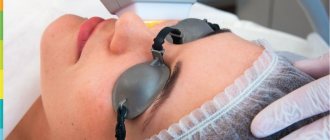
How long does it take for a wound to heal after laser removal?
The duration of the complete healing period depends on many factors:
- from the structure of the tissue itself, for example, the mucous membrane is restored faster than the rough thick skin on the feet or palms;
- depending on the intensity of the blood supply to the area where the removal took place, well-supplied areas heal 2 times faster than, for example, the heels and feet, where there is practically no blood flow in the superficial layers of the dermis;
- depending on the size of the wound that formed after removal, defects of small diameter heal 3 times faster.
Already at the end of the first day, the number of cells involved in regeneration increases along the periphery of the wound (for comparison with other methods of exposure only at the end of 3 days). By 5-7, there is an active process of angiogenesis, the germination of new vessels. And by 14-20 days, the tissue structure is completely restored without the formation of scar changes.
What types of keratomas exist?
There are several types of keratomas:
- seborrheic;
- age;
- senile;
- horny;
- solar;
- follicular.
Seborrheic keratoma is an extremely unpleasant skin disease. In appearance, these are small spots of yellow or brown color, but they tend to increase in size over time, become covered with cracks, and become rough. There may be a feeling of itching and burning, as well as bleeding and pain in the affected areas of the epithelium.
Senile keratomas form in older people. They are yellowish or brown in color and can reach 5–6 centimeters in diameter. Age-related keratomas are usually raised and felt above the skin. They may have an uneven surface, as if covered with pits, resembling a thimble. Most often, these skin growths form on the neck, face and arms.
Senile keratoma usually appears in people over 30 years of age. It becomes covered with a grayish crust. A benign neoplasm is subject to constant inflammatory processes and bleeding.
Recommended articles on the topic:
- Ultrasonic facial peeling is a pleasant and beneficial procedure for your skin
- Redermalization of the skin: all the pros and cons
- Almond peeling for the face: features of the procedure
Horny keratoma gets its name because it looks like an animal horn. Typically, this variant of the skin disease is an advanced form of age-related or seborrheic type keratoma.
Solar keratoma affects those who like to abuse tanning or visiting a solarium. It forms on areas of the skin that are most exposed to ultraviolet radiation - the face, arms, shoulders, chest. Keratoma is colored gray, yellow or brown.
Follicular keratoma appears on areas of the skin covered with hair (head, face). It looks like a nodule, colored gray and reaching 1.5 centimeters in diameter. This is the rarest type of keratoma.