For example, electroagulation destroys a wart or papilloma using electric current. Cryodestruction destroys tumors using extreme low temperatures. Today, laser removal of tumors is the most effective and safe method without the risk of complications. This is explained by the fact that the laser has high accuracy. Laser is a strong antiseptic; it ensures sterility and active restoration of cells and tissues. The impact of the laser is dosed.
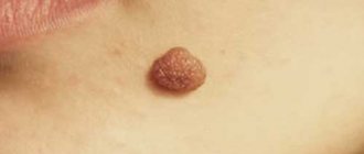
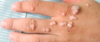
Laser removal of tumors allows you to remove all benign growths (moles, papillomas, etc.)
The laser “evaporates” tissue due to the high temperature and power of the beam. In this case, the doctor fully controls the depth of the laser’s impact on the tumor.
How does laser affect tissue?
Let's figure out how a CO2 laser affects tissue when it cauterizes tumors. The doctor directs the laser beam at the area of removal; when the beam hits the tissue, it is reflected and causes pendulum-like movements of the molecules, which leads to the evaporation of water from the cell and charring of the tissue. Part of the energy is transferred to surrounding cells, in which, under the influence of high temperatures, reversible protein denaturation occurs. It is the latter effect that causes the appearance of redness and swelling around the cauterization site.
But I would like to note that the surface after laser removal has a number of differences from other types of wounds, for example after a scalpel or electrocoagulation:
- The incision is made without touching the surface of the skin, this 100% guarantees the risk of infection;
- The laser coagulates the superficial layers of the dermis, while the deep ones remain undamaged, which is why it does not cause scarring and stenosis;
- Another feature is that after laser exposure in tissues, the temperature rises, this leads to the death of bacteria and viruses, thus it has an antiseptic effect;
- In the area affected by the laser, due to high temperatures, the vessels are sealed, so there is no bleeding, and a dense scab is formed that closes the wound, preventing bacteria from entering it;
- After laser exposure, regenerative processes at the site of exposure are stimulated due to the penetration of a small amount of light quanta into the deep structures of the skin;
- Compared to other types of cauterization, with laser cauterization the swelling around the wound is less pronounced and therefore there is no pain after laser removal.
Thanks to the above symptoms, the wound heals faster after laser, almost never suppurates or causes pain, and most importantly, does not lead to the formation of scars.
Limitations and complications after laser mole removal
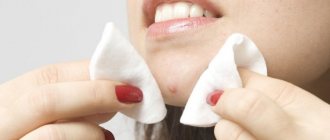
Specific requirements must be strictly adhered to for four weeks after surgery, regardless of how quickly healing occurs. In particular, you should refrain from visiting a solarium and sunbathing, and do not visit the pool or sauna to protect your skin from any infectious diseases. It is also recommended not to drink alcohol, as they dilate blood vessels, often causing bleeding. In case of removal of a mole on the face, you should not apply decorative cosmetics to the skin for 7-10 days after the operation.
Cosmetics with a softening effect speed up the skin restoration process.
As for complications after a laser procedure, they can occur if you do not comply with the doctor’s requirements and do not pay proper attention to the damaged area. In such cases, negative symptoms occur that require immediate contact to the clinic for additional treatment. These complications include:
- prolonged bleeding;
- purulent discharge;
- significant swelling of the skin that does not go away for several days;
- increased temperature of the whole body or area around the wound;
- severe itching.
How long does it take for a wound to heal after laser removal?
The duration of the complete healing period depends on many factors:
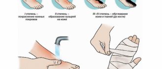
- from the structure of the tissue itself, for example, the mucous membrane is restored faster than the rough thick skin on the feet or palms;
- depending on the intensity of the blood supply to the area where the removal took place, well-supplied areas heal 2 times faster than, for example, the heels and feet, where there is practically no blood flow in the superficial layers of the dermis;
- depending on the size of the wound that formed after removal, defects of small diameter heal 3 times faster.
Already at the end of the first day, the number of cells involved in regeneration increases along the periphery of the wound (for comparison with other methods of exposure only at the end of 3 days). By 5-7, there is an active process of angiogenesis, the germination of new vessels. And by 14-20 days, the tissue structure is completely restored without the formation of scar changes.
A scar appeared after mole removal
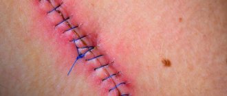
When excision with a scalpel, modern, atraumatic absorbable suture materials are used to eliminate a wound defect and, often, the patient cares for the wound independently.
In this case, the scars remain as aesthetically pleasing and delicate as possible, but nevertheless they are still scars. After removing a mole with a laser or electrocoagulator, the marks are usually invisible on the body (when nevi up to 5 mm are removed), or are visible, but are perceived not as scars and scars, but as minor changes in the skin (there are no associations with surgical procedures).
The appearance of a scar after mole removal depends on the properties of the patient’s skin, the size of the lesion being removed, its location, the duration of healing, and the characteristics of caring for the wound during healing.
The most unpleasant moment is the appearance of a keloid scar at the site of the removed nevus - this is a frequent reason for repeated visits and many questions. A keloid scar after removal of a mole occurs due to the predisposition of the patient’s skin, in certain areas of the body (sternum area, for example), when complications arise in the form of suppuration of the wound, or during prolonged healing due to improper care of the wound.
A keloid scar can be disturbing not only due to its sloppy appearance, but also due to pain or severe itching.
Modern technologies make it possible to correct a keloid scar. For example, we use injection correction in combination with surface laser resurfacing. If treated in a timely manner, one procedure is sufficient.
How long does redness and swelling last after laser vaporization?
As we wrote above, swelling after laser removal is rare and goes away quickly. Most often, swelling occurs during removal of tissues with good blood supply and thin epidermis, for example, when removing papillomas on the eyelid, condylomas on the genitals, or the oral mucosa. Swelling usually appears within a day and completely disappears after 3 days. When removing calluses and warts on the foot and palm, there is no swelling, since the hydrophilicity of the tissues in this area is low.
Redness around the wound after laser removal is not common, since the blood vessels are sealed in the affected area. Like swelling, redness often appears in areas where the skin is very thin and sensitive. Redness should not persist for more than 2 days. If the redness does not go away or begins to increase, you need to rule out inflammation and be sure to see a dermatologist for an examination.
Does the wound hurt after removal?
Laser is a unique method of influencing tissue, which causes complete destruction of neoplasm cells without affecting nearby healthy cells. We have already written above that swelling does not form in the cauterization area, accordingly, peripheral nerve receptors are not irritated and therefore the sore that forms after laser removal does not hurt. Of course, if the scab is in a place of constant pressure (for example, on the foot) or comes into contact with clothing (bra or belt), then discomfort may occur. There is no need to resort to taking painkillers; all sensations in the area are simply unpleasant sensations.
Relapse after removal of skin basalioma WILL BE CURED without surgery!
Afanasyev Maxim Stanislavovich.
Doctor of Medical Sciences, Professor of Sechenov University, oncologist, surgeon, oncogynecologist, gynecologist-immunologist, expert in the treatment of recurrent basal cell carcinoma.
"I am 51 years old. 4 years ago, a basal cell carcinoma was removed with a laser on my face. A year ago she appeared again, in the same place. I really don’t want to cut it out again, the scar is very noticeable, on the cheek near the nose. The size is about 1 cm. I work every day, constantly communicate with people, I need to remove it, preferably in the least traumatic way.”
Patients with repeated basal cell carcinomas after their removal are increasingly turning to me. Indeed, this is a huge problem that is becoming more acute year after year.
In addition to the fact that the incidence of basal cell carcinoma is increasing, most often this tumor occurs on the face, neck or head, where repeated use of any radical treatment method is associated with the loss of an organ or part of it. For this reason, treating relapse with traditional methods is often simply impossible.
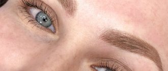
The eyelash growth area is one of the most difficult. Recurrence of basal cell carcinoma in this area will raise very serious questions for the patient and his surgeon. Source: www.tabanmd.com
In my practice, I use photodynamic therapy to treat relapses after radiation or relapses after surgery.
It specifically eliminates altered cancer cells and does not affect healthy tissue. Even against the background of a significant tissue deficit in the relapse area, this allows for almost non-invasive and maximally aesthetic interventions.
In this article I will analyze the causes of relapses. I will give statistics on the effectiveness of different treatment methods and tell you how to save the situation if basal cell carcinoma develops again.
Basalioma of the chin after excision and healing. Source: metro.co.uk
Risks of recurrence of basal cell carcinoma - what is the likelihood of encountering it again after removal?
Cutaneous basal cell carcinoma, or cutaneous basal cell carcinoma, or BCCC, is a skin cancer. It extremely rarely metastasizes, but has a high tendency to relapse.
According to various sources, the incidence of relapses within 3-12 months after treatment is 21%. And over a period of 10 years, the tumor recurs in 40-46% of cases.
Unfortunately, when deciding to remove basal cell carcinoma (and doing the right thing), few doctors and patients plan intervention tactics taking into account possible relapse.
When agreeing to one or another method of treating basal cell carcinoma, you need to take into account one fact that is well known to doctors: any treatment is a provoking factor. The therapeutic effect on the tumor itself stimulates its activity and growth if the intervention is not radical enough.
This approach leads to the fact that treatment of recurrent basal cell carcinoma in the future either turns out to be impossible, or is associated with crippling and disfiguring loss of part of the nose, cheek, eye, ear... And such cases of long-term ineffective treatment of basal cell carcinoma are not uncommon. See for yourself.
The photo was taken immediately after removal of the basal cell carcinoma and after healing. Source: todddunn.files.wordpress.com
Therefore, when deciding on the choice of treatment method for basal cell carcinoma, it makes sense to consider several factors:
- The method guarantees a high degree of radicality of treatment - it allows you to completely remove all tumor cells, including the “root” of basal cell carcinoma.
- Preserves the integrity of the organ - eye, nose, ear, lips, etc.
- Provides minimal cosmetic defect (for example, all surgical methods require excision of an additional 2-5 mm of tissue from the tumor border).
- There are no long-term complications from using the method itself (it is believed that several years after radiation therapy, irradiated cells sometimes degenerate into cancer cells).
These are the properties that the photodynamic therapy (PDT) method has. It allows you to specifically eliminate all tumor cells without affecting healthy tissue, and in the vast majority of cases, obtain a relapse-free result in one procedure.
When treating a relapse, PDT does not require the removal of healthy tissue and preserves the integrity of the organ.
Risk factors for relapse
The high frequency of recurrence is associated with the characteristics of the tumor - basal cell carcinoma does not have a capsule, its real boundaries exceed the visible ones, and it is very difficult to determine the depth of invasion.
Here is a far from complete list of factors for relapse of basal cell carcinoma:
- Tumor shape. The nodular-ulcerative form of BCC recurs more often.
- The depth of tumor “growth” into the tissue. The surgeon sees only the affected area on the skin, but does not understand how deep the tumor goes. Tumors with deep invasion recur more often. Tumors with intradermal spread have the best prognosis.
- Tumor size. Relapses more often develop after treatment of tumors larger than 2 cm, so you should not “grow” moles on your face. In this case, basalioma cells can be detected at a distance of up to several centimeters from the lesion on healthy skin.
- Age and health conditions. Concomitant diseases can reduce immunity, which aggravates the course of the malignant process.
- “Inconvenient” location of basal cell carcinoma in anatomical folds and in close proximity to vital organs. These are the nose, nasolabial folds and ears. The tumor recurs slightly less often in the area around the eyes and in the eyelash growth area. Complete radical removal in these areas is simply impossible due to the risk of organ dysfunction or facial disfigurement.
- Different areas of the face have significantly different thicknesses of the skin and subcutaneous fat, which also affects the nature of the tumor and has a great influence on the choice of treatment method.
- Inadequate choice of treatment method or repeated treatment with different methods. This is the most important risk factor.
Many different methods are used to treat basal cell carcinomas. Unfortunately, none of them guarantee a 100% cure. Below I present a forecast for the development of relapse within 5 years after treatment, drawn from various sources:
- Close-focus X-ray irradiation (close-focus X-ray therapy). Recurrence of basal cell carcinoma after irradiation occurs in 46% of cases.
- PDT produces relapse in 6% of cases.
- Surgical excision. Relapse after removal of basal cell carcinoma develops in 30% of cases.
- Cryotherapy. The inability to control the depth of impact causes the largest number of relapses – 35%.
- Laser coagulation – 31%.
- Micrographic surgery according to Mohs. This is a relatively new method, which is still poorly represented in Russia, so it is still difficult to show the frequency of relapses.
- After previous combination treatment, repeated relapses occur in 47% of cases.
According to research results, vaporization, which is popular today (a method of treating basal cell carcinoma with a laser), turns out to be one of the least effective methods. According to this indicator, it is approaching cryodestruction.
The most effective method is still surgical excision of basal cell carcinoma. But, as we see, the effectiveness of the procedure is accompanied by a noticeable loss of healthy tissue.
What are the dangers of recurrent basal cell carcinoma on the face and head?
Recurrence of basalioma on the nose, as well as recurrence of basalioma on the lip, is fraught with the need for mutilating surgery. Since any surgical excision is carried out with the obligatory capture of 6 mm of healthy tissue, repeated removal of basal cell carcinoma on the nose usually ends with the removal of part of the nose and requires complex plastic surgery of the nose with tissues from other parts of the body.
Scope of surgery for nasal basal cell carcinoma. Source: www.ohniskin.com
The location of basal cell carcinoma in the corners of the eye is generally a contraindication to surgery, since in most cases it ends in the loss of the organ.
Source: www.tabanmd.com
Surgical excision of basal cell carcinoma on the forehead and scalp is complicated by the lack of subcutaneous fat in these areas. Excision of basal cell carcinoma involving healthy tissue leads to the impossibility of suturing the wound and requires the application of a skin flap from the patient's thigh.
Close focus X-ray therapy (FRT) cannot be performed on the ear. This leads to perichondritis - painful suppuration and swelling of the ear. The same procedure cannot be performed on the upper and lower eyelids, or exclusively using a special ocular prosthesis. Many patients report decreased vision after BPRT.
The bottom line, as we see, is that repeated treatment is often simply impossible.
Signs of relapse
80% of relapses occur between 3 months and 1 year after treatment. The remaining 20% - within 5-10 years. Therefore, recovery can be judged no earlier than 5 years after the procedure.
A relapse appears in the same place where the primary basal cell carcinoma was removed. Thus, recurrence of basal cell carcinoma after excision occurs directly in the scar.
What symptoms suggest a relapse? Repeated basal cell carcinoma looks like a non-healing wound or a new formation similar to the previously removed one.
If the formation appears on a new area of skin, it is a new basal cell carcinoma, which is not considered a relapse.
Is relapse treatable?
In the vast majority of cases, basal cell carcinoma is completely curable. The PDT procedure allows me to provide aesthetic and disease-free treatment of basal cell carcinoma in one procedure.
Treatment of recurrent basal cell carcinoma using PDT is performed with tissue preservation and has almost no risk of relapse.
I treat recurrent basal cell carcinoma using PDT. Based on my experience, I can say that basal cell carcinoma can be treated, and is treated extremely successfully.
I strongly recommend that all my patients always choose PDT as a priority treatment method. And that's why:
- This is a gentle, non-surgical and effective organ-preserving method of treating relapses.
- Selectively destroys the tumor while preserving healthy tissue.
- Does not require hospitalization.
- Well tolerated even by older people.
- There are practically no contraindications for people with concomitant diseases, for whom traditional treatment is associated with the development of complications.
- PDT is a non-toxic method: the photosensitizer does not cause significant changes in blood parameters.
- Has no serious complications.
- Can be used multiple times.
- Perhaps the only method of treating patients with a frequently recurrent form of the disease.
- One of the few methods for treating hard-to-reach neoplasms and tumors located in close proximity to vital organs.
- Allowed if there are contraindications to other methods.
- The author's development makes it possible to treat even complex localization of basal cell carcinoma in the eyes without the risk of weakened vision.
- Low risk of relapse.
- In the vast majority of cases, one PDT session is sufficient to eliminate the tumor.
Of course, it is better to use photodynamic therapy as the first method of treating basal cell carcinoma, but even for the recurrent form, my author’s protocols show very good results. Even if the blood flow in the scar is impaired, photodynamic reactions have a reserve in terms of the depth of impact: tumors with a scar of up to several millimeters will still fall under photochemical reactions indirectly through ischemic necrosis around the scar.
To get advice on whether photodynamic therapy is indicated for you, and to calculate the price of the procedure, send your medical history and tests by e-mail or call by phone in Moscow.
The reception is conducted by Afanasyev Maxim Stanislavovich - oncologist, immunologist, oncogynecologist, doctor of medical sciences, professor and member of the academic council of the First Moscow State Medical University named after. THEM. Sechenov Ministry of Health of the Russian Federation, expert in the treatment of basal cell carcinoma.
Reception is carried out in two clinics in Moscow, as well as in St. Petersburg, Makhachkala, Kursk, Stavropol, Barnaul, Samara, Naberezhnye Chelny, Salavat, Chelyabinsk, Surgut and other regions of Russia. You can check the date, location of the appointment in your city and sign up for a consultation with the administrator by calling 8-800-555-77-26.
After treatment, I maintain feedback with all patients and resolve any issues that arise. Hepatitis and positive HIV status are not contraindications for treatment with PDT.
References:
1. Chuprov I.N. Features of recurrence of basal cell skin cancer // Materials of the All-Russian conference with international participation: 100th anniversary of the Russian Society of Pathologists. St. Petersburg, 2009. pp. 345-347.
2. Panova I.E. Recurrence of basal cell cancer of the skin of the eyelids // Breast Cancer. Appendix Clinical ophthalmology. 2006. T. 7, No. 1. P.11-14.
3. Savelyeva A.E. Risk factors for the development of relapses of basal cell skin cancer: abstract. dis. ...cand. honey. Sci. M., 2004. 24 p.
4. Kurdina M.I., Tymchishina M.V. Detection of malignant skin tumors during clinical examination // Abstracts of the 1st Congress of Oncologists. St. Petersburg; M., 1996. Part II. P. 402.
5. Panova I.E., Usova R.A., Semenova L.E. and others. Treatment tactics for various clinical forms of basal cell carcinoma of periocular localization // Standardization in Oncology. Chelyabinsk, 2002. pp. 44–46.
6. Panova I.E. Recurrence of basal cell cancer of the skin of the eyelids // Breast Cancer. Appendix Clinical ophthalmology. 2006. T. 7, No. 1. P.11–14.
7. Chuprov I.N. Features of recurrence of basal cell skin cancer // Materials of the All-Russian conference with international participation: 100th anniversary of the Russian Society of Pathologists. St. Petersburg, 2009, pp. 345–347.
8. Matveeva O.V. Results of photodynamic therapy of basal cell skin cancer with local use of the photosensitizer Radachlorin // Radiation and risk. 2016. T. 25, No. 2. P.79-90.
How to care for a wound after cauterization
After laser removal, the scab is dense and dry; it tightly covers the damaged tissue from bacteria and other substances. Often, dermatologists simply ask not to touch the cauterization area, and not to treat it with anything. Wear natural fabrics to allow air to flow freely to this area, as the healing process in the open air is much faster. Also, for the first week, do not wet the area too much so that the sore does not get wet.
If the damage is localized in hard-to-reach and ventilated areas, it is additionally recommended:
- To reduce pressure on the cauterized area on the foot, wear loose shoes and use special orthopedic gel protectors;
- After removal, treat the mucous membranes with antiseptics (chlorhexidine, miramistin) 2 times a day;
- If the wound gets wet, it is dried with flucarcin for the first three days.
What determines the risk of scarring?
The appearance of a scar and its nature depends on a number of factors:
- Depth and area of damage: if they are small, a scar may not appear at all.
- Tissue healing abilities.
- The presence of chronic diseases (for example, diabetes).
- Time for complete healing of the wound (it may increase if the wound has become infected).
The process of complete wound healing, which lasts for 21 days, ends with pathological scarring in only 33% of cases. If the process lasts more than 21 days, the probability increases to 78%.