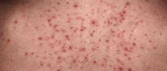
Folliculitis is a common form of staph infection. A sign of pathology is pimples or bumps around the hair on the arms or chest.
Folliculitis is a superficial or deep infection by bacteria that enter from the outside through the sebaceous glands or the hair follicle itself. Particularly prone to this type of inflammation are areas of the body where the skin rubs against clothing, such as the neck or back.
Hair follicles - what are they?
Hair follicles are hollows
in the skin in the form of tubules in which the hair roots are located.
They also serve as an outlet for the sebaceous glands that produce secretions. Their function is to protect the epidermis. Irritation or damage
to the opening of the hair follicle causes germs to enter the hair follicle, leading to infection.
The infection causes inflammation of the hair follicle, which results in painful
and itchy patches in the growing hair area.
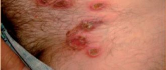
Abscessing undermining folliculitis and Hoffmann's perifolliculitis and follicular occlusion syndrome
Abscessing undermining folliculitis and Hoffman's perifolliculitis is an inflammatory recurrent disease of the scalp, characterized by the formation of abscesses, followed by cicatricial atrophy. The disease occurs predominantly in men and is characterized by a long course [1].
The cause of abscessing undermining folliculitis and Hoffmann's perifolliculitis is not fully known. Pathophysiological changes in the skin in this disease are associated with follicular occlusion, dilation and rupture of the follicular canal, which leads to an inflammatory reaction. Bacterial infection on the rash develops secondarily and is not the cause of this disease [2, 3].
The clinical picture of the disease is represented by multiple painful nodes with purulent discharge, which, merging, form interconnected abscesses and sinuses [1]. The process has typical localization sites with damage to the scalp (usually the parietal and occipital regions), and the back of the neck. The triggering mechanism in the formation of inflammatory elements is associated with occlusion of the sebaceous glands, which explains the characteristic location of localization [4–7]. According to topographic anatomy, the largest sebaceous glands are located on the skin of the scalp, with up to 5 lobules, deep-lying, with long convoluted ducts [8].
After resolution of the inflammatory process, hypertrophic or atrophic scars of the scalp remain, which leads to limited foci of cicatricial alopecia localized at the sites of inflammation [7].
Some authors indicate the possibility of developing complications, such as squamous cell carcinoma and marginal keratitis [2, 3, 7, 9].
Diagnosis of abscessing undermining folliculitis and Hoffman's perifolliculitis is based on the clinical picture, trichological examination, and, if necessary, histology.
The histological picture of the disease depends on the degree of inflammation. At an early stage, follicular hyperkeratosis is observed with blockage and dilatation of the follicle. As a result, apoptosis occurs and the follicular epithelium is destroyed. Subsequently, infiltrates of neutrophils, lymphocytes and histiocytes are formed. Later, an abscess forms, leading to the destruction of the hair follicle. Abscesses can penetrate the dermis and subcutaneous fat. As the process develops, granulation tissue is formed in the dermis and subcutaneous fat, containing lymphoplasmatic infiltrate and histiocytes, as well as multinucleated giant cells around keratin and hair shafts. The healing process ends with extensive fibrosis. In the later stages, scarring, fibrosis, subcutaneous sinuses, and chronic granulomas are noted, which are represented by lymphocytes, macrophages, giant cells of foreign bodies, and blood plasma cells [2, 4, 9, 10].
The trichoscopy method can detect characteristic signs: “3D” (“three-dimensional”) yellow dots in the area of dystrophic hair, yellow amorphous areas, white dots with halos, milky-red areas without follicular openings [4, 11].
The patients we observed with a diagnosis of “abscessing undermining folliculitis” and “Hoffmann’s perifolliculitis” are presented in the photographs (Fig. 1, 2). Clinical examples demonstrate abscesses and hypertrophic scars at the site of former rashes in the scalp in men 23 and 28 years old. In the presented patients, the process was localized only on the scalp, the skin in other areas was not affected, and there was no acne.
Abscessing undermining perifolliculitis may be one of the symptoms included in the follicular occlusion syndrome, which includes a combination of skin diseases similar in pathophysiological manifestations: conglobate acne, inverse acne, abscessing undermining perifolliculitis, pilonidal cyst. Currently, the literature describes double follicular occlusion syndrome, triad of follicular occlusion and tetralogy of follicular occlusion.
Double follicular occlusion syndrome combines abscessing undermining perifolliculitis and acne conglobata. A clinical example is presented in Fig. 3. A 25-year-old man has hypertrophic scars on the skin of the scalp at the site of former rashes in the scalp, post-acne scars on the skin in the face, grade 2-3 acne (Fig. 3).
The triad of follicular occlusion occurs in patients with conglobate acne, abscessing undermining perifolliculitis and inverse acne (or hidradenitis), and in the presence of a pilonidal cyst it is diagnosed as a tetrad of follicular occlusion [2, 9]. The exact pathogenesis of this group of diseases is unknown, but evidence suggests that they share the same pathological process initiated by follicular occlusion. Recent studies have shown that cytokeratin 17 (normally found in the sebaceous duct) is absent from the sebaceous duct epithelium of patients with acne inversis, contributing to epithelial fragility and leading to rupture of the follicular duct wall [12].
Follicular occlusion syndrome occurs more often in the third or fourth decade of life in males. However, this syndrome has been described in adolescence, as well as in women [2, 9, 13]. According to the literature, in rare cases, follicular occlusion syndrome can be combined with spondyloarthritis, osteomyelitis, Crohn's disease, sternoclavicular hyperostosis, Reid's syndrome (a rare combination of keratitis, ichthyosis and deafness) [2, 9, 14].
Inverse acne can be an independent disease, or it can be a symptom of follicular occlusion syndrome. According to the definition of European clinical guidelines, inverse acne is a chronic, inflammatory, recurrent dermatosis, with primary damage to the hair follicle, occurring after puberty and characterized by painful, deep rashes localized in parts of the body where apocrine glands are located (axillary, inguinal, anogenital areas) .
Inverse acne was first described in 1839 by Velpeau, who initially called the disease “hidradenitis suppurativa”, believing that inflammatory changes in this disease occur in the sweat glands [2, 9]. In 1939, Brunsting, based on a histological study, proved that the central link in the pathogenesis of hidradenitis, capitis folliculitis capitis and conglobate acne is hyperkeratosis of the follicular ducts of the pilosebaceous follicle [15]. In 1989, Plewigand and Steger introduced the term “inverse acne”, pointing out the incorrect term “hidradenitis”, since hyperkeratosis of the follicular duct, and not inflammation of the sweat glands, is pathogenetically significant. The difference between inverse acne and acne vulgaris, in addition to localization, is the absence of an increase in the secretion of the sebaceous glands [13, 16].
Currently, diagnostic criteria for inverse acne have been defined, in which basic and additional criteria are identified.
I. Basic:
- Recurrence of rashes two or more times within 6 months.
- The rashes are localized in the axillary, inguinal, anogenital and buttock areas, under the mammary glands (in women).
- The presence of nodes (inflammatory or non-inflammatory), sinuses (inflammatory or non-inflammatory), abscesses, scars (atrophic, hypertrophic or linear).
II. Additional:
- Having a family history.
2. Absence of pathogenic flora in a smear from the discharge (only the presence of normal microbiota) [7].
There are several stages of inverse acne:
- Stage I: abscesses, single or multiple, without sinuses or scars.
- Stage II: Recurrent abscesses with scarring, single or multiple, with areas of unaffected skin.
- Stage III: diffuse or almost diffuse lesions of the scalp, multiple interconnected sinuses and abscesses over the entire area (Hurley, 1989) [17].
Severe variants of follicular occlusion syndrome are associated with metabolic syndrome, obesity, eating large amounts of simple carbohydrates, smoking, and male gender [7].
We present clinical cases of the triad of follicular occlusion in Fig. 4, 5. Patients 43 and 52 years old complained of rashes on the skin of the scalp, face, back, chest, axillary area, and groin (Fig. 4, 5). The patients had similar medical histories and clinical presentations. They consider themselves sick since adolescence, when rashes first appeared on the skin of the face, chest and back. We were treated as an outpatient with a diagnosis of conglobate acne. At the age of about 30, painful rashes began to appear on the skin in the scalp and armpits. Patients were treated on an outpatient basis, doxycycline for 2–3 weeks, and excision of large nodes. The treatment brought temporary improvement, remission lasted less than six months.
Differential diagnosis of abscess folliculitis and Hoffmann's perifolliculitis
Differential diagnosis must be carried out, first of all, with diseases localized on the scalp. Acne-keloid (syn.: papillary dermatitis of the head, sclerosing folliculitis of the back of the head) is also observed in men in the back of the head and on the back of the neck. Initially, groups of small follicular pustules appear, which are located in the form of a cord. The skin around them is significantly thickened, the skin furrows are sharply expressed, which gives the impression of the existence of papillary tumors, and the hair grows in tufts. When the follicles resolve, keloid scars remain. The process proceeds slowly, without the formation of abscesses [1]. The absence of fluctuating nodules, large abscess formations, and fistula tracts is the main difference between acne keloids and abscessing undermining folliculitis and Hoffmann's perifolliculitis.
Often cases of cicatricial alopecia caused by previous folliculitis are usually diagnosed as Hoffmann's abscessive and undermining perifolliculitis. Decalvating (epilating) folliculitis was first described in 1888 by Quinqaud. Folliculitis decalvans also occurs in adults and is more common in men. Lesions in folliculitis decalvans are usually located on the scalp, but can sometimes be localized on other parts of the body covered with hair: beard, armpit area, pubis. The process begins with folliculitis surrounded by a zone of erythema. After inflammation resolves, small round or oval foci of scar atrophy form. Merging with each other, small lesions become larger, usually maintaining rounded outlines. Old areas where the inflammatory process has ended are represented by zones of scar atrophy. Thus, the clinical picture is represented by small foci of cicatricial alopecia with tufts of 5–10 hairs emerging from dilated follicular openings.
Mycotic lesions of the scalp are characterized by the absence of acute inflammatory phenomena and a positive fungal culture.
Other conditions that may mimic abscess folliculitis and Hoffmann's perifolliculitis include malignant proliferating pilar cysts, folliculotropic mycoses fungoides with gross cellular transformations, and erosive pustular dermatosis of the scalp [4, 18, 19].
Treatment of abscessing undermining folliculitis and Hoffmann's perifolliculitis and follicular occlusion syndrome
Therapy for follicular occlusion syndrome, as a rule, begins with treatment with antibacterial drugs prescribed orally (tetracycline, clindamycin, rifampicin) and externally (clindamycin). Some authors recommend ciprofloxacin 250–500 mg or clindamycin and rifampicin 300 mg [20].
However, antibacterial therapy has a short-term effect. According to some authors, it is more effective to combine antibiotic therapy with injections of corticosteroids into the lesions [21]. For the treatment of treatment-resistant cases, dapsone 50–150 mg daily is recommended [13].
However, according to the literature, surgical intervention is often necessary. Surgical treatment is carried out against the background of antibacterial therapy with complete resection of the affected skin of the scalp in step-by-step procedures, followed by skin reconstruction [22].
Currently, most publications devoted to the treatment of follicular occlusion claim the high effectiveness of isotretinoin in the treatment of this dermatosis. Isotretinoin is recommended to be prescribed in the same dosages as for acne vulgaris - 0.5–1 mg/kg for 3–12 months [20, 21].
Some authors recommend a combination of oral antibiotics and topical isotretinoin [20, 21].
In modern literature, there are isolated publications demonstrating the high effectiveness of genetically engineered biological drugs, such as adalimumab, infleximab, which are proposed as a third line of therapy [23, 24].
We observed a 52-year-old patient with a triad of follicular occlusion (acne conglobata, abscessing undermining perifolliculitis and inverse acne), who received treatment with isotretinoin at a dose of 30 mg per day for 6 months. As a result of therapy, long-term remission was observed for a year. The photographs show the results of therapy after 2 months of taking the drug (Fig. 6). The process has completely resolved and is represented by post-inflammatory spots and hypertrophic scars.
Thus, the presented clinical cases demonstrate various variants of follicular occlusion syndrome, as a result of which nodes of various types (inflammatory or non-inflammatory), abscesses and, as a consequence of the inflammatory process, the formation of various scars are formed on the skin. Currently, the most effective drug for the treatment of abscessing undermining folliculitis and Hoffmann's perifolliculitis and follicular occlusion syndrome is isotretinoin in the dosages recommended by the instructions.
Literature
- Berenbein B. A., Studnitsin A. A. Differential diagnosis of skin diseases. M.: Medicine, 1989. 672 p.
- Scheinfeld N. Diseases associated with hidranitis suppurativa: part 2 of aseriesonhidradenitis // Dermatol Online J. 2013, Jun 15; 19(6):18558.
- Badaoui A., Reygagne P., Cavelier-Balloy B., Pinquier L., Deschamps L., Crickx B. et al. Dissecting cellulitis of the scalp: a retrospective study of 51 patients and review of literature // Br J Dermatol. 2016, Feb. 174(2):421–423.
- Gaopande Vandana L., Maithili M. Kulkarni, Avinash R. Joshi, Ashish N. Dhande. Perifolliculitis Capitis Abscedens et Suffodiens in a 7 Years Male: A Case Report with Review of Literature Case Rep Surg. 2016: 21230 // Int J Trichology. 2015 Oct-Dec; 7 (4): 173–175.
- Mihic LL, Tomas D., Situm M., Krolo I., Sebetic K., Sjerobabski-Masnec I., Barišic F. Perifolliculitiscapitisabscedens et suffodiens in a caucasian: diagnostic and therapeutic challenge // Acta Dermatovenerol Croat. 2011; 19 (2): 98–102.
- Karpouzis A., Giatromanolaki A., Sivridis E., Kouskoukis C. Perifolliculitiscapitisabscedensetsuffodiens successfully controlled with topical isotretinoin // Eur J Dermatol. 2003, Mar-Apr; 13 (2): 192–195.
- Zouboulis CC, Desai N., Emtestam L., Hunger RE, Ioannides D., Juhász I. et al. European S1 guideline for the treatment of hidradenitis suppurativa/acne inverse // J Eur Acad Dermatol Venereol. 2015. Apr. 29 (4): 619–644.
- Sobolevskaya I. S. Some morphometric indicators of lipid-accumulating and lipid-synthesizing structures of human skin // Bulletin of VSMU. 2012, vol. 11, no. 2.
- Scheinfeld N. Review Dissecting cellulitis (Perifolliculitis Capitis Abscedense Suffodiens): a comprehensive review on new treatments and findings of the last decade with commentary comparing the therapies and causes of dissecting cellulitis to hidradenitis suppurativa // Dermatol Online J. 2014, May 16; 20(5):22692.
- Branisteanu DE, Molodoi A., Ciobanu D. The importance of histopathologic aspects in the diagnosis of dissecting cellulitis of the scalp // Rom J Morphol Embryol. 2009; 50 (4): 719–724.
- Rakowska A., Slowinska M., Kowalska-Oledzka E., Warszawik O., Czuwara J., Olszewska M., Rudnicka L. Trichoscopy of cicatricial alopecia // J Drugs Dermatol. 2012, Jun; 11(6):753–758.
- Kurokawa I., Nishijima S., Kusumoto K. et al. Immunohistochemical study of cytokeratins in hidradenitis suppurativa (acne inversa) // J Int Med Res. 2002; 30: 131–136.
- Vani Vasanth, Byalakere Shivanna Chandrashekar. Follicular occlusion tetrad // Indian Dermatol Online J. 2014, Oct-Dec; 5 (4): 491–493.
- Kohorst JJ, Kimball AB, Davis MD Systemic associations of hidradenitis suppurativa // J Am AcadDermatol. 2015, Nov; 73(5 Suppl 1):S27–35.
- Brunsting HA Hidradenitis suppurativa: abscess of the apocrine sweat glands // Arch fur Dermatol und Syph (Berlin). 1939. 39: 108–120.
- Plewig G., Steger M. Acne inversa (alias acne triad, acne tetrad, or hydradenitis suppurativa). Marks R., Plewig G., eds. Acne and Related Disorders. London: Martin Dunitz Ltd; 1989. 343–357.
- Hurley H. Axillary hyperhidrosis, apocrine bromhidrosis, hidradenitis suppurativa, and familial benign pemphigus: surgical approach. In: Roenigh RRH, ed. Dermatologic surgery. New York: Marcel Dekker, 1989: 729–739.
- Von Laffert M., Stadie V., Wohlrab J. et al. Hidradenitis suppurativa/acne inversa: bilocated epithelial hyperplasia with very different sequelae // Br J Dermatol. 2011; 164:367–371.
- Torok RD, Bellet JS Tinea capitis mimicking dissecting ce llulitis // Pediatr Dermatol. 2013, Nov-Dec; 30(6):753–754.
- Khaled A., Zeglaoui F., Zoghlami A., Fazaa B., Kamoun MR Dissecting cellulitis of the scalp: response to isotretinoin // J Eur Acad Dermatol Venereol. 2007 Nov; 21 (10): 1430–1431.
- Koudoukpo C., Abdennader S., Cavelier-Balloy B., Gasnier C., Yédomon H. Dissecting cellulitis of the scalp: a retrospective study of 7 cases confirming the efficacy of oral isotretinoin // Ann Dermatol Venereol. 2014 Aug-Sep; 141(8–9):500–506.
- Hintze JM, Howard BE, Donald CB, Hayden RE Surgical Management and Reconstruction of Hoffman's Disease (Dissecting Cellulitis of the Scalp) // Case Rep Surg. 2016; 2123037. DOI: 10.1155/2016/2123037. Epub 2016 Feb 7.
- Martin-García RF, Rullán JM Refractory dissecting Cellulitis of the Scalp Successfully controlled with Adalimumab // PR Health Sci J. 2015, Jun; 34 (2): 102–104.
- Sand FL, Thomsen SF Off-label use of TNF-alpha inhibitors in a dermatological university department: retrospective evaluation of 118 patients // Dermatol Ther. 2015, May-Jun; 28 (3): 158–165.
A. A. Kubanov1, Doctor of Medical Sciences, Professor, Corresponding Member of the Russian Academy of Sciences Yu. A. Gallyamova, Doctor of Medical Sciences, Professor T. A. Sysoeva, Candidate of Medical Sciences
FGOU DPO RMAPO, Moscow
1 Contact information
Causes of folliculitis
The cause of folliculitis is its superficial or deep infection
, causing inflammation.
Most often, bacteria are responsible for them, mainly Staphylococcus aureus
, which is physiologically found on the skin under normal conditions.
Folliculitis - risk factors
Factors that contribute to the appearance of folliculitis include: - wearing tight clothing; - frequent depilation;
- obesity;
- staying in one position for a long time;
- decreased immunity;
- diabetes;
- use of certain medications; - use of inappropriate cosmetics for skin care; - prolonged exposure of the body to the sun.
Men are at risk for chronic inflammation
hair follicles on the face in areas that are covered with hair. The disease has a long course. This is a special form of purulent inflammation of the hair follicle. It is known as sycamore.
Nutrition
Diet is not necessary when folliculitis occurs. But there are concomitant diseases for which dietary adjustments are necessary. Patients who are obese or have diabetes should follow a low-carbohydrate diet.
It is important to adhere to the following dietary rules:
- The consumption of animal fats should be limited.
- Eliminate flour products, spices, chocolate, sweets, alcohol, strong coffee and tea from your diet.
- Eat as many fresh vegetables as possible to provide your body with the required amount of fiber. You can also eat bran.
- You should include a sufficient amount of protein in your diet, including animal protein. Preference should be given to lean meats such as veal, chicken and turkey.
- It is worth eating more foods rich in vitamins. Carrots, beets, rose hips, and blueberries will be especially useful.
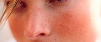
Symptoms of folliculitis
Symptoms of folliculitis - small yellow pustules
or papules, which are usually pierced by hair.
A red border around the lesion indicates inflammation. Over time, it may take the form of a purulent vesicle. Rashes occur singly or in groups
.
If the lesions extend into the deeper layers of the skin, soreness, pain and itching also develop. Facial folliculitis in men is usually very
severe, and the rash appears in the form of bumps.
Areas of the body where inflammation may occur
most often present as hairy skin that is removed by epilation or frequently rubbed against clothing.
This is why folliculitis occurs most often on the labia, buttocks, legs, face and armpits.
Inflammation of hair follicles on the head
Folliculitis of the scalp is usually
occurs in other dermatological diseases, mainly seborrheic dermatitis.
This also often applies to people who wear a hat for a long time during the day. Because of this, the scalp is constantly damp and warm, which promotes the growth of bacteria. Symptoms include pimples and redness around the hair shaft,
often filled with pus.
Proper care is important when treating an illness
behind the scalp.
It is recommended to wash your hair every day to prevent the accumulation of sebum, which is a breeding ground for bacteria. Shampoo for folliculitis on the head
should be gentle, natural, free of chemicals and have antibacterial or antifungal properties.
Complications
In most cases, this disease does not cause serious consequences for human life and health. But if there is no proper treatment, the infection penetrates deeply and the disease becomes more complicated. The situation is aggravated if the patient neglects the rules of hygiene, and also if he has a significant weakening of the immune system.
Complications of the disease include the following:
- furunculosis and carbuncles;
- dermatophytosis;
- formation of scar tissue;
- rotting of tissues.
Scars and cicatrices are formed as a result of attempts to squeeze out pus from inflammatory elements.
How to cure folliculitis
Folliculitis should be treated
under the supervision of a dermatologist.
Therapy is based on the use of topical antibiotics in the form of ointments. In case of serious changes, general antibiotic therapy is recommended. Medicines for folliculitis are available with a doctor's prescription. You can also use urea creams for irritated skin, which will soften and moisturize the epidermis.
Herbs such as chamomile and sage are also effective against folliculitis.
What can you use at home for folliculitis?
Home treatment for folliculitis is primarily about hygiene and infection prevention measures.
To wash your body, use gentle lotions
that contain only natural ingredients.
Underwear should be airy and clothing should not be too tight
. To avoid inflammation of the hair follicles on the legs after depilation, it is necessary to disinfect shaving accessories or use disposable razors.
Diagnostics
Dermatologists must examine the rash and conduct a dermatoscopy, which will show the depth of the lesion. The specialist can also refer the patient to donate discharge from the pustules, which will be required for:
- microsporia;
- research on fungi;
- bacteriological culture;
- tests for Treponema pallidum.
There is also a need for differential diagnosis. Folliculitis should be distinguished from pathologies such as syphilis and gonorrhea. In this case, the doctor prescribes:
- RPR test.
- PCR diagnostics.
If necessary, the specialist will prescribe blood sugar testing and an immunogram. In addition to gonorrhea and syphilis, the disease should be differentiated from the following:
- phrynoderma;
- streptococcal impetigo;
- Hoffmann's perifolliculitis;
- drug toxicoderma;
- furunculosis;
- pink lichen of Zhiber;
- nodular cystic acne.
The treatment required for hair folliculitis, as well as ailments on the legs, pubis, face, back, and skin, is described below.
Pathogenesis
The mechanism of penetration of pathogenic microflora into the follicle (most often staphylococcal) is based on a violation of the outflow of secretions from the sweat and sebaceous glands. Also, the gates of infection can form as a result of scratching the skin due to itching , if a compress was applied incorrectly, when clothing, straps rub against the body and other minor superficial injuries to the skin as a result of bruises, weeping, etc.
The process of development of hair disease begins with redness and the appearance of a red spot around the hair, then the development of a pustule - a small cone-shaped vesicle that usually protrudes above the surface of the skin, has a rim of hyperemia, and upon palpation the accumulation of a small amount of infiltrate is determined.
Healthy and infected hair follicle
The nodule turns into an abscess with a yellowish-green filling of pus, which can open and dry out. If inflammation covers a large area, then folliculitis can develop into a boil . The inflammation subsides after 2-3 days or a week and is accompanied by the formation of a purulent crust, which, when it disappears, leaves a small shiny red wound, and then, after epithelialization, a bluish-pink one. To better understand the pathogenesis of folliculitis, it is necessary to consider the structure of the hair follicle.
Information about the structure of the hair follicle
The structure of the hair follicle is quite complex, because for the life of the hair it is necessary:
- The nutritional system - for which the papilla, consisting of connective tissue and combined with a network of blood vessels, is responsible; if it dies, the entire hair dies; if the papilla survives, then a new one grows in place of the lost hair.
- Protection system - at the point where the hair root passes into the shaft there is a hair funnel into which the duct of the sebaceous gland opens, which allows you to moisturize and lubricate the skin and protect against the penetration of microbes; sweat glands also help form a protective film.
- Ensuring hair growth - thanks to the keratinization of the inner layer of the root sheath, hair formation and growth occurs; the outer and middle layers are also distinguished.
- Innervation and tactile function - thanks to the levator pili muscle and nerve endings, “goose bumps” are formed in conditions of fear or cold.
The structure of the hair follicle
Clinical picture of folliculitis on the penis
The clinical picture of the development of the pathological process is clear and characterized by rapidity.
Most of the symptoms cannot be missed, as they cause significant discomfort, especially when folliculitis is localized in the groin area.
In dermatology, the symptoms of penis folliculitis develop in the following sequence:
- In the area of the affected follicle, a distinct hyperemia of the skin occurs with noticeable traces of infiltration. At this stage, most patients experience severe itching; scratching can aggravate the pathological process and provoke complications.
- The area of hyperemia increases in diameter; in place of the follicle (at the base of the hair), a pustule is formed, filled with serous or whitish purulent contents. This formation also itches, but when touched it responds with strong painful sensations.
- After spontaneous, accidental (when scratching, rubbing on clothes) or intentional (it is recommended to do this only at a doctor’s appointment) opening of the pustule, pus flows out of it (or is cleaned out by a doctor). At the site of the pathological formation, an ulceration remains, covered with a purulent-bloody crust.
Subsequently, upon receiving adequate treatment, the crust comes off, and a scar remains at the site of ulceration.
When the entire body of the follicle is affected, the scar is more pronounced, and traces of hyperpigmentation may be present.
Before any signs of the disease appear, patients often experience itching and soreness at the site of future development of folliculitis.
It is noteworthy that even in the area of the penis, on the body of which there is a small number of hairs, folliculitis has a multiple course (in most cases).
Folliculitis on the penis – which doctor should I contact?
The disease is characterized by pathological damage to various layers of the skin; accordingly, diagnosis and treatment are carried out by specialists associated with dermatology.
If the disease is caused by poor hygiene, chafing and other everyday causes, even a dermatologist can help.
If we are talking about factors related to sexual activity and STDs, you should contact a venereologist.
However, the best solution to receive complete treatment would be a visit to a dermatovenerologist.
In some cases, it is possible to prescribe treatment after the initial examination.
If a severe form of the disease is suspected, for example, provoked by a syphilitic infection, a full diagnosis is required, on the basis of which drugs are selected.
Folliculitis on the penis after sexual intercourse
The development of penis folliculitis after sex is possible in two scenarios, in each of which sexual intercourse takes place:
- During friction (during vaginal, anal or oral sex), swellings may form on the penis, which will allow pathogenic pathogens access to the follicle cavity. This probability increases significantly if there is insufficient production of pre-ejaculate (lubricant produced during sexual arousal) or the use of low-quality condoms.
- Infection of the follicle with sexually transmitted sexually transmitted infections. This is possible even when having sex with a regular partner, if the latter is a latent carrier. But the chance of infection is many times higher in cases of promiscuity and neglect of contraception.
Treatment with folk remedies
Natural remedies and medicinal plants can help in the fight against various skin infections:
- Turmeric – as a natural antibiotic, it helps well with staphylococcal folliculitis; it is enough to regularly apply the spice mixed with olive oil (to form a paste) on the affected areas for a couple of hours.
- Apple cider vinegar - should be used diluted with water in a ratio of 1 to 2, after moistening a tampon with the solution, apply it to the inflamed follicle for at least 10 minutes, then rinse with clean water.
- Tea tree essential oil - also used in the form of lotions no more than 1-2 times a day, it should be 1-2 drops on a swab moistened with water or olive oil, applied for 1-2 hours.
- Chamomile officinalis - used in the form of an infusion (30 g of plant material per 300 ml of boiling water), which should be used to regularly wash the inflamed areas.
- Aloe vera - the juice of this plant has an antibacterial and anti-inflammatory effect; it should be applied to the skin for about 1 hour once a day for a week.
Folliculitis on the penis due to poor hygiene
The most common factor contributing to the development of folliculitis on the penis.
Obviously, the likelihood of penile folliculitis due to insufficient intimate hygiene is many times higher than for people who regularly take care of themselves.
Poor hygiene also leads to the development of other pathological processes of infectious origin.
The main factor for folliculitis in this case is irregular washing, bathing or showering.
The skin on the genitals quickly becomes contaminated; this is facilitated by a number of physiological processes:
- urination
- secretory functions
- increased sweating in the intimate area
- having sex and stuff
If a man showers irregularly, the chances of developing folliculitis increase significantly.
Since a favorable environment is created for the development of pathogenic microorganisms.
Another factor related to hygiene is wearing underwear for more than 1-2 days, which happens especially often.
In addition, it is important to select suitable personal hygiene products (body gels, soaps, etc.), without neglecting their regular use.
Compliance with the rules of intimate hygiene allows you to eliminate pathogenic and conditionally pathogenic bacteria from the surface of the skin, controlling their population.
It also removes sebum, cleanses pores and layers of dead cells that feed bacteria.
The use of hygiene products dries out the skin, creating an unfavorable environment for the life of various microorganisms that love moisture.
Intimate hygiene also includes removing hair from the groin area and directly from the body of the penis, which is increasingly used by men today.
This is a useful procedure from a hygiene point of view, but it is important to carry it out correctly, preferably in a trusted salon.
Otherwise, especially when removing hair on your own, there is a high probability of damage to the follicles, which can cause folliculitis.
Folliculitis on the penis due to enlarged sebaceous glands
Anatomically, hair follicles are associated with endocrine glands.
Some representatives of the stronger sex experience an increase in sebaceous glands.
This anatomical feature caused by increased production of sex hormones is called hypertrophy.
Folliculitis of the penis with enlarged sebaceous glands occurs due to the increased secretory function of the latter.
As a result, the follicle becomes clogged with sebaceous secretions and epithelial cells.
An artificially created isolated environment promotes the development of pathogenic microorganisms that provoke the occurrence of folliculitis.
This factor occurs especially often among young men who have increased synthesis of androgens.
They stimulate the function of the sebaceous glands, causing hypertrophy and increasing sebum production.
Prevention
Most often, folliculitis is diagnosed in southern latitudes and in disadvantaged sections of the population, therefore, as preventive measures it is very important:
- compliance with hygiene standards;
- selection of clothing and linen according to size, made from natural materials, models that do not restrict movement, do not rub or squeeze;
- regular hair care on the head, face, body using high-quality products;
- use of personal protective equipment when working with hazardous substances;
- avoiding minor skin injuries and timely solving the problem of itching to prevent scratching;
- avoiding hypothermia, visiting water bodies of questionable condition;
- maintaining immunity at a sufficiently high level with the help of good nutrition and a healthy lifestyle.
Principles of treatment of folliculitis on the penis
Treatment of penis folliculitis is conservative, based on drug therapy.
Drugs are selected based on diagnostic results.
The key factor in choosing medications is the etiology of the disease and identification of the pathogen.
So, for fungal infections, antifungal agents are used, for bacterial ones, antibiotics are used.
In the early stages of development of the pathological process, local drugs are prescribed.
How to anoint penis folliculitis, there are different variations:
- Bacterial etiology, for example, common staphylococcal infection - penis folliculitis is treated with antibiotic ointments. One option would be to use Zinerit to treat penis folliculitis.
- Fungal infection - drugs with a wide spectrum of antifungal activity are prescribed (Terbinafine, Fluconazole).
- Herpetic form - acyclovir-based products are used.
- Demodicosis - the best option is to use Parmethrin cream.
Examples can be given for a long time; it is important that each medication is prescribed by the attending physician.
In severe cases of the disease, with deep damage to the follicle, systemic therapy is added to local therapy.
The doctor will answer what antibiotics to take for penis folliculitis.
Most often these are drugs of the erythromycin or tetracycline series.
In order to reduce the spread of infectious lesions to nearby tissues, doctors recommend the auxiliary use of boric alcohol.
The solution is used to treat the affected and surrounding areas of the skin.
In some cases, physiotherapy using ultraviolet radiation is performed.