A burn is an injury that leads to disruption of tissue integrity. They are distinguished by the type of traumatic factor and the depth of damage.
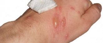
According to the type of damaging factor, the following are known: thermal, chemical, electrical, wave types of damage. According to the degree of penetration of the burn, 4 stages are known. In the second and higher stages of the burn, blisters appear on the dermis, which burst after a while. After this, lesions remain on the skin that are susceptible to getting wet.
Why does a wound after a burn not heal and become wet?
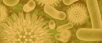
An open burn wound is a site of accumulation of pathogenic microorganisms. With proper treatment and normal functioning of the human immune system, the body successfully fights bacteria, viruses and fungi. However, if the protective forces are weakened, then active proliferation of microorganisms occurs.
One of the reasons for a weeping wound after a burn is the active reproduction and activity of pathogenic microflora.
Wetting of the burn also occurs due to the accumulation of lymph in the surrounding tissues and the penetration of fluid from the blood vessels into the wound cavity. This is the norm in the first 2 to 3 days after the burn, but then the wound should begin to dry out. When the fluid-filled blister bursts, the wound also becomes moist. But this is a temporary phenomenon.
There are groups of patients who are prone to developing weeping burns:
- If you have diabetes. In patients with this diagnosis, the healing of any wounds is difficult and long,
- For immunodeficiency conditions (rheumatoid arthritis, HIV and others),
- Aged people. In this case, regeneration occurs much slower.
Treatment of local radiation damage
Local radiation damage, which develops mainly during radiation therapy of malignant tumors, is characterized by its resistance to treatment with various medications. Radiation damage to integumentary tissues (skin, mucous membranes) and internal organs, as a result of exposure to ionizing radiation in a total focal dose of 60-70 Gy, manifests itself in the form of chronic epitheliitis and dermatitis, with their progression to radiation skin ulcers, radiation proctitis, cystitis, etc. etc. In the pathogenesis of such damage, along with disruption of microcirculation, the direct effect of radiation on cells and the suppression of reparative processes play a leading role. In the future, the addition of infection of damaged tissues and the aggravation of negative healing processes of damaged tissues come to the fore [1]. That is why the complex of medications for the treatment of local radiation damage includes substances whose effects are aimed at improving tissue microcirculation, increasing reparative processes and suppressing the infectious process. Almost all known medications that meet the listed requirements have been tested for the treatment of local radiation damage. The low therapeutic effectiveness of existing drugs has become the basis for the search for new treatment methods. In the department for the treatment of radiation injuries of the MRRC RAMS, a significant number of patients with radiation ulcers of the extremities and other areas of the body, radiation damage to the intestines, bladder, etc. are treated annually. The main component of local treatment is the drug dimexide (dimethyl sulfoxide, or DMSO), used in the form of solution dressings 5-10% or 10% ointment. This basic treatment, prescribed taking into account the specific characteristics of each patient, can be supplemented by the prescription of other antiseptics (dioxidine, chlorhexidine, etc.), proteolytic enzymes, and agents that stimulate reparative processes (curiosin, fortified oils, etc.). The developed local and general treatment regimens allow achieving favorable results in 57% of patients [1, 2].
Since September 2002, we have studied the therapeutic effectiveness of the drug Gepon for the treatment of patients with local radiation damage (see Table 1).
| Table 1. Use of Gepon in the treatment of local radiation damage. |
Radiation ulcers in patients developed after radiation therapy for malignant tumors (skin cancer - 16 patients, breast cancer - six, sarcomas - four). The total focal dose (FOD) was 45-70 Gy. Radiation proctitis resulted from radiation therapy for cervical and uterine cancer (13), bladder (3) and rectal cancer (2). Radiation cystitis has also been observed after radiation therapy for cervical and uterine cancer (13) and bladder cancer (4). Pneumofibrosis is a consequence of radiation therapy for lymphogranulomatosis (6) and breast cancer (5 patients).
In the treatment of radiation ulcers, Gepon was used at the first stage (7-10 days) in the form of irrigation of the ulcer with a solution. Gepon (0.002) was dissolved in 5 ml of sterile saline before use. Irrigation with the resulting solution of 0.04% Gepon was carried out daily. At the second stage, as granulation developed, 0.04% ointment was used (10-18 days). The results of treatment of radiation ulcers with Gepon were compared with the dynamics of the course of the wound process in more than 800 patients who were treated with the treatment methods adopted in the department, consisting of local application of a solution of 10% dimexide (applications or electrophoresis), electrophoresis of proteolytic enzymes and heparin, the use of levomikol ointments, iruxol, curiosin and eplan.
The effectiveness of Gepon was assessed clinically by the state of the wound surface (reduction of exudation, rate of development of granulations and rate of epithelization of the ulcer according to L.N. Popova (see Table 2)), calculated using the formula:
SD = (S-St)/St x 100, where SD is the healing rate S is the area of the radiation ulcer (mm2 before the start of treatment) St is the area of the ulcer (mm2) on the day of measurement t is the time in days from the start of treatment
| Table 2. Healing rate of radiation ulcers. |
In assessing the dynamics of healing, the study of the microflora of radiation ulcers and its sensitivity to antibiotics turned out to be informative. Before the use of Gepon in the wound discharge, 67.5% of cultures were found to have a monoinfection, mainly staphylococcus associations, and in 16.3% other microbes were also detected (Escherichia coli, gram-negative associations of microbes and Candida). After 12–15 days of use of Gepon, sterility was detected in 18.9% of cases or saprophytes (27%) characteristic of normal skin were detected. Compared to the initial level, 107-8 microbes per gram of tissue, by the end of treatment with Gepon, the contamination rate was reduced to 102-3, and the sensitivity of the flora to antibiotics significantly increased. All of the above indicates the undoubted effectiveness of the treatment.
We tend to associate the positive therapeutic effect of using Gepon primarily with its beneficial effect on the microflora, which helped reduce the inflammatory process and its negative consequences (swelling of surrounding tissues, impaired microcirculation, etc.). In addition, an important aspect of the action of Gepon is its immunomodulating effect, manifested in the activation of secretory immunoglobulin, a decrease in the level of anti-inflammatory cytokines, activation of a-interferon, a decrease in the adhesive function of cells and their apoptosis, cessation of viral replication and an increase in the body's resistance to bacterial flora.
Currently, when the wound-healing effect of Gepon has been proven, treatment of patients with radiation ulcers begins with the use of Gepon, and then is supplemented, according to indications, with other drugs. Treatment of radiation rectitis (18 patients) and radiation cystitis (17 patients) was carried out in the form of daily double microenemas or instillations of an aqueous solution of 0.04% for 12-18 days. The results of using Gepon were also compared with the results of “traditional” treatment practiced in the department over the past 25-30 years (microenemas of dimexide 5-10%, syntozone emulsions, fortified oils, etc.). Intracavitary administration of Gepon reduced the intensity of pain and hemorrhages and shortened the duration of treatment from 28-36 to 15-23 days. The use of Gepon activated immunity in this group of patients.
Thus, the immunomodulator Gepon in the treatment of patients with local radiation injuries (radiation ulcers, radiation rectitis and cystitis) has proven to be an effective medication that helps to quickly reduce the severity of the inflammatory process in radiation-damaged tissues and accelerate the reparative processes in them.
Literature
- Bardychev M. S., Tsyb A. F. Local radiation damage. - M.: "Medicine", 1985. - 240 p.
- Bardychev M.S., Katsalap S.N., Kurpesheva A.K. et al. Diagnosis and treatment of local radiation injuries // Medical Radiology, 1992, 12. - P. 22-25.
- Dudchenko M. A., Katlinsky A. V., Ataullakhanov R. R. Complex treatment of trophic ulcers // Journal “Attending Physician”. - 2002, No. 10. — P. 72-75.
- Perlamutrov Yu. N., Solovyov A. M., Bystritskaya T. F. et al. A new approach to the treatment of infections of the skin and mucous membranes // Bulletin of postgraduate medical education. - 2001, 2. - pp. 21-23.
- Kladova O.V. Kharlamova F.S., Shcherbakova A.A. et al. Effective treatment of Croup syndrome using the immunomodulator "Gepon" // Russian Medical Journal. - 2002, 10, 3. - P. 138-141.
M. S. Bardychev , Doctor of Medical Sciences, Professor Medical Radiological Research Center of the Russian Academy of Medical Sciences (Obninsk)
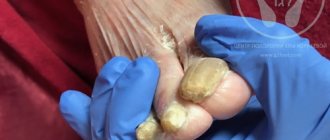
Symptoms of a weeping burn wound
When a weeping burn develops, local symptoms of inflammation come to the fore :
- Pain that increases with palpation of the edges of the wound,
- The skin around the wound changes color. They turn red
- Slight swelling of the wound
- Loose granulation, fills the wound,
- Exudate is constantly released from the wound. Because of this, the bandage constantly gets wet.
It should be noted that the severity and severity of pathological symptoms depends on the severity of the burn. With 2nd degree burns, the signs are not clearly expressed. With deep injuries, there is an abundant release of exudate, which is often purulent in nature.
The pain in this case can be very pronounced. If proper treatment is not carried out in this case, the infection spreads beyond the burn wound. In severe cases, a general infection of the body develops - sepsis.
Rules for treating wound surfaces
If a weeping burn occurs, frequent dressings with appropriate treatment are required. Scheme for treating a weeping wound:
- Dressing and treatment are carried out in the appropriate room (dressing room),
- Instruments and dressings must be sterile,
- The old bandage is removed carefully without causing pain to the patient. After which the used dressing material is placed in a container filled with a disinfectant,
- Dressing is carried out by a doctor and a nurse,
- If the wound is deep, then it is washed to thoroughly remove purulent exudate,
- If necessary, the doctor performs a revision of the wound,
- The burn is treated with antiseptic solutions that do not irritate tissue (Chlorhexidine, Furacilin, Hydrogen Peroxide, etc.),
- The edges of the wound are treated with alcohol antiseptics (Iodine, medical alcohol, etc.),
- If necessary, ultraviolet irradiation of wounds is carried out,
- Medicines are placed into the wound depending on the stage of the inflammatory process. If the wound is very wet, use drying agents in the form of powders that contain antibacterial agents,
- A bandage is applied.
It should be noted that dressing is done several times a day, depending on the degree of wetness and contamination of the dressing. At the very beginning of the inflammatory process, dressings are carried out up to 4 times a day.
What not to do with a weeping wound after a burn:
- Touch the surface of the burn with your hands,
- Use cotton wool to apply a bandage. When cotton wool is directly applied to the wound, its fibers will linger on the open surface. This will lead to even greater separation of exudate and the development of severe inflammation,
- Wash the burn wound with water,
- based on fat and oils when dressing Such drugs create a greenhouse effect, which only aggravates the pathological process. A weeping wound must “breathe”, that is, oxygen must be provided to it, otherwise the inflammation intensifies,
- Injure the wound surface when performing dressings,
- Tear the bandages off the wound with force. If the dressing material has dried to the wound surface, then it is necessary to pour it generously with any non-alcohol antiseptic. As the material gets wet, it will come unstuck from the wound,
- Self-medicate and independently prescribe and discontinue medications.
ANTIBACTERIAL THERAPY IN COMPLEX TREATMENT AND PREVENTION OF INFECTIOUS COMPLICATIONS IN BURNS
Infectious complications of burns are the most common cause of death of the burned. Addition to life threats, infection retards burn wound healing. The paper considers the antibacterial therapy of infectious complications in the burnt and its role in the multimodality treatment of burns.
A.A. Alekseev, M.G. Krutikov, V.P. Yakovlev Institute of Surgery named after. A.V. Vishnevsky RAMS, Moscow AA Alekseyev, MG Krutikov, VP Yakovlev
AVVishnevsky Institute of Surgery, Russian Academy of Medical Sciences, Moscow
Introduction
Currently, the incidence of burns in developed countries reaches 1:1000 population per year, and the mortality rate from burns ranges from 1.5 to 5.9% [1]. At the same time, the most common cause of death in burn victims is infection, which, according to some authors, accounts for 76.3% [2] in the structure of mortality in burn victims. In addition to the immediate danger to the patient's life, the prolonged existence of infection leads to a delay in the healing process of burn wounds and contributes to excessive scarring, which continues as a result of chronic stimulation of inflammatory cells [3]. Infection creates difficulties for timely autodermoplastic closure of burn wounds [4], and issues of infection remain relevant during early excision of a burn wound [5]. Significant difficulties are caused by infection when using such modern methods of closing the burn surface as keratocyte transplantation [6] and allofibroblast cultures [7]. Necrotic tissues formed in the burn area are a favorable environment for invasion and proliferation of microorganisms. Thus, any burn injury of any severity creates conditions for the development of wound infection. With extensive and deep burns, a number of pathological processes occur in the body, manifesting themselves in the clinical picture of burn disease and creating additional prerequisites for the development of the infectious process and its generalization. In addition to the loss of protective skin over a large area of the body surface, which creates an entrance gate for microbial invasion, this is the disintegration of the most important neurotrophic and metabolic functions of the body, leading to a violation of anti-infective defense factors.
Course of burn disease
The course of burn disease is divided into several periods: burn shock, acute burn toxemia, septicotoxemia and convalescence [1].
Such a division, although it can be considered quite arbitrary, facilitates understanding of the pathogenesis and contributes to the development of systematic treatment tactics. Thus, during the period of burn shock, microcirculation disorders, plasma loss and associated protein loss lead to alterative-dystrophic changes in the organs of immunogenesis. Immunosuppression is aggravated during the period of burn toxemia, which is associated with the accumulation in the body of medium molecular peptides and other toxic products of histiogenic, bacterial origin, nonspecific metabolites and biologically active substances. The waste products of microorganisms support immunosuppression during the period of septicotoxemia. Prolonged existence of burn wounds leads to the development of exhaustion, progression of protein deficiency and, as a consequence, immunodeficiency. A decrease in the body’s protective and compensatory capabilities predetermines the development of infection and its generalization. The above aspects of the pathogenesis of burn wounds and burn disease make the development of a set of methods for the prevention and treatment of infection and infectious complications in burn patients one of the priority areas for the development of modern combustiology. Antibacterial therapy plays an important role in the complex of measures aimed at preventing and treating infections in burned patients.
Antibacterial therapy
Experience in the treatment of burnt patients at the Scientific and Practical Center for Thermal Injuries of the Institute of Surgery named after. A.V. Vishnevsky RAMS allowed us to develop basic approaches to antibacterial therapy for these patients. The prescription of antibacterial drugs to burnt people should be based on a comprehensive assessment of their condition, taking into account the extent of the damage, its depth, the stage of the burn disease, its complications, the degree of microflora contamination of burn wounds, immune status, as well as the age of the patient, the nature and severity of concomitant pathology. For victims with II - III A degree burns, as well as patients with limited deep burns occupying no more than 10% of the body surface, the prescription of systemic antibacterial drugs in most cases seems inappropriate.
The exceptions are elderly and senile patients suffering from diabetes mellitus, chronic infections, as well as victims admitted for treatment late after injury with pronounced general and local signs of infection, when local antibacterial drugs have no effect.
The remaining patients are prescribed local antibacterial therapy: dressings with a 1% solution of iodovidone or iodopirone, water-soluble ointments containing chloramphenicol or dioxidine, silver sulfadiazine preparations. The combination of Levomekol ointment dressing with gentamicin or neobacitracin powder for gram-negative flora has proven itself to be effective. The use of synthetic coatings containing antibacterial drugs is promising. When treating such patients in regional bacterial isolation centers, daily treatment of wounds with a solution of iodopirone or iodovidone is sufficient. Performing early surgical necrectomy with simultaneous autodermoplasty in patients with limited deep burns requires prophylactic administration of systemic antibacterial drugs for a course of 3 to 5 days, starting from the day of surgery. In this case, it is enough to take the drugs orally or intramuscularly. Preference should be given to synthetic penicillins or cephalosporins of the first and second generations, and in case of gram-negative microflora - aminoglycosides. With the development of burn disease in victims with extensive deep burns, antibacterial therapy is an integral part of a whole range of measures aimed at preventing and treating burn wound infections and infectious complications. In the complex of measures to prevent infectious complications of burn disease, timely and effective treatment of burn shock occupies an important place. Adequate detoxification therapy and correction of homeostasis disorders should continue during periods of acute burn toxemia and septicotoxemia. One of the important measures is early immunotherapy and immunoprophylaxis. Local conservative treatment of burnt patients is of great importance. This should mean not only the use of medications, but also treatment in an abacterial environment using Clinitron beds or abacterial isolators, as well as physical methods of treatment: ultraviolet irradiation, laser therapy, ozone therapy, etc. The use of these methods of general and local treatment of severely burned patients is intended ultimately ensure necrectomy and plastic restoration of the skin. Antibacterial therapy with this chain is carried out in two directions: local use of antibacterial drugs and systemic antibacterial therapy. Among the local antimicrobial drugs, solutions of polyvinylpyrollidone iodine (iodopyrone or iodovidone), ointments on a polyethylene glycol (PEG) basis (levosin, levomekol, dioxykol, 5% dioxidine ointment), silver sulfadiazine preparations, etc. have proven themselves to be effective. Features of the wound process require an individual approach to the choice of drug for local treatment. Thus, with a wet scab, it is preferable to use wet-dry dressings with an antiseptic solution, of which 1% solutions of iodopirone or iodovidone are the most effective. These solutions are also used to treat the wound surface in the open method of treating burnt patients. After chemical or surgical necrectomy, treatment is carried out with PEG-based ointments; After autodermoplastic closure of burn wounds, a bandage with furacillin solution and a cotton-gauze bandage with PEG-based ointment are applied to the transplanted flaps. Indications for systemic antibiotic therapy in patients with extensive deep burns depend both on the severity of the injury and on the period of the burn disease.
Patients with deep burns covering more than 10% of the body surface are usually treated with systemic antibacterial therapy, strictly individually, depending on the contamination of the burn wound, the degree of intoxication and indicators of the body’s immunological reactivity. It should be emphasized that in victims with burns of 10–20% of the body surface, one can limit oneself to taking drugs orally or intramuscularly, resorting to intravenous infusions only in severe cases of the infectious process.
With an increase in the area of deep damage, the risk of developing generalized infectious complications of burn disease increases significantly. In this regard, for victims with extensive deep burns of more than 20% of the body surface, antibacterial therapy for the purpose of prevention, and then treatment of complications of burn disease, is included in complex therapy immediately after removing the patient from a state of burn shock. All antibacterial drugs are administered intravenously to these patients. The absolute indication for immediate and intensive antibacterial therapy is the development of infectious complications of burn disease. The use of antibacterial drugs in severe and extremely severe burn shock seems inappropriate due to difficult-to-correct circulatory disorders and impaired excretory function of the kidneys and liver. The exception is cases of combined skin damage with thermal inhalation damage, which in most cases leads to the rapid development of purulent diffuse tracheobronchitis and pneumonia and requires immediate initiation of antibiotic therapy. In this case, preference should be given to drugs with minimal nephrotoxic effects, and therapy should be carried out under strict control of drug concentrations in the blood serum. As a rule, the causative agents of infection in burnt patients in the early stages after injury are gram-positive microorganisms, mainly S. epidermidis and S. aureus, therefore, during the period of burn shock, it is advisable to prescribe cephalosporins of the first or second generations. During the period of acute burn toxemia
The main importance in the prevention of burn wound infection and infectious complications of burn disease belongs to detoxification therapy using forced diuresis or extracorporeal detoxification.
The absolute indications for antibacterial therapy during this period of burn disease are the early development of infectious complications and the onset of purulent melting of the burn scab, in most cases associated with the development of Pseudomonas aeruginosa infection. For deep, extensive burns of more than 20% of the body surface during the period of toxemia, prophylactic administration of systemic antibacterial drugs is possible. Antibiotic prophylaxis during the period of acute burn toxemia is preferably carried out with broad-spectrum drugs from the group of semisynthetic penicillins or cephalosporins of the first and second generations, using aminoglycosides for pseudomonas infection. The use of more modern drugs, such as fluoroquinolones and cephalosporins of the third and fourth generations, during the period of acute burn toxemia is inappropriate, except in cases of generalized infection. Our experience shows that in approximately 50% of cases, the use of these highly effective antibiotics does not prevent the development of suppuration under the scab. At the same time, the further selection of antibacterial drugs in connection with the selection of multidrug-resistant strains of microorganisms becomes significantly more complicated. During the period of septicotoxemia,
systemic antibacterial therapy in victims with deep and extensive burns is carried out both to combat wound infection and to prevent its generalization, which is inextricably linked with one another. Indications for the prescription of systemic antibacterial drugs during this period of burn disease depend on the area of the deep burn, the nature of the infectious process, the threat or development of infectious complications and are determined according to clinical data, taking into account the level of microbial contamination of burn wounds and the state of the immune status. As a rule, antibacterial therapy should be carried out in all patients with developed immunosuppressive syndrome, as well as in burns with a contamination of burn wounds exceeding the critical value of 10 CFU per 1 g of tissue. Usually, during the period of septicotoxemia, a change in pathogen occurs in burn wounds; the wounds are colonized by multidrug-resistant, hospital strains of microorganisms, represented in most cases by associations of gram-positive and gram-negative flora. In this regard, it is especially important to carry out antibacterial therapy during this period strictly on the basis of antibiograms, taking into account the sensitivity of the isolated microflora. If the microflora is multicomponent and not all of its representatives are sensitive to antibacterial drugs, therapy should be carried out based on the sensitivity of the main causative agents of burn infection, using the principle of sequential action on the components of the association. This makes it possible to achieve the elimination of some of the components of the association and reduce the level of microbial contamination of the wound, and, accordingly, the risk of possible complications. Subsequently, antibacterial therapy is carried out based on the sensitivity of the isolated microflora with broad-spectrum drugs. The drugs of choice are semisynthetic penicillins (ampicillin, carbenicillin) and their combination with beta-lactamase inhibitors (amoxicillin + clavulanic acid, ampicillin + sulbactam), third generation cephalosporins (cefotaxime, ceftazidime, ceftizoxime, ceftriaxone, cefoperazone), a combination of cefoperazone with sulbactam, amino glycosides (gentamicin, tobramycin and sisomycin), fluoroquinolones (ofloxacin, pefloxacin and lomefloxacin). For deep burns with damage to bone structures, it is advisable to prescribe lincomycin; if an anaerobic non-clostridial infection is detected, it is advisable to prescribe clindamycin and metronidazole.
| In the absence of antibiograms, antibacterial therapy for patients with extensive deep burns should begin with intravenous administration of gentamicin 80 mg 3 times a day. Ineffectiveness of therapy against the background of severe infection and suspicion of gram-negative pathogens requires immediate initiation of combination antibacterial therapy - gentamicin (80 mg 3 times a day) in combination with carbenicillin (3 - 4 g 4 times a day). |
In recent years, the frequency of infections caused by fungi of the genus Candida has been increasing in foreign burn centers [8], and the number of such infections in domestic hospitals is also increasing. Detection of a fungal infection requires the administration of nystatin, amphotericin B, or fluconazole. Prophylactic administration of nystatin is necessary for all burned patients who are undergoing systemic antibacterial therapy with broad-spectrum drugs. Some antimicrobial agents should be considered as reserve drugs that can be used if the above antibacterial drugs and their combinations are ineffective. These drugs include: ureidopenicillins - piperacillin, meelocillin and aelocillin; combination of piperacillin with tazobactam; IV generation cephalosporin – ceflirom; aminoglycosides – amikacin and netilmicin; fluoroquinolones – ciprofloxacin; rifampicin; ristomycin and vancomycin; dioxidin and fusidine. The infectious process that begins in a burn wound can generalize and lead to the development of such severe complications of burn disease as pneumonia and sepsis. The likelihood of this increases in patients with extensive, deep burns. In addition to severe generalized infection, the course of burn disease can be complicated by tracheobronchitis, urinary tract infection, purulent arthritis, myocarditis, endocarditis, lymphangitis and lymphadenitis, etc., Sepsis
is the most serious infectious complication of burn disease.
The etiology of sepsis in burned patients is diverse: all types of microorganisms inhabiting a burn wound can cause its development. The most common causative agents of sepsis are S.aureus and P.aeruginosa, which are isolated from burn wounds in 70–80% of patients
, also predominant in the blood cultures of patients with sepsis [9].
When studying blood cultures, most researchers note the “advantage” of gram-positive flora: the ratio of inoculation of S.aureus and P.aeruginosa strains in blood cultures of patients with burn sepsis is 2: 1. Less commonly, the causative agent of sepsis is E.coli, Acinetobacter spp., Citrobacter spp., Enterobacter spp., beta-hemolytic streptococcus, non-sporogenic anaerobic bacteria. When these microorganisms are isolated from wounds, and especially in blood culture, the prognosis is usually unfavorable. In recent years, cases of sepsis caused by pathogenic fungi, mostly of the genus Candida, and less frequently Actinomycetes, Phycomycetes, Zygomycetes, have become more frequent. The most severe course of sepsis is observed when an association of microorganisms is isolated in a blood culture. Established sepsis or a high risk of its development requires the immediate initiation of complex intensive therapy, taking into account all parts of the pathogenesis of this complication [9]. The main form of lung damage in burn disease is pneumonia.
According to our data, as the area of deep burns increases, the frequency of infectious complications, especially pneumonia, increases significantly: the incidence of pneumonia with deep burns of more than 40% of the body surface reaches 65%. Pneumonia was detected in 205 (76.5%) of 268 deceased severely burned patients. The etiological factor of pneumonia, as well as sepsis, can be any microorganisms inhabiting a burn wound. With the development of pneumonia in the early stages after injury, mainly against the background of thermal inhalation injury, endogenous infection with microorganisms from the oral cavity, nasopharynx, etc. is possible. A targeted study of the microflora of burn wounds showed that the main causative agents of infection are P.aeruginosa and S.aureus, isolated from patients with pneumonia and thermal inhalation injury in 84.3 and 81.8% of cases, respectively. In the fight against generalized infection, rational systemic antibacterial therapy is of primary importance. The choice of antibacterial drug should be based on antibiogram data with mandatory consideration of the sensitivity of microflora isolated from blood or burn wounds. Antibacterial therapy should be carried out for a long time, in maximum doses with timely change of drugs. All drugs are administered intravenously. Microbiological monitoring is carried out every 7–10 days. In case of severe infection, combined antibacterial therapy with two or three drugs is carried out. For sepsis or pneumonia caused by gram-positive flora, the antibiotics of choice are broad-spectrum semisynthetic penicillins, 1st or 2nd generation cephalosporins, lincomycin, as well as fusidine and dioxidine. Reserve antibiotics are fluoroquinolones and vancomycin. For gram-negative microflora, treatment is carried out with carbenicillin, gentamicin, tobramycin or sisomycin in maximum doses. A combination of carbenicillin with an aminoglycoside is preferable. Reserve antibiotics are piperacillin, meelocillin, ciprofloxacin, amikacin and netilmicin. Combinations of ciprofloxacin with metronidazole or dioxidine, carbenicillin with gentamicin and dioxidine or metronidazole have proven themselves to be effective. Sepsis caused by nonsporogenous anaerobic bacteria requires treatment with clindamycin or metronidazole. For fungal sepsis, the drugs of choice are amphotericin B and fluconazole.
| Treatment of pneumonia includes a set of measures aimed at normalizing hemodynamic disorders and microcirculation, improving tracheobronchial patency and bronchial drainage function, fighting infection and reducing inflammation. For this purpose, patients undergo treatment and rehabilitation bronchoscopy, physiotherapeutic procedures, including inhalation of antibiotic or antiseptic solutions, exercise therapy, as well as systemic antibacterial therapy, according to indications. |
“Local” antibacterial therapy in the form of inhalations is carried out for all patients with pneumonia. The inhalations include solutions of an antiseptic (dioxidine 10 ml of 1% solution) or semisynthetic penicillins (100 - 200 thousand units/ml), bronchodilators (aminophylline 3 ml of 2.4% solution, proteolytic enzymes (trypsin, terrilitin or panhipsin), heparin , as well as anti-inflammatory drugs (prednisolone, hydrocortisone). In case of severe lesions, treatment and sanitation bronchoscopy is performed, ending with the introduction of antibacterial drugs into the tracheobronchial tree in combination with anti-inflammatory and bronchodilator drugs. Systemic antibacterial therapy for severe pneumonia is carried out on the basis of a microbiological study of sputum cultures or swabs from the tracheobronchial tree. If it is impossible to obtain material, based on the fact that the infectious process in the lung tissue is caused by the same pathogen that is found in the wound contents, antibiotic therapy is carried out based on the study of wound microflora. In this case, control of the microflora of burn wounds with determination of the sensitivity of bacteria to antibiotics should be carried out at least once every 7–10 days. This approach makes it possible to promptly change the antibacterial drug if necessary, taking into account the sensitivity of the microflora. Tracheobronchitis is mainly a consequence of thermal inhalation lesions of the respiratory tract; less often they occur as exacerbations of chronic processes in the tracheobronchial tree. Treatment is similar to that of pneumonia. Issues of prevention and treatment of urinary tract infections
in burnt patients has hardly been discussed in recent years. On the one hand, this is due to their relatively low frequency (1 – 4.5%), on the other hand, to the objective difficulties of diagnosing them in this category of patients [10]. Inflammatory changes in the kidneys in most cases occur during the period of burn septicotoxemia and are manifested by pyelonephritis associated with an ascending urinary tract infection (usually in the form of cystitis). The most common cause of ascending urinary tract infection is prolonged bladder catheterization and poor catheter care. Our observations confirm the low incidence of pyelonephritis in burned patients. Thus, in the period from 1990 to 1995, the detection rate of pyelonephritis ranged from 0.5 to 1.2%. At the same time, a clinical picture of cystitis was observed in 5–9% of those burned. When a urinary tract infection develops, complex therapy is carried out depending on its nature and severity. For acute nonspecific urethritis and cystitis, complex therapy includes furagin 0.1 g 3–4 times a day or 5-NOK 0.1 g 4 times a day, antispasmodics and plenty of fluids are prescribed. In most cases, this set of measures leads to rapid relief of symptoms of the disease. If these drugs are ineffective, fluoroquinolones may be included in complex therapy. The development of pyelonephritis requires longer targeted antibacterial therapy, the use of antispasmodics, diet, and correction of metabolic acidosis that develops in some cases. In the past, infectious joint lesions or purulent arthritis occupied a significant place in the structure of infectious complications of burn disease. The frequency of these complications ranged, according to various authors, from 1 to 7%. In most cases, purulent arthritis developed 2–4 months after injury against the background of burn exhaustion or sepsis. Currently, the widespread introduction of active surgical tactics into the practice of combustiology has led to a significant reduction in the frequency of purulent arthritis. In the treatment of purulent arthritis, the main importance is antibacterial therapy and daily punctures of the joint with washing of its cavity with solutions of antiseptics or antibiotics. If therapy is ineffective, the joint is drained and a system is installed for continuous flow lavage. From the beginning of treatment, the joint is immobilized. When prescribing systemic antibacterial drugs, preference should be given to drugs with targeted osteotropic action.
Conclusion
The rational use of antibacterial therapy in the complex treatment of burn victims can reduce the frequency and severity of infectious complications of burn disease, but to this day they are a serious threat to the lives of victims with thermal injury. That is why continuous improvement of methods for preventing and treating infection remains one of the priorities of combustiology.
Literature:
1. Yudenich V.V. // “Treatment of burns and their consequences.” Moscow. “Medicine” 1980, 191 p. 2. McManus WF. Arch Surg 1989;124(6):718–20. 3. Hunt TK. J Trauma 1979;19(11):890–3. 4. Atyasov N.I., Matchin E.N. // “Restoration of severely burned skin with mesh grafts.” Saransk. 1989. 201 p. 5. Deitch EA. Burns 1985;12(2):109–14. 6. Teepe RGC, Kreis RW, Koebrugge EJ, et al. J Trauma 1990;30:269–775. 7. Sarkisov D.S., Alekseev A.A., Tumanov V.P. and others // Surgery. 1993,3,8–12. 8. Sapata-Sirvent RL, Xue-Wei-Wang, Miller G, et al. Burns 1985;11:330–6 9. Alekseev A.A. // “Burn sepsis: diagnosis, prevention, treatment.” Author's abstract. diss. doc. honey. Sci. Moscow. 1993. 10. Alekseev A.A., Krutikov M.G., Grishina I.A. and others // Clinical pharmacology and therapy. 1996,2,40–4.
Treatment of a weeping burn
Treatment of weeping wounds after a burn depends on the stage of the pathological process. There are 3 stages in the treatment of a weeping wound :
- Inflammation . At this stage of the pathological process, the clinical picture is clear. There is copious discharge of exudate,
- Regeneration . When the burn dries, active work begins on the formation of new cells (epithelialization). The wound surface is covered with delicate and thin young skin,
- Scarring. The final stage of burn healing is when the new skin becomes strong. In some cases, large scars form.
The duration of each stage depends on:
- The age and general health of the patient,
- The correctness of the treatment,
- Timely seeking medical help.
Drying ointments
Ointments that have a drying effect are used in the phase of active inflammation, when exudate is released in large quantities.
Medications to help dry a weeping burn:
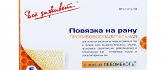
- Levomekol. This ointment has an intense antibacterial and anti-inflammatory effect. The advantage of this Levomekol ointment is that it remains effective even in the presence of purulent and sanguineous exudate. The ointment should be applied to the damaged surface under a bandage up to 2 times a day,
In addition to ointments, hydrogen peroxide will help dry a wet wound. It is recommended to use it with every dressing.
- Zinc ointment not only dries out the burn, but also prevents pyogenic bacteria from multiplying. The ointment is applied to the wound treated with an antiseptic up to 4 times a day. For minor burns, no bandages are required over the ointment. If the burn wound is large and deep, then a bandage is applied over the ointment,
- Argosulfan - ointment with silver. It is used in the treatment of weeping burns of 2 and 3 degrees of severity up to 3 times a day. The ointment is applied to the burn both under a bandage and without it.
Wound healing agents
Wound healing agents are used in the form of ointments and powders:
- Levosin. This ointment has the following effects: antibacterial, anti-inflammatory, wound healing, analgesic. You need to smear a weeping burn once a day,
- Solcoseryl . Solcoseryl ointment and gel helps improve nutrition and respiration of damaged tissues. Tissues are restored faster. In the inflammation phase, it is recommended to use the drug in gel form; when the wound dries, you can use an ointment,
- Baneocin (in ointment and powder form). Treatment with Baneocin powder is carried out during the active phase of inflammation. The powder is evenly distributed over the surface of the weeping burn once a day. The ointment can also be used both during the period of active exudate secretion and during the healing period. The ointment is used up to 3 times a day under a bandage,
- Povidone-iodine in the form of a gel and solution for topical use. This iodine-based drug has regenerating and anti-inflammatory properties. The solution is diluted in water and the wound surface is treated with it.
Antiseptic solutions
Antiseptics are widely used in the treatment of weeping wounds after a burn. You can treat a weeping wound after a burn using the following means:
- Miramistin is a universal antiseptic that can cope with a large number of pathogenic microorganisms. Miramistin solution is sprayed onto the wound with each dressing and treatment,
- Chlorhexidine. This is a broad-spectrum antiseptic that is also active against fungi. The solution is used for washing and irrigating the wound surface,
- Streptocide in powder form. This is an antibacterial agent that is distributed on the wound surface after pre-treatment of the wound with one of the antiseptic solutions,
- Hydrogen peroxide dries out and has a detrimental effect on anaerobic bacteria. Peroxide is used to wash and treat wounds with each dressing.
Regeneration drugs
During the healing process, drugs are used that promote:
- Improving tissue nutrition,
- Acceleration of epithelization,
- Healing of damaged tissues,
- Moisturize the affected areas.
The drug of choice in this case is Panthenol. During the period of active regeneration, ointment or cream is used.
The drug is applied to the wound 1 – 2 times a day. The duration of treatment is individual and determined by the attending physician. Ointments and gels also used for burns:
- Bepanthen ointment. Eliminates inflammation, helps restore damaged tissue. Bepanten ointment is applied to a cleaned wound, both under a bandage and without it.
- Actovegin in the form of tablets and solution for injection. It helps improve blood circulation at the site of injury. In the form of ointment, gel and cream, the drug is used topically. Moreover, this drug not only accelerates healing, but also dries the wound surface.
- Olazol is a wound-healing drug that is used at any stage of the inflammatory process and regeneration. Olazol also helps eliminate signs of inflammation and infection.
Remedies for skin scarring
During the scarring process, drugs are used that prevent the growth and formation of scars. Such drugs include:
- Contractubex. This drug has a number of medicinal properties: anti-inflammatory, regenerating, antithrombic, fibrinolytic. The gel is applied under the occlusion, based on an area of 20 centimeters 0.5 centimeters of gel,
All anti-scar medications are effective for small, shallow scars. They only slightly smooth out large scars. This is due to deep soft tissue damage.
- Vitamin complexes that contain vitamins A, C, E. They help improve the body’s production of collagen, which is so necessary for proper healing of a wound after a burn,
- Dermatix (gel). This drug can be used when the burn site has healed well. It helps to even out the color of the skin at the burn site, improve blood supply to the wound surface, and moisturize the tissue.
How to treat a weeping wound after a burn
It is better to entrust the choice of drug to a doctor. The following are the most effective drugs.
Solcoseryl is one of the most effective medications for healing the skin. It is most often prescribed by doctors for burns. The formula of the medication includes active components that nourish the skin, help deliver oxygen to cells and regenerate cells. Solcoseryl is available in the form of a gel or ointment. For weeping wounds, it is better to use a gel consistency.
Lioxazine is a high-tech medicine that provides pain relief after injury. It is able to accelerate regeneration processes and prevent the penetration of microorganisms into the wound.
Combination drugs
Amprovisol is a medicine in the form of an aerosol. It is very convenient to use for burns, since there is no need to contact the affected area. This remedy helps relieve inflammation in burns, disinfect and anesthetize the wound. Also ensures quick recovery.
Olazol is a local drug with a healing effect. Available in aerosol form. Contains sea buckthorn fruit oil. Due to the antimicrobial, analgesic effect, the process of epithelial restoration is significantly accelerated.
Traditional methods of treatment
If the burn becomes wet, you should seek help from a doctor. There are, of course, many traditional medicine recipes that promise to cope with this disease. However, it should be remembered that self-medication is unacceptable.
Before using traditional methods, you must consult a specialist and make sure there are no contraindications. Let's look at the most effective folk remedies that are used for weeping burn wounds:
- Aloe juice. The fleshy leaves of the plant must be washed and dried. After that, the juice should be squeezed out of the pulp of this plant, which is used to soak gauze or a bandage and make bandages with this product,
- Chamomile decoction. This solution has an antiseptic and drying effect. To prepare a chamomile decoction, you need to take 2 tablespoons of dry chamomile herb and 250 milliliters of boiling water. After 30 minutes, the solution must be filtered,
- Fresh potato juice. The root vegetable must be washed, peeled and grated. The juice is squeezed out of the resulting slurry, which is used to soak dressing material (bandage, gauze),
- Calendula decoction. This solution is used to treat and wash the wound surface. To prepare this decoction, you need to take 2 tablespoons of dried calendula and 1 cup of boiling water. After 30 minutes, strain the solution and you can use it for its intended purpose,
- Onion . The onion head is crushed to a pulp and applied to the weeping burn, first wrapped in a bandage or gauze.
When to see a doctor
If the patient notices that the burn does not heal for a long time or begins to get wet, then you should immediately contact a specialist for help. Only a doctor can prescribe the correct treatment, which will prevent the development of severe complications. Symptoms for which you should consult a doctor :
- Isolation of serous exudate and pus from the wound surface,
- Increasing pain or its reappearance after subsiding,
- Hyperemia and swelling,
- Chills and increased local and general body temperature.
A surgeon treats a weeping and purulent wound after a burn. You should contact the clinic. If necessary, the doctor will give a referral to a surgical hospital. If you have a weeping wound of a 2nd degree burn, you can also consult a dermatologist.
Wound healing time
Wound healing after a burn depends on several factors:
- Presence of complications. With purulent, necrotic and inflammatory complications, healing is delayed for a long time,
- Patient's age. The younger the person, the faster the healing occurs,
- Timely consultation with a doctor,
- The treatment being carried out
- The presence of chronic pathologies and immunodeficiency pathological conditions.
For small and shallow burns, healing occurs in 1 to 2 weeks. If the burn begins to go silent, the recovery time increases by 7 to 10 days.
With deep and weeping wounds, healing is long and difficult. Restoration of damaged tissue occurs within several months. Often in this case, scarring changes in the skin are formed.
Description
A person faces various types of trauma throughout his life, starting from childhood. Before treating a lesion, you need to know everything about this type of injury. The damage is a violation of tissue trophism, which can be localized in various parts of the body. Weeping damage can cover different areas and depths of penetration. In this case, various structures from the dermis and blood vessels to bones and internal organs can be affected. Non-healing weeping lesions occur when the skin humidity is high. The mechanism of occurrence of such burns is similar to the way of formation of ordinary injuries.
There are several phases in it:
- inflammatory process;
- regeneration;
- scarring.
Restoration and healing of weeping type wounds should be carried out according to the scheme. It is necessary to constantly bandage the open wound, use restorative and disinfecting medications.
Possible complications
Burn wounds are dangerous due to the development of a number of complications:
- Wound infection. An open and weeping burn is a favorable environment for the development of pathogenic microorganisms. An infected burn wound does not heal for a long time and is characterized by the presence of severe symptoms (pain, increased temperature in the wound area, swelling and discharge of pus from the wound surface),
- Formation of ulcers and gangrene of the limb. This complication occurs when there is no adequate treatment, medications are selected incorrectly,
- Sepsis. General blood poisoning leads to infection of the entire body. As a result, multiple organ failure occurs. This complication is dangerously fatal. The patient experiences a sharp and persistent increase in body temperature, severe weakness, dizziness, loss of consciousness, and chills.