In the classification of skin damage, a 2nd degree burn refers to the partially disrupted integrity of the epidermis under the influence of chemical or temperature exposure. The damage covers the epidermis and dermis, capillaries, horn cells and nerve endings. With such a lesion, the germ layer is not affected, which allows you to count on independent skin regeneration. The speed of recovery and the severity of the consequences depend on competent and timely provision of first aid for second-degree burns.
Associated symptoms
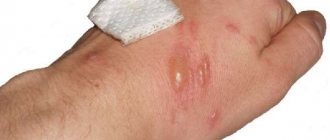
With a 2nd degree burn, hyperthermia, swelling, and redness of the affected area most often occur. Liquid bubbles may form. When the blisters break, sores form. They will take about 2 weeks to heal. The listed symptoms may include pain and increased sensitivity at the site of the lesion. If the area of damage is extensive, vomiting, weakness, dizziness, and fever may occur. Particularly severe symptoms of 2nd degree burns occur in children.
Signs of a purulent process
Pus on a burn is a characteristic sign of an inflammatory reaction. However, the lesion begins to fester gradually, the process manifests itself after some time. Inflammation with the formation of pus can grow from the inside; it is not always possible to detect it visually.
If the burn festers, the following signs appear:
- high pain at the slightest touch;
- swelling and redness of the skin around the affected area;
- discharge of pale yellow liquid when pressed;
- feeling of pressure, tension in the problem area;
- local increase in temperature around the wound;
- general malaise, increased sweating.
Removal of pus and subsequent therapy are within the competence of the doctor. Self-medication is fraught with serious complications, primarily for high-degree burns.
Diagnostic measures
Before treating a 2nd degree burn, it is necessary to assess the extent and depth of tissue damage. You should definitely consult a doctor in the following situations:
- More than 5% of the skin surface is affected (in children - more than 2%).
- Localization - on the face or perineum, or the respiratory tract and esophagus are affected.
- The pain only gets worse over time.
- Swelling appears and foci of suppuration develop.
With a 2nd degree burn, bubbles with transparent contents usually appear. To determine the affected area, use the palm rule, the essence of which is that the area of the palm is counted as 1%. Based on this, they determine how much the affected area has spread and decide whether hospitalization is necessary. Even with a small degree of burn, but a significant area of damage, there is a risk of painful shock.
Why can a burn fester?
The appearance of an infection in a wound provokes the development of a purulent process, which is always a consequence of inflammation. An inflamed wound usually festers when immune defense reactions are triggered. After the penetration of pathogenic microflora, a huge number of white blood cells - leukocytes - rush to the site of inflammation. In the fight against the pathogen, leukocytes die en masse. The burn surface festers - this is how the “victims” of this confrontation accumulate.
A burn can fester only due to infection, that is, penetration of pyogenic bacteria into the wound.
Sometimes pus appears almost immediately, sometimes after a certain time. There are primary and secondary stages of the appearance of pus. In both cases, the penetration of infection with subsequent suppuration is facilitated by:
- use of non-sterile instruments and bandages for dressing;
- unskilled wound treatment with antiseptic agents;
- carrying out primary treatment of the affected area under non-sterile conditions;
- applying cold compresses based on tap water to the injury;
- an attempt at self-medication with homemade drugs.
The larger the affected area, the higher the risk that the injury will fester. Immediate consultation with a doctor significantly reduces the risk of infection.
Proper first aid
Further recovery depends on how competent first aid is provided for a 2nd degree burn. The procedure is as follows:
- Remove the source of damage.
- Rinse the damaged area under running cold water.
- Treat the skin with an alcohol-free antiseptic.
- Apply a sterile bandage.
- Give pain medication as needed.
But here is what you should absolutely not do in case of a 2nd degree burn:
- tear off clothing stuck to the wound;
- cool the burn with ice;
- bandage the wound tightly or use cotton wool in the dressing;
- Lubricate the damaged area with oil, sour cream or alcohol.
After providing first aid, you should consult a doctor. Only a specialist will determine how to treat a 2nd degree burn and give individual recommendations. As a rule, such lesions are treated at home. The exception is burns of the respiratory system and esophagus.
Possible consequences and prevention
Injuries caused by burns do not heal quickly and are always fraught with complications. This is due to direct contact between the wound and the environment. Lack of complete sterility at any stage of treatment can cause suppuration after a burn. With all the ensuing consequences:
- a long healing period as a result of the spread of inflammation to other areas and extensive intoxication of the body;
- surgical intervention for deepening of the purulent focus and gangrenous tissue changes;
- irreversible deformations of tissues and organs when the injury festers;
- general blood poisoning as a complication of an advanced form of suppuration.
Successful treatment of burn injuries is possible with timely medical care, strict adherence to sterility rules and knowledge of some subtleties regarding the medications used:
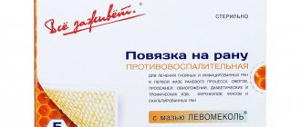
- If the burn festers and does not heal for a long time, use ointments with regenerating properties such as Levomekol, Nitacid.
- Avoid creams and ointments that are too greasy, as they do not allow the wound to “breathe.”
- Do not use improvised materials instead of sterile dressings. If there are no special bandages, any material for dressing can be sterilized by ironing it with a hot iron.
To prevent the burn from festering, it is initially recommended to use drugs with a pronounced antimicrobial effect when treating the wound. Along with Vishnevsky ointment, the following ointment preparations are effective:
- Ichthyol ointment;
- Erythromycin;
- Levomycetin;
- Sintomycin;
- Oflomelid.
Self-medication may be justified only in the case of minor superficial injuries. Timely contact with specialists is the best way to avoid complications from burns.
Stages of treatment
The choice of therapy depends on the presence of concomitant diseases and the nature of the skin lesion. The standard approach involves solving specific problems at each stage of wound healing:
- Rejection of necrotic tissue. Treatment is carried out with an antiseptic and complex anti-inflammatory agents.
- Granulation. The blisters with liquid open, the inflammation gradually subsides, but the wound is open. At this stage, treatment with an antiseptic continues, dressings with wound-healing compounds are indicated.
- Epithelization. There is almost no pain, the wound is covered with fresh skin. Compositions are used that improve metabolism and stimulate regeneration.
It is not recommended to wet the wound until it has completely healed. Dressings are carried out according to schedule. The doctor monitors the situation in time to notice the worsening of the situation. Skin restoration lasts about 2 weeks.
How to treat a festering burn
Primary processing does not always guarantee 100% sterility. If the burn begins to fester, the first thing to do is go to the clinic, since self-medication can lead to irreversible deformation of the affected organ.
Measures to treat a festering burn are aimed at:
- cleansing the inflamed area from pus;
- preventing further “spreading” of inflammation;
- creating conditions for healing of the wound surface;
- reducing the time for complete recovery of affected tissues.
The effectiveness of therapeutic measures depends on a systematic approach to treatment. When the burn festers, treatment and dressings are carried out according to the following scheme:
- Wash the purulent surface using Chlorhexidine or Miramistin.
- Wound sanitation is completed with hydrogen peroxide.
- The edges of the burn are treated with brilliant green without touching the wound itself.
- Apply a bandage moistened with an antiseptic such as Furacilin to the surface.
- After 2 hours, the napkin with Furacilin is replaced with a new one treated with Vishnevsky ointment. It disinfects the area that is festering and promotes healing without scarring.
Frequent changes of dressings, antiseptic treatment of the lesion, application of ointment applications every 3-4 hours guarantee a good result. The injury stops getting wet and festering, and healing processes begin.
Treatment methods
A 2nd degree burn with an affected surface area of less than 10–15% responds well to treatment in an outpatient setting and at home. The success of treatment and rapid healing largely depends on how quickly and competently first aid was provided for second-degree burns.
Outpatient treatment is aimed at creating optimal conditions for wound healing, protecting the affected area from mechanical damage and infection, as well as stimulating regeneration processes.
First aid for a 2nd degree burn involves the following sequence of actions:
- remove the source of damage;
- Cool the burn area under running cold water for 10–15 minutes. This prevents the burn from spreading deep into the dermis;
- carefully clean the burn site using gentle antiseptics, such as hydrogen peroxide solution;
- cover the burn site with a sterile bandage;
- If necessary, give the victim pain relief, ensure rest and drink plenty of fluids.
Local treatment can be carried out using open and closed methods. The first is most often used when the lesion is localized on the face and groin area, so as not to impede physiological processes.
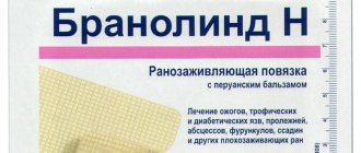
Treatment with a closed method involves the application of atraumatic dressings, for example a dressing with the Peruvian balsam "Branolind-N". For 2nd degree burns, usually a single application of a bandage is sufficient to begin the epithelization process.
Drug therapy involves the use of antihistamines, anti-inflammatory and antiseptic drugs. Antiseptics are used in the first days of treatment; they prevent secondary infection of the wound. Antihistamines eliminate itching and help relieve swelling. Only a doctor can prescribe any medications for you; do not select medications on your own, consult your doctor first!
With proper care, complete healing occurs within two to three weeks. Whenever processing a burned surface, you must pay attention to its condition. If the pain intensifies, the swelling does not subside, but rather increases, and pus is released, it is necessary to seek medical help as quickly as possible.
Mild burns do not require treatment with antibiotics, except in cases where the affected area occupies a large surface. Antibiotics can also only be prescribed by a doctor.
Thermal burns
The course and outcomes of burn injury largely depend on the timeliness of first aid and rational treatment throughout the course of the disease. With burns of more than 10%, and in small children 5% of the body surface, there is a real danger of developing shock, therefore, even when providing first aid, measures must be taken to prevent shock and infection in the wound. For this purpose, painkillers are administered (50% analgin solution with 1% diphenhydramine solution or 2% promedol solution). If the epidermis is preserved, it is advisable to cool the burned surface with a stream of cold water or other available means, and apply a sterile bandage to the wound. Early (within the first hour after injury) cryotherapy with liquid nitrogen helps reduce tissue hyperthermia, inflammatory reaction, edema, depth of necrosis, resorption of toxic substances from burned tissues, and intoxication of the body. When burns are localized on the extremities involving functionally active areas, it is necessary to provide transport immobilization. The victims are evacuated to a surgical medical facility, treatment in which necessarily includes the administration of anti-tetanus serum and cleaning of wounds.
For first degree burns , a bandage is not applied; topical application of weakly disinfectant preparations is sufficient. Irrigation from aerosol cans with medicinal mixtures containing corticosteroid hormones is advisable.
For second degree burns, the wound is cleaned while painkillers are administered (2% solution of promedol or pantopon). It consists of cleaning the wound and surrounding skin with warm soapy water, 0.25% ammonia solution, antiseptic solutions (ethacridine lactate, furatsilin, chloracyl, detergent solutions), removing foreign bodies and scraps of epidermis. If the epidermis is not desquamated, then the burned surface is treated with alcohol. Whole blisters are trimmed or punctured, removing the contents. The preserved epidermal film protects the wound from external irritants, healing under it proceeds faster and less painfully.
Second degree burns can be treated in an open manner, as well as third degree burns, in the absence of copious purulent discharge and the creation of optimal conditions for reparative processes in the wound. In the absence of conditions for abacterial treatment, after cleaning the wounds, to prevent secondary infection and suppuration, bandages with antiseptic solutions are applied to the wounds (rivanol 1:1000; furatsilin 1:5000; 0.1-1% solution of dioxidine, etc.) or use aerosol anti-inflammatory drugs ( panthenol, vinylsol, light fir, olazol, oxycort). In the cold season, it is preferable to cover the wound with a bandage with low-fat creams or ointments (synthomycin liniment, 0.5% furacilin and 15% propolis ointments, balsamic liniment according to A.V. Vishnevsky).
In conditions of mass lesions, after careful cleaning of the burn wound, it is advisable to use aerosols with film-forming polymers (Furoplast, iodovinisol, Lifusol, Plastubol, Akutol, Acrylasept, etc.). Their advantage lies in a significant reduction in the duration of treatment of the burned surface and savings in dressing material. The film protects the wound from infection, prevents the loss of fluid through the wound, facilitates monitoring the course of the wound process (if it is transparent), which allows, if necessary, timely adjustments to the treatment of the wound. With a smooth course of the wound process, healing occurs under the initially applied film. Film coverings eliminate the possibility of them becoming saturated with liquids and protect the wound from contamination and infection more reliably than bandages. If necessary, primary toileting of the burn wound can be postponed. It should not be performed in the presence of shock in victims with severe burns. In such cases, burn wounds are covered with a slightly warmed bandage with ointment, and the toilet is postponed until the patient’s condition is stabilized and he is brought out of shock. The same applies to mass influx of patients.
The initially applied bandage is not changed for 6-8 days. The indication for its replacement in case of second degree burns is suppuration, as evidenced by pain in the wound and specific wetting of the bandage. When a burn wound suppurates, after cleaning it using hydrogen peroxide or an antiseptic solution, apply wet-dry bandages containing antiseptics or antibiotics, to which the wound microflora is sensitive.
Healing of second degree burns occurs within 10-12 days. Recovery of patients with first degree burns occurs 3-5 days after injury.
With third-degree burns, partial necrosis of the dermal layer of the skin occurs, so suppuration is more often observed, which can lead to the death of skin derivatives and the formation of granulating wounds. The main task in the treatment of III degree burns is to prevent their deepening. This is achieved through timely removal of dead tissue and targeted control of wound infection. During dressings, which should be done after 1-2 days, the wet necrotic scab is gradually removed (starting from the 9-10th day). If the scab is dry, then you should not rush to remove it, since epithelization may occur underneath it.
For dressings for IIIA degree burns, it is advisable to use dressings with antiseptics (ethacridine lactate, furatsilin, 0.25% chlorocil solution, 0.5% silver nitrate solution, etc.) or antibiotics. Infrared and ultraviolet irradiation of wounds helps prevent wet necrosis, helps reduce purulent discharge and faster epithelization.
As the exudation subsides at the final stage of treatment of burns after rejection of necrotic tissues, one should move on to ointment and oil-balsamic dressings (5-10% syntomycin liniment, 0.5% furacilin, 0.1% gentamicin, 5-10% dioxidine, 15% propolis ointment , levosin, levomekol, olazol, etc.), which help accelerate healing and have a pronounced bactericidal effect. Ointments do not irritate the wound and provide a softening and analgesic effect. The dressings are changed as they become wet with purulent discharge (after 1-2 days).
Treatment of II-III degree burns can be carried out in local isolation wards with a controlled environment in an open manner, which reduces the level of bacterial contamination of wounds and promotes their faster spontaneous epithelialization.
Infusion and transfusion therapy of extensive and deep burns. Infusion-transfusion therapy occupies one of the leading places in the complex treatment of patients with extensive burns. With extensive burns, significant energy costs occur, reaching 5000-6000 kcal, or 60-70 kcal per 1 kg of body weight, and the loss of nitrogen from the wound surface is 20-50% of the total losses, which leads to a negative nitrogen balance. In this regard, in the treatment of burn disease in all its periods, exceptional importance is attached to infusion-transfusion therapy, the correct and timely implementation of which determines the possibility of surgical treatment of patients with deep burns, as well as the outcome of the disease.
All patients with deep burns of 10-15%, and children - 3-5% of the body surface from 1 year after injury require intensive infusion and transfusion therapy. In case of severe intoxication, intravascular infusions are performed daily according to an individual program in accordance with the severity of the thermal injury.
In case of burn shock, infusion-transfusion therapy involves replenishing liquid volumes of electrolytes, proteins and red blood cells in the vascular bed, improving metabolic processes, kidney function and detoxification of the body.
In patients with burns of less than 10-15% of the body surface, if they do not vomit, fluid loss can be replenished by ingesting a 5% glucose solution with vitamins C and group B, alkaline solutions. Replenishment of fluid volumes in the vascular bed is achieved through intravascular fluid administration, as well as by returning deposited blood for active circulation using hemodilution.
For the purpose of anti-shock therapy and detoxification of the body, saline solutions (Ringer-Locke, lactasol), plasma and colloidal plasma-substituting drugs (reopolyglucin, hemodez, polydes, gelatinol, etc.) in an amount of 4-6 l, 5-10% glucose solution with vitamins C and group B in a dose of 500-1000 ml on the 1st day after injury in adults. For mild burn shock, therapy is carried out without blood transfusions. In the case of the development of severe and extremely severe shock, blood transfusions (250-1000 ml) are carried out at the end of the 2nd or 3rd day, depending on the severity of the condition, hematological parameters and renal function. In order to combat acidosis during shock, use a 4% solution of sodium bicarbonate, which is prepared before use and administered taking into account the deficiency of bases in quantity.
In elderly and senile people, the volume of intravenous fluid should not exceed 3-4 liters, and in children - 2-3 liters per day. The volume of infusion and transfusion therapy for burn shock in children can be approximately determined using the Wallace scheme: triple the child’s body weight (in kilograms) multiplied by the area of the burn (in percent). The resulting product is the amount of fluid (in milliliters) that must be administered to the child during the first 48 hours after the burn. It does not include the physiological need for water (700-2000 ml per day depending on the age of the child), which is satisfied by additionally giving a 5% glucose solution.
The ratio of colloidal (protein and synthetic) and crystalloid solutions is determined by the severity of burn shock. Approximately, for mild burn shock, the ratio of colloidal, saline solutions and glucose should be 1:1:1, for severe burn shock - 2:1:1, and for extremely severe - 3:1:2. Two-thirds of the daily amount of infusion media is administered in the first 8-12 hours. The total volume of intravascularly administered fluid is reduced by 2 times on the 2nd day after injury.
After replenishing fluid volumes in the vascular bed, as evidenced by an improvement in blood volume, osmotic diuretics are used. Mannitol in the form of a 20% solution is administered at the rate of 1 g of dry substance per 1 kg of the victim’s body weight, urea solution (20%) - in a volume of 150 ml at a rate of 40-60 drops per minute. An effective diuretic is Lasix, which is prescribed at a dose of 60-250 mg/day after eliminating the deficiency of blood volume.
When carrying out infusion therapy for burn shock, you can use a 20% sorbitol solution, which is administered at the rate of 1.5-2.5 g of dry matter per 1 kg of patient body weight per day. A pronounced diuretic effect usually occurs 40-60 minutes after the administration of osmotic diuretics. If necessary, after 3-4 hours they can be reintroduced.
Infusion and transfusion therapy for burn shock is carried out in combination with measures aimed at pain relief, prevention or elimination of oxygen deficiency, dysfunction of the cardiovascular, respiratory systems and other organs. For this purpose, cardiotonic drugs, antihypoxants, and antihistamines are used. Corglicon and cordiamine are administered intravenously 1-2 ml 2-3 times a day, oxygen is prescribed for inhalation. The effect of cardiac glycosides is enhanced by the administration of cocarboxylase 50-100 mg 2 times a day, which has a beneficial effect on carbohydrate metabolism. Aminophyllin, which also has a diuretic effect, which is administered in the form of a 2.4% solution with a 5% glucose solution, 5-10 ml up to 4 times a day, contributes to a significant improvement in blood supply to the heart muscle and kidneys.
For pain relief, a 1% solution of morphine or a 2% solution of promedol is administered intravenously in combination with a 50% solution of analgin. The use of the neuroleptic droperidol in the form of a 0.25% solution eliminates psychomotor agitation.
In severe and extremely severe burn shock, when infusion and transfusion therapy is not effective enough, corticosteroids have a normalizing effect on hemodynamics and renal function. They increase cardiac output, improve blood supply to the heart muscle, eliminate spasm of peripheral vessels, restore their permeability and increase diuresis. In the presence of burns of the respiratory tract, they help reduce swelling of the bronchial tree. Patients are prescribed intravenous hydrocortisone 125 mg as part of infusion media or prednisolone at a dose of 30-60 mg 3-4 times during 1 day of anti-shock therapy until hemodynamics and diuresis are normalized.
Due to the disruption of redox processes in burnt patients and the deficiency of vitamins in their body, when carrying out infusion-transfusion therapy, it is necessary to administer ascorbic acid 5-10 ml of a 5% solution up to 2-3 times, vitamins Bi, Be 1 ml and vitamin Bc 100-200 mcg 3 times a day, nicotinic acid 50 mg.
Sodium hydroxybutyrate (GHB, sodium salt of hydroxybutyric acid) has been successfully used as an antihypoxic agent. Sodium hydroxybutyrate neutralizes changes in CBS, reduces the amount of under-oxidized products in the blood, and improves microcirculation. For burn shock, the drug is prescribed intravenously at 2-4 g 3-4 times a day (daily dose 10-15 g).
In order to inhibit proteolysis and enzymes of the kallikrein system, it is advisable to introduce contrical at 100,000 units or trasylol at 500,000 units per day into the infusion media, which helps to normalize the permeability of the vascular wall.
In patients with burn shock, 6 hours after injury there is a significant increase in the histamine content in the blood. In this regard, the use of antihistamines is pathogenetically justified: 1% solution of diphenhydramine, 1 ml 3-4 times a day, 2.5% solution of pipolfen, 1 ml 2-3 times a day.
Infusion and transfusion therapy is carried out under the control of central venous pressure and blood pressure, pulse rate and its filling, hourly diuresis, hematocrit, hemoglobin level in the blood, potassium and sodium concentrations in plasma, CBS, blood sugar and other indicators.
A relatively low central venous pressure (less than 70 mm H2O) indicates insufficient replacement of blood volume and serves as a basis for increasing the volume and rate of administration of infusion media (if there is no danger of developing pulmonary edema). High central venous pressure is a sign of heart failure, and therefore it is necessary to reduce the intensity of infusion therapy or temporarily stop it.
When monitoring hourly diuresis, they focus on a level of 40-70 ml/hour. When conducting infusion therapy, it is necessary to ensure that the plasma sodium concentration is not lower than 130 mmol/l and not higher than 145 mmol/l. Plasma potassium concentration should be maintained at 4-5 mmol/l. Rapid correction of hyponatremia is achieved by infusion of 50-100 ml of 10% sodium chloride solution, which usually eliminates hyperkalemia. Otherwise, administration of 250 ml of a 25% glucose solution with insulin is indicated.
Transfusion media for burn disease are administered by venipuncture or venesection of accessible saphenous veins. In this case, it is necessary to strictly observe the principles of asepsis and antiseptics. If those conducting therapy have experience in catheterization of the subclavian, jugular or femoral vein, then it is preferred. Catheterization of the central veins more reliably provides the required volume of infusion and transfusion therapy during the entire period while the victim is in a state of shock.
When catheterizing central veins, in order to avoid thromboembolic complications, the catheter inserted into the vein must be systematically washed with an isotonic solution of sodium chloride with heparin (2-3 times a day). After the end of the infusion, the catheter is filled with heparin solution (2500 units per 5 ml of isotonic solution) and closed with a stopper. If signs of phlebitis or periphlebitis appear, infusions into this vein should be stopped immediately. If a purulent process develops in burn wounds, especially in the later stages of a burn disease, the catheter from the vein should be removed so that it does not become a conductor of purulent infection and the cause of purulent-septic complications.
Monitoring the adequacy of infusion and transfusion therapy in the absence of laboratory test results can be carried out based on clinical signs of burn shock. Pale, cold and dry skin indicates a violation of peripheral circulation, to restore which rheopolyglucin, gelatinol, hemodez, polydesis can be used. Strong thirst is observed in a patient with a lack of water in the body and the development of hypernatremia. In this case, it is necessary to administer a 5% glucose solution intravenously, and in the absence of nausea and vomiting, increase oral fluid intake. Collapse of the saphenous veins, hypotension, and decreased skin turgor are observed with sodium deficiency. Infusions of electrolyte solutions (lactasol, Ringer's solution, 10% sodium chloride solution) help eliminate it. Severe headache, convulsions, blurred vision, vomiting, salivation, indicating cellular hyperhydration and water intoxication, are indications for the use of osmotic diuretics. The main signs indicating that burnt people are recovering from shock are persistent stabilization of central hemodynamics and restoration of diuresis, elimination of spasm of peripheral veins, warming of the skin and the onset of fever.
During the period of burn toxemia, infusion and transfusion therapy is continued in a volume of 2-4 l, or 30-60 ml per 1 kg of body weight. In order to combat alkalosis in patients with severe burns, it is advisable to infuse 20% glucose solution up to 500-600 ml per day with insulin at the rate of 1 unit per 2-4 g of glucose and 0.5% potassium chloride solution up to 500 ml under the control of potassium content and sodium in the patient's blood serum.
For the purpose of detoxification and prevention of anemia, hypo and dysproteinemia, systematic transfusions of freshly preserved Rh-compatible single-group blood or its components (erythrocyte mass, native and dried plasma, albumin, protein) 2-3 times a week, 250-500 ml for adults and 100-200 ml are advisable children under the control of hematological parameters (hemoglobin level, number of red blood cells), which must correspond to the age norm. A particularly pronounced detoxification effect is exerted by direct blood transfusions, transfusions of freshly heparinized blood or convalescent blood and plasma, no more than 1 year has passed since recovery from burns.
The reduction of intoxication is facilitated by osmotic diuretics included in the complex of infusion media (mannitol, Lasix, 30% urea solution), the infusion of which is advisable to alternate with the intravenous administration of low molecular weight plasma-substituting solutions (hemodez, rheopolyglucin), which ensures forced diuresis.
For the purpose of detoxification for burns and acute surgical infections, hemodialysis, hemosorption, plasma and lymphosorption are used. One of the mechanisms of the therapeutic effect of hemosorption is a decrease in the level of proteasemia and peptidemia, a decrease in plasma toxicity and the severity of metabolic disorders. Sorption allows you to reliably and quickly rid the body of burned people of toxic metabolites. However, hemosorption is accompanied by loss of blood cells (platelets, leukocytes, erythrocytes), chills, and changes in the physicochemical properties of erythrocytes. The positive effect of hemosorption lasts no more than 2-3 days. To ensure effective detoxification, there is a need to carry out repeated hemosorption with an interval of 24-48 hours. In this regard, hemosorption is justified primarily in cases where other therapeutic measures are ineffective. Hypovolemia and hemodynamic instability, observed with extensive burns, are a contraindication to the use of hemosorption.
During the period of septicotoxemia, intensive infusion and transfusion therapy is especially necessary in preparation for surgical operations and during their implementation, when increased replenishment of the body's energy costs is required. During this period, blood transfusions of 250-500 ml 2-3 times a week, alternating with transfusions of protein blood products and plasma-substituting detoxification solutions, are the main component of infusion-transfusion therapy.
Along with blood transfusion, to replace ongoing protein losses, improve colloid-osmotic and transport functions of the blood, transfusions of dry and native plasma 250-500 ml 2 times a week are essential, which allow stabilizing the levels of total protein and albumin in the blood serum. If blood transfusions do not provide an improvement in the albumin fraction of serum proteins, then it is advisable to use a 5-10% albumin solution of 200-250 ml for 3-4 days, especially in elderly and senile patients. Albumin solution is highly effective in compensating for the loss of extracellular protein and eliminating hypo and dysproteinemia, maintaining normal plasma colloid osmotic pressure, and treating toxic hepatitis in burnt patients. Maintaining the level of total serum protein 6.5-7 g% and albumin 3.5-4.0 g% is necessary to ensure a favorable course of the wound process, successful preparation for surgical intervention to restore the skin and its implementation.
High energy costs in the body of burned people are due to the destruction of lipids, carbohydrates and proteins. This consumes whey proteins and tissue proteins, especially skeletal muscles. The most pronounced disturbances of protein metabolism occur during the first weeks of burn disease in patients with severe burns. Mainly albumins and only some of the globulins are subject to catabolism; hypo and dysproteinemia, deficiency of intracellular and extracellular proteins, and protein deficiency develop. Clinically, this is manifested by exhaustion, muscle atrophy, and loss of body weight.
To replenish energy costs and restore nitrogen balance in the late period of burn disease, parenteral nutrition is of great importance, which makes it possible to provide the patient with easily digestible nutrients and compensate for profound disorders of all types of metabolism. For parenteral nutrition, protein hydrolysates are used at a rate of 15 ml/kg (average 800 ml), amino acid preparations (10 ml/kg), which are administered at a rate of no more than 45 drops per minute, and energy components (glucose, fat emulsions).
For severe burns, glucose is administered in the form of a 10-20% solution with insulin. To reduce insulin resistance, which often develops in patients with severe burns, and improve the processes of glucose utilization, it is advisable to use tocopherol in the form of a 10% solution, 1 ml once a day. For parenteral nutrition, sorbitol and fat emulsions can be used.
In many patients, parenteral nutrition can be successfully replaced by enteral nutrition - using a probe inserted through the nasal passage into the stomach or duodenum. For enteral tube feeding, mixtures containing glucose, proteins and fats are used, which are administered by drip (20-30 drops per minute). They can be administered only after the absorption and motor function of the intestine has been restored.
During the period of acute burn toxemia and septicotoxemia, infusion-transfusion therapy should be carried out against the background of a balanced diet using high-calorie food containing 120-140 g of protein, mineral salts, vitamins A, C, group B, the energy value of which is at least 3500-4000 kcal.
Purulent demarcation inflammation in a burn wound leads to melting and rejection of necrotic tissue. At the same time, intoxication of the body increases due to the absorption of products of purulent melting of tissues and microbial toxins. Intoxication of the body can be significantly reduced when treating patients in a controlled abacterial environment using an open method of wound management in isolators with infrared irradiation on a mesh bed with constant blowing of wounds with warm air and oxygen therapy. The constant unidirectional movement of heated sterile air significantly reduces energy losses in burnt patients, reduces exudation and microbial contamination of burn wounds, converts wet necrosis into dry necrosis, thereby reducing protein losses, reducing the activity of proteolytic enzymes in the wound, accelerated epithelization is noted in superficial burns, it becomes possible remove the burn scab at an earlier date and prepare the wound for restoration of the skin.
The presence of damaged tissue is the main cause of the development of burn disease, therefore, removal of necrotic tissue and restoration of the skin is the main task of treating patients with deep burns. All other activities carried out in the process of complex general and local treatment are aimed at preparing for skin plastic surgery.
Surgery
Indications, choice of method and timing of skin grafting. The general condition and age of the victim, the extent of the lesion and the localization of deep burns, the availability of donor skin resources and the condition of the receiving bed are decisive in determining the timing and choice of the method of surgical intervention, as well as the method of restoring the skin.
For limited deep burns, the most rational method is complete excision of necrotic tissue in the first 2 days after injury with simultaneous suturing of the wound, if its size and the condition of the patient and surrounding tissues allow. If it is not possible to bring the edges of the wound closer together, then primary free or combined (a combination of free and local skin grafting) skin grafting is performed.
Early excision is possible only in the presence of a dry scab. It is especially necessary when localizing limited deep burns in the joints, hands and fingers. Due to the high functional activity of the hand and fingers and the complexity of their functions, it is advisable to excise the necrotic scab in cases where skin derivatives are preserved (degree III burns) and epithelization of wounds is possible, usually accompanied by scarring.
For burns accompanied by osteonecrosis in functionally active areas, it is advisable to perform early excision of non-viable areas of the bone, without waiting for its spontaneous sequestration, with simultaneous replacement of the defect by combined skin grafting, if the condition of the surrounding tissues allows. In this case, the bone tissue is covered with a rotational flap of skin with subcutaneous fat or a flap on a pedicle, and the newly formed defect is eliminated using free skin grafting.
At the same time, as our experimental and clinical observations have shown, for burns in the area of the cranial vault with bone damage, treatment is quite possible while preserving non-viable areas of the bone. In the absence of suppuration in the wound, non-viable soft tissue is removed, ultrasonic cavitation is performed and multiple craniotomy is performed with a conical and spherical cutter to the bleeding layer of bone and the focus of osteonecrosis is covered with a well-supplied skin-fascial flap from local tissues or from distant areas of the body. In such cases, osteonecrotic areas are not sequestered and resorption of non-viable bone elements occurs with its gradual new formation.
Early necrectomy, performed in the first 4-10 days after injury in conditions of abacterial keeping of patients, is the most optimal method of surgery. By this time, the border of a deep burn becomes most distinct and a certain stabilization of the patient’s condition with extensive lesions is noted. The exception is for patients who have circular deep burns of the torso, when there is a threat of severe respiratory distress due to compression of the chest or the same extremities, in which the blood supply to their distal parts and deeper tissues is disrupted. In such cases, emergency multiple decompressive necrotomy or partial necrectomy is indicated, which eliminates the compression and the disorders caused by it.
Tactics and technique of necrectomy. When performing early necrectomy, it is most advisable to perform layer-by-layer excision of the burn scab using an electrodermatome until a continuous, evenly bleeding wound surface appears. Such excision of the burn eschar makes it possible to preserve viable tissue to a greater extent, significantly reduce the duration of the most traumatic stage of the operation and create a smooth surface of the wound, which ensures a better fit of the grafts during skin grafting and more favorable conditions for their engraftment.
Hemostasis during surgery is achieved by applying gauze pads with a solution of hydrogen peroxide or aminocaproic acid. Large blood vessels are ligated. Due to the difficulties encountered in stopping bleeding, in some cases the operation is performed in two stages. At the second stage, carried out 2-3 days after necrectomy, free skin grafting of the previously prepared bed is performed. By this time, reliable hemostasis has occurred after applying a tight aseptic dressing, and areas of necrotic tissue that were not removed at the first stage are also identified. Additional removal of nonviable tissue contributes to a more successful outcome of skin graft surgery. In an aseptic wound formed after early excision of necrotic tissue, optimal conditions are created for the engraftment of skin grafts.
Primary and early skin plasty, if successful, can prevent the progression of intoxication from the lesion, the development of infection in wounds and the further development of burn disease, which leads to primary healing of burn wounds in the shortest possible time. Early restoration of the skin leads to a reduction in treatment duration and provides more favorable functional and cosmetic results of free skin grafting.
Extensive necrectomy with simultaneous skin grafting is a traumatic operation accompanied by significant blood loss. After the operation, the patient's condition deteriorates if the wounds are not completely replaced with skin autografts or complete engraftment occurs. The use of a carbon dioxide laser to excise a burn eschar can reduce blood loss, but difficulties encountered in determining the depth of tissue damage and the traumatic nature of the operation hinder its use. In this regard, early necrectomy is performed mainly for burns of no more than 10-12% of the body surface.
Extensive necrectomy and skin grafting in the early stages can only be performed in specialized burn departments by surgeons with experience in plastic surgery, subject to adequate compensation for blood loss during surgery and anesthesia.
Indications for secondary skin grafting. In severe condition of the patient and deep burns of more than 10-15% of the body surface, there are indications for performing secondary skin grafting on the granulating surface after rejection of necrotic tissue. To remove these tissues, it is advisable to use staged bloodless necrectomy as they begin to be rejected. This is facilitated by the use of enzymatic and chemical necrolysis. Removing a burn scab using 40% salicylic ointment, benzoic acid or an ointment containing 24% salicylic and 12% lactic acid allows you to reduce the duration of preoperative preparation by 5-7 days. A more rapid rejection of necrotic tissue is facilitated by the systematic use of hygienic baths, rational general treatment aimed at increasing the body's reactivity, preventing anemia and severe disorders of protein metabolism. These measures and careful cleaning of wounds during dressings after rejection of a burn scab in order to reduce bacterial contamination make it possible to prepare patients for skin grafting for bright, juicy and clean granulations within 2.5-3 weeks after injury.
Timely, thorough preparation of wounds eliminates the need to excise granulations before skin grafting, if they are not clearly pathological in nature and there is no perversion of the wound process. In clinical practice, difficulties often arise in determining the readiness of granulating wounds for skin grafting. A large contamination of the wound surface with pathogenic microflora in weakened patients usually coincides with a poor appearance of granulations, distortion of reparative processes and pronounced inflammation in the wound, which in turn aggravates their general condition and leads to generalization of the infection. Free skin grafting is contraindicated in these conditions. In such cases, vigorous restorative treatment and thorough local antibacterial therapy are necessary, which are carried out until the patient’s condition improves and regenerative processes in the wound intensify.
Irrigation of wounds with antiseptic solutions, hygienic baths with detergents, local application of magnetic therapy, ultrasound, scattered laser irradiation, treatment on the bed and the use of the most accessible method - frequent changes of dressings with antiseptic solutions have a beneficial effect on the course of the wound process. In patients with burn exhaustion and a sluggish course of the wound process, hormonal therapy with glucocorticoids and anabolic steroids is advisable along with the use of antibiotics under the control of the sensitivity of the wound microflora to them.
The presence of uniform, granular, juicy, but not loose or bleeding granulations with moderate discharge and a pronounced border of epithelialization around the wound is a good indicator of its suitability for skin grafting.
The most favorable receptive bed for skin grafts is young granulation tissue, rich in blood vessels and with a small amount of fibrous elements, which usually favors a period of 2.5 to 6 weeks after the burn. This is the optimal time to perform free skin grafting on a granulating surface.
Planning free skin grafting. In modern conditions, the main method of treating deep burns is dermatomal skin grafting using mainly an electrodermatome. Skin-plastic operations for deep burns of no more than 10-15% of the body surface using an electrodermatome can be performed in one stage. For more extensive lesions and in weakened patients, multi-stage skin grafting is performed. Repeated operations of free skin autoplasty should be carried out every 5-7 days, covering from 7 to 10% of the body surface simultaneously with grafts 0.2-0.3 mm thick in children and 0.3-0.4 mm in adults, if
Features of home therapy
After providing first aid for a 2nd degree burn, it is necessary to take a responsible approach to completing the course of treatment prescribed by the doctor. Rules that are recommended to be followed:
- eliminate heavy physical labor, since excessive stress provokes inflammatory processes in the body;
- avoid working in dusty and dirty objects to avoid infection of an open wound;
- Monitor the sterility of the dressings and follow the schedule for their replacement.
New tissues regenerate quickly if there are no complications due to external irritants. Recovery will take about 2 weeks, but may take up to 3 weeks for some. Healing slows down in older patients with chronic diseases. With successful therapy, recovery occurs without scars or scars.
Why are gum burns dangerous?
Despite all the external harmlessness, any gum burn can lead to serious consequences. Firstly, after an injury it is easy to introduce an infection into the soft tissues and cause complications. Secondly, with severe burns and significant lesions, necrosis of the mucous membrane occurs, which requires immediate intervention by a specialist. It is important to remove dead tissue as soon as possible to prevent necrosis from spreading further.
Do not forget that the thin layer of epithelium in the mouth is a huge number of capillaries. It is the gums that ensure the correct and strong fastening of the teeth; it is the gums that keep our smiles beautiful and even for many years.
Prevention measures
Preventing burns involves careful handling of chemicals, household electrical and heating appliances. If we are talking about a child, he needs to be protected from danger - potentially unsafe objects and liquids should be kept away. If an injury occurs, you must immediately provide assistance and, if necessary, call a doctor.
Prevention of complications is careful adherence to the doctor’s recommendations during the treatment process, timely wound care, and the use of sterile materials. It is better to use specialized ointment dressings that accelerate healing and do not stick to the wound surface. You should also limit mechanical and physical irritation of the injured area, maintain hygiene, establish a drinking regime and eat a balanced diet, and the body will do the rest itself.
How to diagnose the cause
The cause of inflammation of the wound surface is often pyogenic bacteria. Identifying the pathogen is the main task of specialists at the stage when the burn has festered.
Further treatment is carried out based on the results of microbiological examination.
To do this, a culture of the contents of the wound, which is festering, is done in the laboratory. Colonies of harmful bacilli grow in a Petri dish on a nutrient substrate. Based on their identification, treatment tactics are built.
Traditional methods
Traditional medicine offers its own methods of treating an infected burn. However, it should be remembered that this type of treatment cannot replace drug therapy. In some cases, the use of traditional methods is dangerous. You should first consult with your doctor.
Let's look at a few folk recipes that will help cope with suppuration of a burn.:
- Chamomile decoction is used to wash an infected burn. It has antiseptic and anti-inflammatory effects. Chamomile is poured with boiling water and boiled for some time in a water bath. After which the solution should be filtered,
- Aloe juice and pulp . This plant has anti-inflammatory and regenerating properties. The juice is applied to napkins and applied to the infected surface. If you don’t have time to squeeze the juice, you can apply an aloe leaf cut lengthwise to the burn. It draws out pus from an infected burn wound that accumulates on a napkin or leaf surface,
- Onion juice has antiseptic and bactericidal properties.
Symptoms of burn infection
With an infected burn, the following pathological symptoms occur::
- Increased pain. Moreover, the pain sensations are pulsating, bursting, intense,
- Hyperemia intensifies, a vascular pattern appears around the burn injury,
- Swelling of soft tissues occurs or sharply increases,
- Local, and in severe cases general hyperthermia,
- Purulent contents in the bladder, as well as discharge of pus from the wound,
- Chills,
- Headache, severe weakness,
- Nausea.
The inflammatory process occurs in 3 phases:
- Phase 1, suppuration. Changes occur in the wound, the first signs of inflammation appear (pain, hyperemia and swelling), gradually purulent exudate forms in the wound,
- Phase 2, granulation. In this case, the wound is cleaned and filled with granulation cells. Without treatment, this phase does not occur,
- Phase 3, regeneration (healing). New cells are formed that close the wound.
Infection is more severe with extensive and deep burns, as well as in weakened individuals suffering from immunodeficiency (HIV, diabetes, hormonal disorders, etc.).
Basic rules for applying bandages for burns
To maximize the effect of using an anti-burn patch or dressing, you must follow the rules. The size and location of the burn are important when choosing appropriate dressings. As a rule, medicinal compounds are applied to the wound, covering them with a sterile bandage or cloth. The following recommendations are taken into account:
- before dressing, the wound must be treated with an antiseptic and washed;
- Do not apply strong pressure to damaged skin;
- if the injury is deep, urgent medical attention is needed;
- When treating minor burns at home, use only sterile materials.