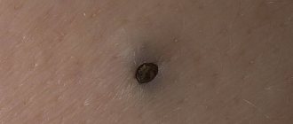
A blocked duct is usually a white dot (or bubble) on the nipple. Thickened milk or curds clog one duct and prevent the rest of the milk from coming out. Sometimes the plug is not visible, but it is noticeable that milk does not flow from one duct or barely oozes, and above it it stagnates. Often, blockage leads to lactostasis (see the selection Mastitis, lactostasis). How to deal with a blocked duct? What are its reasons?
Among the reasons are an insufficiently deep latch on the breast (due to which one of the ducts is emptied worse) - sometimes one or two unsuccessful feedings are enough. Also - an increase in the break between feedings for various reasons (travel, ate a lot of complementary foods, etc.). First aid is to simply feed the baby from this breast, ideally with the chin towards the problem point (because the lower jaw is better at expressing the breast). It is important that the baby sucks this breast for a long time and well, perhaps several times in different positions.
If this does not help, then they usually advise 1) preparation: pre-steam the chest, or rub the nipple with a towel. It may also help to rub such a point with a sterile cotton swab moistened with warm sterilized oil.
After this, 2) the baby is applied and/or the mother can gently press “upstream” on the blocked duct, along it towards the nipple, to express milk in this lobule and push the plug. Some also use a breast pump, although this is less effective. When the plug pops out, a fountain of milk often appears. And before that, milk may not come out at all or ooze droplets. If milk has stood in such a duct for some time, it often tastes saltier - this can help determine that this particular duct is problematic.
3) If you can’t cope quickly on your own, you can consult a doctor. If a bubble has formed on top of the blockage, the doctor may puncture the bubble with a sterile needle (you can explain the situation to the doctor and what is usually done in this case, see the quote below from doctor Jack Newman).
If the situation repeats , then it would be good to check the attachment to the breast (it can deteriorate due to cutting teeth, using a pacifier, bottle, or sippy cup). Ultrasound or taking lecithin may also help (see below for details). The reason may also be an increased interval between feedings (traveled somewhere, slept all night, ate a lot of complementary foods and did not latch on). In this case, you just need to watch and not allow long breaks; if you don’t want or can’t breastfeed your baby, express a little milk yourself. Occasionally, the cause may be some combination of foods in the mother's diet, especially an abundance of calcium (dairy products), fats, especially animal fats and nuts, some medications - you can track what changed during the period when blockages began to appear. Some mothers noted that frequent blockages occurred in breast segments prone to mastopathy. You need to review all unusual products in your diet, including vitamins and dietary supplements. One mother said that the blockages stopped when she began to drink noticeably more clean water. Another doctor ordered a vitamin D test, the test showed a severe deficiency of the vitamin, taking it eliminated the problem of constant blockages.
4) Another possible problem is that for some mothers, despite the actions described above, the white spots do not go away for a long time. Moreover, the duct may not become clogged with “curd” or solid material, but it still closes. This happens due to the fact that the chest is injured again and again, and in this place the wound heals (its edges grow together). Due to this, the duct becomes “clogged” with the overgrown skin itself and milk does not come out or does not come out well. The whitish dot in this case may not be due to the plug, but to the fact that the milk partially penetrates the tissue. The main idea of treatment in this case is to maintain patency of the duct. In [1], it is proposed to first take baths for the nipples with salt water for 5-7 days before and after feedings, and empty the breasts of milk. If the baths do not help, then it is suggested to pierce the duct and/or use antibacterial and hormonal creams.
Such treatment should be carried out under the guidance of a physician. For example, one method of treating a duct from which milk cannot flow and which constantly becomes overgrown involves “piercing” a hole in the duct with a blunt needle treated with an antiseptic, for example, the blunt end of a sewing needle treated with alcohol, expressing, and then lubricating with hormonal cream, so that the edges of the wound do not grow together, and the sore itself heals. You have to pierce it as needed, up to 2 times a day, if the duct is “clogged” again. This treatment is carried out under the supervision of a dermatologist or a knowledgeable doctor of another specialty, experienced with breastfeeding mothers (for example, a mammologist). If you pierce or “pry” the sore sharp needle, there is a much higher chance that the wound will heal again. Some mothers even have to stop feeding their baby for this period if sucking causes retraumatization of the nipple. Hormonal cream, if prescribed by a doctor, accelerates healing, and antibacterial cream protects against infection. If the tactics are correct, the situation goes away or at least improves greatly within a week.
The differences between a milk plug and skin overgrowth at the exit of the duct are summarized in a convenient table in the article What’s that whitening there? (Article by Tatyana Kondrashova on the New Level).
example of working with blockage in a medical protocol
[1] (p. 6)
information about a scientific publication describing the treatment of a duct overgrown with skin https://subscribe.ru/archive/home.children.gvconsultant/201211/09004306.html
information about nutrition that contributes to the formation of blockages https://www.instagram.com/p/B5E47yTA9hU/ (Irina Ryukhova)
Causes of purulent discharge from the nipples
Mastitis
Purulent discharge is most often caused by inflammation of the mammary gland.
In 90% of cases, this is associated with breastfeeding (lactation mastitis). Droplets of purulent discharge from the nipples appear after pressing on the chest; their release can be facilitated by squeezing the breast gland with a tight bra. Mastitis is characterized by damage to only one breast, which is manifested by sharp asymmetry due to tissue swelling. Women note a combination of discharge with redness of the skin, severe pain in the affected area, and an increase in temperature. Non-lactation mastitis occurs with a similar clinical picture. Some patients associate the presence of purulent discharge with chest bruises and skin injuries (cuts, abrasions). The flow of pus from one nipple begins when pressure is applied to the areola area. The symptom is accompanied by unbearable pain in the mammary gland, the skin in this area becomes red and very hot to the touch. Suppuration from the nipple is a sign of severe bacterial damage to the glandular tissue, so the patient should seek medical help as soon as possible.
Breast abscess
Without treatment, the inflammatory disease often develops into a breast abscess - a massive formation filled with pus. This condition is combined with the periodic spontaneous appearance of purulent discharge from the nipples; blood clots and admixtures of necrotic tissue can be found in the discharge. The mammary gland is very swollen, women complain of sharp pain at rest, which intensifies with movement. Suppuration with the release of scanty thick yellow-green contents also occurs with the formation of fistulas due to chronic inflammation.
There may be shocking photos of medical operations hidden here that show blood and guts.
Are you over 18 years old?
Yes
No
Local purulent processes
The cause of pus discharge from the nipples can be inflammatory skin lesions - boils and carbuncles, if they are localized in the areola area. Pathological discharge appears when an abscess is opened into the lumen of the milk ducts. Patients note the sudden appearance of thick greenish pus with a strong unpleasant odor; the amount of purulent discharge varies from a couple of drops to several milliliters. On the skin of the chest, closer to the nipple, a painful hyperemic elevation is found. After the pus drains, the condition improves and the pain subsides.
Rare causes
- Surgical complications
: infection of mammary tissue after surgery. - Suppuration of neoplasms
: intraductal papillomas, adenocarcinoma. - Damage to the nipples
: non-compliance with the technique of expressing milk, presence of piercings. - Purulent breast cyst
.
Thrush on the chest
Breastfeeding is a very important process, which is sometimes fraught with difficulties. One possible problem is nipple candidiasis, which can cause discomfort for mother and baby.
Cause of candidiasis
The causative agent of thrush is the fungus Candida albicans. After it penetrates the damaged skin of the nipples, inflammation begins. One of the most common causes of candidiasis is the reaction of the female or child’s body to taking antibiotics. In addition to pathogenic microflora, such drugs also destroy beneficial bacteria, resulting in favorable conditions for the active proliferation of fungi. Often, nipple thrush occurs in parallel with oral candidiasis in infants.
How to treat
Before prescribing a nursing mother a course of drug therapy, the doctor may take a smear to confirm the infection. If the diagnosis of candidiasis is confirmed, the woman will be prescribed antifungal treatment. At the same time, medications will have to be given to the child, since pathogenic microorganisms multiply rapidly, and thrush easily spreads by touching.
Despite the fact that a woman needs to use medications, breastfeeding can and should be continued. This is due to the fact that in mild cases, it is sufficient to use external medications for the lesions, which is carried out after feeding. In advanced forms of candidiasis, mothers may be prescribed medications for systemic use (tablets). In this case, the issue of breastfeeding should be discussed with your doctor.
To prevent the spread of fungal infection during treatment, you must adhere to the following rules:
1. Mom should wash her hands thoroughly after any contact with the mammary glands, changing diapers, or applying medicine to the breast.
2. All family members must use personal towels.
3. Breast pads should be changed as often as possible.
4. All toys, pacifiers, pacifiers, and objects that the baby can put in his mouth must be sterilized or thoroughly washed.
5. Bed linen should be washed at the highest possible temperature, and after drying it should be ironed.
6. If a mother with nipple thrush expresses milk, it should not be given to the baby until the course of therapy has been completed, as there is a risk of re-infection. Low temperatures cannot destroy the fungus, so milk should not be frozen.
Main symptoms of candidiasis
You can suspect the development of thrush based on the characteristic symptoms. There is pain in the nipples or breasts, which does not go away for a long time after feeding. Visually, redness of the skin in the nipple area, rash and peeling are detected, and itching is disturbing.
In children, candidiasis manifests itself in the form of a whitish, cheesy coating on the oral mucosa, which cannot be removed with a napkin. A white film may appear on the lips, and anxiety increases during breastfeeding. In some cases, thrush is accompanied by diaper dermatitis.
If a woman experiences pain in one breast or nipple, an increase in body temperature, or a red spot on one of the mammary glands, she should immediately go to the hospital for examination, since these symptoms are not typical for candidiasis.
Diagnostics
A specialist mammologist or surgeon examines patients with suppuration from the nipples. During the diagnosis, the doctor collects a detailed medical history, conducts an external examination and palpation of the mammary glands to make a preliminary diagnosis. To establish the causes of purulent discharge, a set of laboratory and instrumental methods is prescribed, the most informative of which are:
- Ultrasonography
. Sonography is a non-invasive diagnostic method that is recommended in all cases of nipple discharge. Using ultrasound of the mammary gland, the structure of the alveolar tissue and milk ducts are visualized, deformations, cysts, and other voluminous neoplasms are detected. - X-ray examination
. Mammography is more informative in patients over 40 years of age, whose breast tissue is less dense. Using an x-ray, the doctor evaluates the structure of the glandular tissue, its homogeneity and echogenicity. To clarify the diagnosis, if necessary, a CT scan of the mammary glands is performed. - Laboratory methods
. Accurate determination of the type of pathogenic microorganism that caused purulent inflammation is necessary to select a treatment regimen. Bacteriological culture is supplemented by rapid bacterioscopy of pus smears and a study of the sensitivity of the isolated microflora to antibiotics. To detect nonspecific signs of the inflammatory process, general and biochemical blood tests are indicated.
If a space-occupying lesion is detected, ultrasound-guided puncture is necessary to differentiate abscesses from suppuration of a tumor formation. Cytological examination of purulent discharge from the nipple is carried out in doubtful cases to exclude oncological pathology.
Nipple smear examination
Our specialists
Dr. Rodriguez
Doctor specialist
Certified oncologist, mammologist, antihomotoxicologist, ultrasound diagnostic specialist, treatment of all types of cancer, traditional medicine doctor
Kuznetsova Elena Eduardovna
Doctor specialist
Certified onco-mammologist, onco-gynecologist, ultrasound diagnostic specialist, RTM
Tipsin Denis Sergeevich
Doctor specialist
Certified oncologist-mammologist, ultrasound diagnostics specialist, head of the “Doctor’s Second Opinion” department, rehabilitation specialist, bioresonance diagnostics specialist
Volkova Tatyana Nikolaevna
Doctor specialist
Certified physician, chief physician, chairman of the medical commission, rehabilitation specialist, endocrinologist
Avturkhanova Diana Akhmedovna
Doctor specialist
Certified specialist, obstetrician-gynecologist, mammologist, specialist in ultrasound diagnostics, pregnancy management, member of the scientific council
Wall Tatyana Evgenevna
Doctor specialist
Certified specialist, rehabilitation doctor, diagnostician, therapist, cardiologist, pulmonologist, member of the scientific council.
In the mammology center, we use the latest diagnostic equipment from European manufacturers to diagnose mammary gland diseases, and the treatment of mammological diseases is carried out necessarily using antihomotoxicological medicine drugs - using non-chemical techniques based on the principles of cybernetic medicine. We provide treatment using naturopathic drugs created on the basis of natural plant extracts, minerals, vitamins, and nutrients.
Please do not self-medicate!
In our clinic, for each case of treatment of nipple discharge after an established diagnosis, an individual treatment regimen is selected depending on the form, type, course, statute of limitations, stage of development and other individual characteristics of the manifestation of this disease.
By delaying seeing a doctor, you are harming not only your health, but also risking your life!
Treatment
Help before diagnosis
The appearance of purulent discharge from the nipples indicates a massive inflammatory process in the mammary glands, so self-medication can lead to serious complications and generalization of the infection. You should not specifically squeeze the breasts and areola to squeeze out pus; it is undesirable to wear tight bras, as they increase the discomfort. For unbearable pain in the affected chest, various analgesics are used. Self-administration of antibiotics without a doctor’s prescription risks the disease becoming chronic, which is less treatable.
Conservative therapy
Medical tactics depend on the degree of tissue damage, the presence of encapsulated purulent foci, and the general condition of the woman. Drug treatment as an independent method is used only in the early stages of the disease. During the treatment period, the mammary gland is given an elevated position to reduce pain. Nursing mothers need to regularly use a breast pump to prevent lactostasis. For suppuration from the nipples, the following is prescribed:
- Antibiotics
. Etiotropic drugs are selected taking into account the sensitivity of the pathological microflora. The primary drugs administered are cephalosporins or penicillins; in severe bacterial cases, combinations of antibiotics are used. - NSAIDs
. The medications reduce the concentration of inflammatory molecules in the breast tissue, which reduces pain in patients. For severe pain, anti-inflammatory drugs that have a powerful analgesic effect are selected. - Infusion formulations
. Intravenous administration of glucose-salt and protein solutions is recommended for detoxification of the body in case of extensive purulent inflammation. The drugs can be used together with antihistamines and immunomodulators.
Surgery
The accumulation of pus in the mammary gland is an indication for minor surgical intervention - opening of purulent foci and installation of drains. In the presence of a single abscess of small size, it is usually limited to puncture of the formation under ultrasound guidance followed by aspiration of the contents. When a large abscess forms, purulent melting of glandular tissue requires opening and drainage of mastitis, extended operations, including excision of the affected areas (sectoral resection, quadrantectomy).


