This article uses excerpts from the chapter of the same name in the Face Anatomy atlas. It is of particular importance for specialists involved in the correction of age-related changes and contouring using fillers. The title “Dangerous triangle of the face” was not chosen by chance; it is in this zone that there are prerequisites of an anatomical and functional nature that can interfere with medical manipulation and cause side effects, including severe ones. Let's look at the topography of this triangle and the reasons for its danger.
Introduction
So, a dangerous triangle of the face. We can describe this zone in this way: the apex of the triangle is located in the glabella area, its legs enclose the nasolabial folds and reach the base, which is located under the lower lip [Fig. 1].
Rice. 1. Dangerous triangle of the face.
The area within this zone is often corrected with filler: just think about glabellar lines, nasal hump correction, nasolabial folds and lip remodeling. The anatomical feature, or originality of this zone, which makes it so “insidious”, lies in its blood supply and especially in the topography of the arteries.
Triangle and arteries
One of the main arteries in this area is the facial artery, a branch of the external carotid artery. The facial artery, immediately after its origin, goes upward and passes to the face in front of the masticatory muscle, where its pulsation can be determined by palpation. Next, it is directed medially and towards the lips, giving off branches - the superior and inferior labial arteries, then passes under the muscle that lifts the upper lip, and reaches the wing of the nose, where its course becomes superficial and ends with two terminal branches - the arteries of the wing of the nose and the angular . Up to this point, everything is clear and clear, but in reality this is not always the case [Fig. 2].
Rice. 2. Facial artery - angular artery.
I will explain more clearly: there are many works with cadavers that emphasize the fact of the existence of frequently encountered anatomical variations and the place of origin of the facial artery, the course of this vessel and its branches. Often such variations are more the norm than the exception. Therefore, it can be argued that even with in-depth knowledge of anatomy, there is a possibility of complications due to the abnormal arrangement of blood vessels. Therefore, it is necessary to use techniques that help to avoid possible side effects as much as possible.
Types of lip diseases
Cheilitis can be divided into several categories. Here are their names:
- exfoliative;
- glandular;
- meteorological;
- actinic;
- atopic;
- eczematous;
- candida.
In addition, there are other diseases that are not related to cheilitis. Therefore, dryness, peeling, crust formation, and a red border near the lips are causes for serious concern and a reason for examining the entire body.
Atopic cheilitis
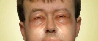
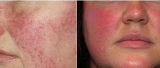
Atopic cheilitis is sometimes called allergic because it is caused by various irritants. The cause of the disease can be food or cosmetics. It turns out that the allergen affects the skin of the lips both from the inside and outside.
The disease manifests itself as inflammation of the red border of the lips. The skin becomes dry and peels. Cracks, itching and burning may occur.
Most often, children and adolescents suffer from lip allergies. Often it is the only symptom of neurodermatitis or atopic dermatitis.
Glandular cheilitis
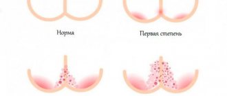
Glandular cheilitis is an inflammation of the salivary glands, which are located on the surface of the mucous membrane of the lips.
The disease is more common in men over 50 years of age and is characterized by the following symptoms:
- noticeable red dots appear on the lower lip;
- copious secretion of saliva from the inflamed glands, the appearance of “dew drops”;
- dryness, cracks and erosion;
- Bacteria can enter the irritated canals, leading to the formation of pus.
There are primary and secondary forms of glandular cheilitis. The primary disease develops due to genetic predisposition. Secondary lip disease can be caused by lupus, oral leukoplakia, or lichen planus.
Meteorological cheilitis
People have to deal with this disease all the time. Standard chapping, which more often appears in winter, is meteorological cheilitis.
The first sign of the disease is a feeling of skin tightness. In advanced cases, it turns red, dries out, and becomes covered with cracks. This lip inflammation can be treated at home. It is enough to isolate yourself from harmful factors, moisturize and nourish the skin until it is completely restored.
Eczematous cheilitis
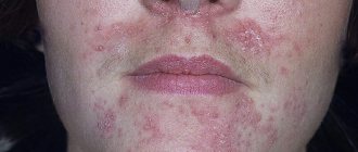
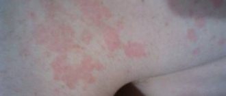
Eczematous cheilitis is one of the manifestations of eczema - an inflammatory process of a neuroallergic nature, which most often manifests itself on the face or dry areas of the body. Usually the disease is accompanied by constant dryness and redness. In advanced cases, the skin begins to peel off and become covered with blisters.
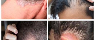
The disease often affects the tissue around the lips, so the patient may develop a red border, as in the photo on the right. This area of skin is constantly itchy and itchy.
If the disease is chronic, the symptoms are less pronounced. But in this case, seals appear on the skin.
Actinic cheilitis
Many people believe that lips need to be protected only in winter. Therefore, it is at this time of year that people stock up on moisturizing and nourishing balms to relieve dry and itchy lips. However, delicate skin must be protected not only from frost, but also from the burning sun.
With increased sensitivity to ultraviolet radiation and prolonged exposure to open areas, actinic cheilitis forms. His symptoms are standard:
- dryness, flaking;
- redness and swelling;
- compaction of individual areas.
If you do nothing, your lips become crusty. This is how the body tries to somehow protect the vulnerable part of the face. This symptom appears less frequently than others.
In advanced cases, ulcers, erosions and small compactions occur around the oral cavity. This condition is precancerous.
Exfoliative cheilitis
The exfoliative form of the disease occurs due to stress and disturbances in the functioning of the immune system. Genetic predisposition plays a major role. If your parents had this disease, there is a high risk that you will develop it too.
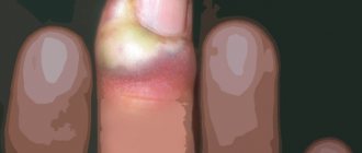
Exfoliative cheilitis on the lips occurs in two forms: exudative and dry. In the first case, you can observe the so-called yellow lips (pictured). A dense crust of this shade forms on the skin.
The yellow crust is easy to tear off; this process does not cause much discomfort. There are no erosions or other damage under the crust.
In the dry form of the disease, a crust also forms on the lips, but not yellow, but a lighter shade. The patient is concerned about dry skin, which explains the name of the disease. There is a desire to lick your teeth, but it is better not to do this: you can cause an infection and provoke more irritation.
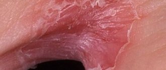
Candidal cheilitis
If the lips are red, inflamed and crusted with a cheesy coating of white or yellowish color, it means that the cause of these symptoms is candidiasis. If you clean off the plaque, inflamed areas of skin will be exposed. How such a lip disease manifests itself is shown in the photo.
The disease occurs due to the excessive development of the Candida fungus. The pathogen lives on the human mucous membranes constantly, but the active development of the fungus begins only under favorable conditions, which include:
- decreased immunity due to past illnesses or lack of nutrients;
- long-term use of antibiotics;
- a sharp change in climate to a hotter and more humid one.
Fungal inflammation of the lips begins externally, but can spread to the internal tissues of the oral cavity, thus leading to candidal stomatitis.
Lip cancer
Many of these diseases, if not given proper attention to their treatment, lead to cancer. Perhaps this is the most terrible disease that can affect the lips and oral cavity.
At first, the symptoms of cancer are unremarkable. The lips will turn red and there will be slight inflammation. The skin may become dry and cracked. If the patient moisturizes and nourishes the affected tissues, but the lips remain sore for several weeks, he should sound the alarm and consult a specialist. Later, ulcers and lumps may appear.
Usually, if treatment is started correctly and on time, the cancer recedes completely. Only in rare cases are relapses possible.
There is another disease that is compared to cancer - Manganotti syndrome. The disease manifests itself in the form of a noticeable ulcer on the lip, which is precancerous. However, due to the fact that in most cases the syndrome still develops into a tumor, it is more often classified as oncological diseases.
Possible side effects
Basically, complications arise not so much due to rupture of a venous vessel, but due to interruption of arterial blood flow due to compression or when the drug is administered into the lumen of the vessel with subsequent embolization of the terminal branches with small fragments [Fig. 3].
Rice. 3. Reasons for the development of skin necrosis. Intravasal injection (top), vessel compression (bottom).
What could happen next? Stopping arterial flow leads to necrosis and tissue death in the blood supply zone of a given vessel. Cases of necrosis of the skin of the wing and tip of the nose, lip tissue and glabella area have been described [Fig. 4–5].
Rice. 4. Necrosis of the tip or wing of the nose.
Rice. 5. Zones of necrosis.
An even more serious risk factor is associated with the fact that the facial artery represents the communication between the external and internal carotid arteries. Its terminal branch, the angular artery, anastomoses with the ophthalmic artery, a branch of the internal carotid artery. It is this connection between the vessels that can lead to embolization of the ophthalmic artery with small fragments of filler and further penetration into the central retinal artery with a possible decrease in vision up to blindness [Fig. 6–7].
Rice. 6. Iatrogenic retinal artery occlusion caused by the injection of fillers.
Rice. 7. Microembolism of the ophthalmic artery: etiopathogenesis.
Blue discoloration of the nasolabial triangle
Blue discoloration of the nasolabial triangle.
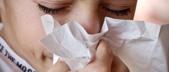
Very often, parents turn to the pediatrician with complaints about blue discoloration around the lips, or, as it would be more correct to say, cyanosis of the nasolabial triangle. What is this and is it worth worrying about this issue, we will now consider.
The nasolabial triangle is an area on the face limited by the nasolabial folds on the sides, the nose above and the lips below. Cyanosis can be caused by physiological (i.e., occurs in absolutely healthy babies) and pathological (various diseases) conditions of the body.
Physiological conditions.
In the area of the nasolabial triangle, the blood supply is highly developed: both arterial and venous vessels are present. And the skin of infants and young children is very thin and delicate, so the vascular plexuses seem to “shine through” through it and it appears bluish.
Baby crying . Sometimes mothers note that cyanosis appears when crying or prolonged screaming. This is due to the peculiarity of the pulmonary system in newborns. At this time, the level of oxygen in the body decreases (its amount can decrease by up to 92%), and the capillaries expand from tension and are therefore clearly visible through thin skin. In medicine, this phenomenon is called pulmonary cyanosis, and does not pose any threat to health. It disappears when the baby calms down. As the child grows up and lung function improves, this symptom goes away.
Feeding. Sucking at the breast is a big burden for the baby, during which capillaries appear and become visible near the surface of the skin. After feeding, the blueness goes away immediately.
Low air temperature . When your baby is cold, some parts of the body, including the nasolabial triangle, may turn blue. This is due to the imperfect heat exchange system of the child’s body. As soon as the baby warms up, the skin will take on a natural color. The same thing happens while walking. If you notice a blue nasolabial triangle, it’s time to leave the street, because this may be the first sign of hypothermia in a child.
We have reviewed with you the physiological conditions of cyanosis of the nasolabial triangle in children, which are not associated with the manifestation of any diseases. What pathological conditions can lead to the appearance of this symptom?
Pathological conditions.
Respiratory diseases. Respiratory diseases that can lead to cyanosis of the nasolabial triangle include ARVI, pneumonia, bronchial asthma, obstructive or allergic bronchitis, that is, those that lead to impaired air exchange. Of course, along with cyanosis, other symptoms of these diseases should be present (fever, nasal discharge, prolonged dry cough). In this case, you cannot do without consulting a pediatrician, who in turn can refer you to other specialized specialists.
Foreign objects. This is another common cause of cyanosis of the nasolabial triangle. Little children are very curious. Their taste buds are best developed, and therefore they experience the whole world through their mouth. Everything that interests them ends up in the mouth - one careless breath and a tiny object ends up in the respiratory tract. The child begins to cough heavily and gasp for air. It is necessary to provide first aid and call an ambulance.
First aid for a foreign body in the respiratory tract : Turn the child face down and tap the back with force, but not excessively. It is better to carry out manipulations over a sofa or armchair so that the baby does not fall on the floor and accidentally slip out of your hands.
Second option. Sit in a chair or chair. Place your baby face down on your left knee. Your left palm should be on his chest and support his neck. With your right hand, make strong pushes with the edge of your palm between the shoulder blades towards the mouth. Additionally, induce vomiting in the child by pressing your fingers on the root of the tongue. Follow all steps until the ambulance arrives.
Neurological pathology. Very often, cyanosis can be observed in children who experienced hypoxia or asphyxia during birth (for example, when entwined with the umbilical cord), and were also born premature. We see a blue nasolabial triangle in children with increased intracranial pressure and immature brain structures. In any case, consultation with a neurologist is necessary.
Congenital heart defects . Cyanosis of the nasolabial triangle in a child is one of the first signs of congenital heart disease and heart failure. To clarify the diagnosis and timely treatment, it is necessary to conduct an echocardiogram (ultrasound of the heart), an ECG and consult a cardiologist.
Problems with the development of the respiratory system. Malformations of the bronchopulmonary system (tracheal stenosis, bronchial hypoplasia, etc.) can also lead to cyanosis of the nasolabial triangle. Such conditions require urgent treatment.
Poisoning with drugs or chemicals. Poisoning causes tissue hypoxia, which is manifested by impaired absorption of oxygen by cells, hence cyanosis of the nasolabial triangle. Cyanosis will also be observed in children in whose presence their parents smoke, as a consequence of nicotine intoxication.
If the doctor, after examining the baby, did not find any pathologies and recognized cyanosis of the nasolabial triangle as normal, the following measures should be taken so that the blue discoloration disappears as soon as possible.
Be sure to take your child for a walk in the fresh air every day. Long walks will not only strengthen the baby’s body, but also saturate it with oxygen. And if there is no deficiency, then the nasolabial triangle will not turn blue.
Don't let your baby cry for too long. This is harmful not only to the physical health, but also to the emotional comfort of the child. Crying is a form of communication accessible to the baby to the mother. By crying, the child tries to draw her attention to some factors that cause him discomfort (wet diaper, hunger, loneliness and others). Provide him with everything he needs.
Monitor the air temperature in your baby's room. It should not be too hot or cold. Ideally - 21-25 degrees. If your child starts to feel cold, just dress him warmly (in clothes made from natural fabrics so that the skin can breathe).
Therapeutic strategies to prevent and manage complications
It is obvious that it is necessary to use techniques that minimize the risk of developing these dangerous complications. The text of the atlas describes, area by area, all the precautions and manipulations that must be taken to reduce this risk: the use of a cannula, the depth of injection, the quantity and quality of filler injected, and so on. Pallor of the skin and patient complaints of sudden pain in the injection area are signs that blood flow has stopped in this area. We must be able to control this situation.
All measures are aimed at restoring blood flow: urgent dissolution of the filler (if hyaluronic acid was used), warm compresses, massage, etc. Then there are prescriptions that need to be followed at home: antibiotic therapy to prevent bacterial superinfection, antiplatelet agents, topical medications.
In the introduction to the atlas I placed the inscription
“Only non-practitioners do not make mistakes; only through practice does it become possible to reduce the risk of error.”
If all measures are carried out on time and correctly, the spread of the necrosis zone will be minimal, a large area of skin will be preserved and, therefore, the chance of restitutio ad integrum will be higher.
Features of cheilitis in children
Lips suffer due to minimal protection. In children it is even weaker, so cheilitis worries them somewhat more often. In addition to children, the risk group includes the elderly and pregnant women.
The main causes of cheilitis in childhood:
- allergic reaction to food;
- use of products for the care of the skin of the lips and around them, not intended for children;
- genetic predisposition;
- infectious and fungal infections;
- weather.
Typically, infantile cheilitis does not develop until critical stages. When children's lips turn red, parents immediately begin to treat them. After all, people pay more attention to the health of the younger generation than to their own. If cheilitis in children still requires treatment, therapy should not be delayed longer than a few weeks. The main thing is to remove all allergens from the children's diet and balance the diet.