This article presents a list of the most common diseases and pathological conditions of the nail apparatus and skin.
Deviation of the appearance of nails from the norm usually indicates the presence of a nail disease.
Lesions can be primary or secondary, that is, they are symptoms of diseases of the internal organs and skin.
The same changes in appearance occur in different conditions. Also, changes may be associated with incorrect and unsafe work of the manicurist. In addition, diseases are divided into infectious and non-infectious. Nail technicians are only allowed to work with non-communicable diseases.
Doctors without medical education do not make diagnoses on their own.
But familiarity with the symptoms of nail damage will allow you to suspect these diseases in clients and refer them to a doctor, as well as competently plan and predict your work.
| Infectious | Non-infectious |
|
|
Causes
The main reason for the development of such defects is not only vascular diseases. Often, trophic ulcers occur against the background of neoplasms localized in the axillary, subclavian and mediastinal lymph nodes. In addition, injuries of various types (stabbed, cut, chopped, bitten), infectious and inflammatory diseases (chickenpox, helminthic infestations), pathologies of the immune system, systemic connective tissue diseases (lupus erythematosus, vasculitis), sepsis cause aggravation of the skin syndrome and the development ulcerative defects on the hands.
Education mechanism
As a result of exposure to the causative factor, the pressure in the venous capillaries of the upper extremities increases, and they begin to change their shape and direction. They become crimped and thinned. Their permeability increases, fibrin and blood clots are deposited between the venous vessels. This leads to hypoxia (low oxygen saturation of the blood vessels). The level of blood circulation decreases sharply, causing ischemia (circulatory failure). A prolonged absence of normal blood circulation provokes the development of necrotic processes. Plaques of various shapes form on the hands, which over time either dry out and turn into a dry form of necrosis or begin to quickly increase, reaching large sizes.
Felon
Diabetes
22431 December 23
IMPORTANT!
The information in this section cannot be used for self-diagnosis and self-treatment.
In case of pain or other exacerbation of the disease, diagnostic tests should be prescribed only by the attending physician. To make a diagnosis and properly prescribe treatment, you should contact your doctor. Panaritium: causes of appearance, symptoms, diagnosis and treatment methods.
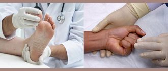
Definition
Panaritium is an acute purulent inflammation of the tissues of the fingers of the upper and, less commonly, lower extremities. The disease is considered one of the most common in purulent surgery and accounts for up to 46% of all cases requiring surgical treatment in a clinic.
Causes of panaritium
Panaritium can form as a result of visible or invisible microtrauma. In 33% of cases, small puncture wounds and splinters are responsible for the development of inflammation, in 25% - abrasions and minor scratches. The cause of panaritium can be injuries received during a manicure or injection, hangnails, or ingrown nails (due to the peculiarities of its anatomical structure or improper trimming).
Some chemicals, when they come into contact with the skin of the fingers, have a toxic effect on it, creating conditions for the penetration of infectious agents. Bacteria penetrate through the damaged skin of the finger and cause a purulent-inflammatory process - most often we are talking about Staphylococcus aureus, less often - streptococcus, Escherichia coli, Proteus. The microflora in a purulent focus can be represented by microbial associations of three or more microorganisms.
Type 2 diabetes mellitus, vitamin deficiencies, and circulatory disorders predispose to the occurrence of panaritium.
Classification of the disease
There are superficial and deep forms of panaritium.
Surface forms:
- cutaneous panaritium,
- subcutaneous panaritium,
- subungual panaritium,
- panaritium of the periungual fold.
Deep forms of felon:
- bone panaritium (acute and chronic),
- tendon panaritium,
- articular panaritium,
- osteoarticular panaritium,
- pandactylitis.
According to the stages of development of the disease, serous and purulent stages are distinguished.
The serous stage is reversible and can be treated with conservative methods - drug therapy, dressings, physiotherapy. It is characterized by swelling, redness, and pain. At the purulent stage, there is pronounced inflammation caused by the formation of a purulent focus. This stage requires surgical treatment. Symptoms of felon
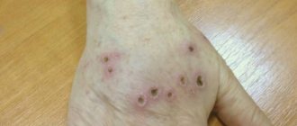
The main symptom of the disease is always intense throbbing pain. Sometimes the pain intensifies at night and deprives the patient of sleep, serving as a sign of the need to urgently seek medical help and, possibly, surgery.
The abscess can spontaneously open, in which case the pain subsides, which creates a false impression of improvement.
The pain is accompanied by soft tissue swelling and redness, but with deep panaritiums, redness may be absent or insignificant. As the inflammatory process develops, movement in the finger is limited, especially in deep forms with damage to joints or tendons. The patient may experience increased body temperature, weakness, and malaise.
The mildest form of the disease is considered to be cutaneous panaritium. A purulent focus forms in the thickness of the skin, under the epidermis, while other tissues are not affected. The resulting serous, serous-hemorrhagic or purulent exudate leads to detachment of the epidermis and the formation of a bladder. About 80% of cases of cutaneous panaritium are recorded on the palmar surface of the fingers.
One of the most common forms of the disease is subcutaneous felon, manifested by an inflammatory process in the subcutaneous fatty tissue. In 80-90% of cases, the process is localized on the palmar surface of the distal phalanx, most often the 1st, 2nd and 3rd fingers. Inflammation of the fiber causes swelling, which compresses the blood vessels and causes severe throbbing pain.
The affected phalanx becomes red, swollen, and hot. With this form, the temperature can rise to 38°C.
With subungual felon, inflammation develops under the nail plate as a result of injury or a splinter getting under the nail. The patient experiences constant bursting pain, which intensifies with pressure on the nail; the finger swells and becomes hot. Accumulating purulent exudate can peel off the nail. Possible increase in body temperature.
Paronychia is an inflammation of the periungual fold that occurs when it is damaged. The roller becomes swollen, hyperemic, pus accumulates under the nail fold and can be visible through the skin.
Tendon felon (purulent tenosynovitis) is a deep, severe form of the inflammatory process, which most often develops as a result of late or ineffective treatment of subcutaneous felon or as a consequence of injury. With tendon panaritium, patients note intense tugging and bursting pain along the entire tendon, which intensifies with flexion and extension of the finger, passive movements are severely limited. The finger may be in a forced half-bent position and swell throughout, sometimes the inflammation spreads to the hand and forearm.
Articular felon occurs as a consequence of the progression of the subcutaneous and tendon felon, as well as as a result of trauma to the back of the finger with primary damage to the capsule of the interphalangeal joint. With articular panaritium, the interphalangeal or metacarpophalangeal joints become inflamed, in which inflammatory exudate accumulates. As a result, the finger acquires a flask-shaped or spindle-shaped appearance with maximum volume in the joint area. The skin over the joint becomes smooth, shiny and hyperemic. The pain intensifies when trying to make any movement. A long course of the disease is fraught with destruction of articular cartilage and the spread of the process to bone tissue.
Bone felon is an inflammatory process that occurs in the bones of the fingers. It occurs as a result of the transfer of infection from surrounding tissues to the bone or as a consequence of extensive trauma. Bone panaritium is characterized by bursting, throbbing pain. When the process is localized on the nail phalanx, swelling occurs, and the phalanx becomes like a flask. The skin is very hyperemic and hot. Signs of general intoxication appear: fever, chills, headache, weakness. The formation of a purulent fistula indicates the development of chronic panaritium.
The most severe purulent pathology of the finger is pandactylitis. It is characterized by damage to all anatomical structures of the finger (skin, tissue, tendons, bones and joints) and extends to at least two phalanges.
Pandactylitis is most often considered as a consequence of delayed/insufficient treatment of other forms of panaritium or as a result of extensive finger injury. Throughout the inflammation, swelling is observed, the finger is sharply thickened, has a purplish-blue color, is very painful, active and passive movements are impossible. Multiple fistulas often form, from which purulent discharge oozes. The finger takes a forced half-bent position. There is general intoxication, regional lymph nodes are enlarged and painful. With this form there is a very high risk of losing a phalanx or the entire finger.
Diagnosis of felon
A doctor can diagnose panaritium based on a clinical examination. Recommended examinations:
- general blood analysis;
Diagnostics
Diagnosis of trophic ulcers on the hands consists of carefully collecting data on the development of the underlying disease. Upon examination, the doctor determines the nature of the ulcerative defect, linking it with the cause of its development. After this, laboratory and instrumental research methods are carried out. Laboratory methods include a complete blood count. It is used to determine the presence of blood diseases (anemia), the presence of inflammation (the level of leukocytes and erythrocyte sedimentation rate increases), the presence of an oncological focus (young and mature forms of blood cells). In addition to a general blood test, microbiological cultures are performed on various media. This is necessary to determine the infectious pathogen and prescribe appropriate antimicrobial drugs. Also, in the presence of immunological pathologies, an enzyme-linked immunosorbent assay is performed. It is used to determine the presence of foreign particles. Instrumental methods include: ultrasound diagnostics and computed tomography. With their help, blood flow in the vessels, the presence of pathological neoplasms and areas of ischemia are determined.
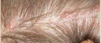
Cutaneous candidiasis: causes of appearance
Patients often ask the question: why doctors often send them either for tests or for consultations with “related” specialists. It would seem simpler: if there is a rash on the skin, treat the skin, but, unfortunately, not everything is so simple...
There are diseases and skin conditions that, in addition to burdening the patient with their own symptoms, are also markers, signs of some more serious pathology of the internal organs. One of these pathologies is skin candidiasis.
Case from practice
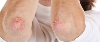
A young, slightly over 30, plump lady, who had previously complained of dry skin on her hands, began to feel itching between the fingers of her hand. Then the itching was joined by redness, and the skin began to peel off. Since she often had to do laundry and wash dishes by hand, both she and the doctors who treated her interpreted the incident as an “allergy to washing powder.”
However, the prescribed treatment (topical corticosteroids and antihistamines) brought only temporary relief, the symptoms never completely went away, and cessation of treatment caused a severe exacerbation of the dermatosis. Repeated changes of washing powder and detergents did not help either.
And here is the dermatologist's office. After examining the sufferer, I recommended interrupting treatment for a while and getting tested for fungi. Having received the results and additionally interviewed the patient, I referred her for a consultation with an endocrinologist, where, after an additional examination, diabetes mellitus was diagnosed for the first time.
The prescribed antifungal treatment brought a quick effect, and diet correction compensated for the initial manifestations of diabetes. I also recommended a more gentle regimen for the skin of the hands, softening creams... 2 years have passed since then. The dermatosis no longer recurred, and the lady, who went to the dermatologist, was able to prevent further development of the severe endocrinological disease in time.
Where does cutaneous candidiasis come from?
Fungi of the genus Candida are found almost everywhere: in the environment, on the skin, in the intestines of healthy people, birds, animals... However, most of us do not have skin candidiasis; only those who are very “lucky” get sick.
Most often, lesions of the skin and its appendages are caused by Candida albicans, which under normal conditions can be part of the normal microflora of the skin and intestines of humans and animals. Significantly less common pathogens are other candida (C. stellatoidea, C. tropicalis, C. parapsilosis, C. guillermondi, C. kursei, C. pseudotropicalis, etc., more than 160 species), however, recently they have become more common.
Factors influencing the possibility of developing cutaneous candidiasis
, can be divided into two main groups - internal and external.
Internal:
- metabolic disorders, especially diabetes, obesity;
- severe, debilitating diseases, often cancer;
- conditions after surgical interventions;
- chronic infectious diseases (tuberculosis);
- conditions associated with increased sweating (from prolonged elevated temperature to neuroses);
- circulatory disorders of the extremities;
- primary and secondary immunodeficiency conditions, including viral etiology;
- long-term treatment with corticosteroids (systemically), cytostatics, antibiotics (more often with self-prescription of the latter);
- alcoholism, drug addiction;
- chronic intoxication.
External:
- direct and prolonged exposure to harmful substances (detergents, acids, alkalis, cement, etc.) on the skin and mucous membranes;
- increased temperature and humidity in the room;
- clothing that is too warm, tight, occlusive (not breathable);
- erosions, burns, skin damage during manicure;
- lack of sufficient hygiene;
- bad habits (for example, lip biting in children, nail biting);
- ionizing radiation, etc.
Most often, these factors are combined, as in the above example, when an aggressive environment (solutions of washing powder and dishwashing detergents), which previously only caused dry skin, with the development of internal pathology served as an impetus for the development of a fungal infection.
I will tell you in one of the following materials how the symptoms of skin candidiasis manifest themselves, how contagious this disease is and what to do if you are diagnosed with it.
Good health!
Leonid Shchebotansky
Photo istockphoto.com
Products by topic: (glucometer), (glucometer test strips), (fluconazole), [product strict=”PIMAFUTSIN 2%”](pimafucin cream), [product strict=”nystatin”](nystatin ointment), [product strict ="clotrimazole"](clotrimazole)
Treatment
After identifying the main cause that led to the development of trophic ulcers on the hands, adequate therapeutic measures are selected. For antibacterial purposes, antibiotics are prescribed - Ceftriaxone, Amoxicillin, Tetracycline. Venarus and Detralex are used to strengthen blood vessels and restore impaired blood flow. For shallow/superficial ulcerative defects, the use of healing ointments is recommended for topical application. These include Zinc ointment, Argosulfan, Solcoseryl and Levomekol.
Surgical treatment consists of excision of the edges of the ulcer and rinsing with antiseptic drugs (Betadine, Furacillin). If purulent contents are present, drainage is installed.