Effective procedures for the treatment of hyperkeratosis
Phototherapy
Plasma therapy
NeoGen procedure
Mesotherapy
Peeling
Medical pedicure
One of the fairly common types of skin diseases is hyperkeratosis. This is an excessive thickening of the stratum corneum of the epidermis. With the development of such a pathology, horn cells begin to rapidly divide and desquamation is impaired, which ultimately leads to thickening of the skin. Moreover, the thickening can reach several centimeters. According to statistics, hyperkeratosis is observed in approximately 20% of men and 40% of women. As a rule, the problem occurs in individual areas and leads to a significant deterioration in the appearance of the skin.
In children, the pathology can be observed at the age of 1–3 years, and it mainly affects the face and hands. In adults, changes in the stratum corneum are more common on the feet and toenails, as well as on the face and scalp. There are several types of the disease, which have their own symptoms and causes of development. Let's figure out what hyperkeratosis is and whether such a pathology is dangerous for health.
Classification
The classification of hyperkeratoses is based on a number of factors, according to which the following are distinguished:
- By origin: hereditary and acquired hyperkeratosis .
- By area of damage: local (calluses/warts) and diffuse, occupying large areas of the skin.
According to clinical forms:
- Follicular hyperkeratosis - in which exfoliating scales of the stratum corneum clog the ducts of the follicles, which is manifested by the appearance of numerous small tubercles on the skin.
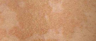
- Seborrheic hyperkeratosis is localized mainly on the scalp, less often on the facial skin. It manifests itself in the formation of areas of peeling in the form of an easily detachable greasy crust, after which reddish spots remain.
- Warty hyperkeratosis . It is characterized by the appearance on the skin of formations that outwardly resemble warts, but without the etiological factor of true warts - papillomavirus .
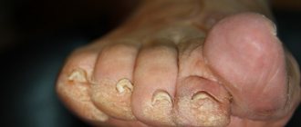
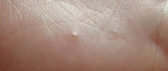
- Hyperkeratosis of the feet . It manifests itself as thickening of the skin of the feet with the appearance of corns and calluses.
- Disseminated hyperkeratosis . It appears as polymorphic elements on the skin of the trunk and limbs, reminiscent of thick/short hair, located separately and not merging with each other.
- Lenticular hyperkeratosis . It is characterized by the appearance of horny papules on the hair follicles of the lower extremities, and after removal, small indentations remain on the skin.
- Senile hyperkeratosis . The appearance of dark keratinized spots on the skin of older people is typical.
Features of the pathology
The concept of hyperkeratosis comes from two Greek words: hyper – “many” and keratosis – “formation of keratin”. In fact, hyperkeratosis is not an independent disease - most often it is just a symptom of other diseases. For example, such a problem occurs with lichen, ichthyosis or erythroderma. However, thickening of the stratum corneum can sometimes appear in a healthy person: for example, the feet, elbows and knees are often susceptible to this problem.
Hyperkeratosis is considered a potentially life-threatening disease, but treatment is still recommended. Pathology can cause serious cosmetic defects, which often lead to social maladaptation and decreased self-esteem.
The basis of the stratum corneum of the skin is made up of horny plates, which contain the protein keratin. Typically, the layer includes 15–20 layers of horny scales; when the epidermis thickens, their number can reach 100.
Essentially, the stratum corneum is the end result of the division of keratinocytes of the epidermis, which is why it has a specific structure. It is based on horn cells held together by intercellular lipids. Normally, this layer of the epidermis is constantly renewed: the horny scales are peeled off and replaced with new cells.
The desquamation process starts in the basal layer of the skin, where keratinocytes are produced. After this, the cells gradually move to the upper layers, losing their nucleus and organelles and turning into flat scales, which develop into the stratum corneum.
Normally, the epidermal renewal cycle lasts about 21–28 days; with age it slows down and can reach 45–72 days. In addition, the state of cellular metabolism is influenced by various factors such as lifestyle, nutrition, and hormonal levels of the body.
With hyperkeratosis, the patient's production of proteolytic enzymes, which regulate the processes of destruction of protein bonds between the cells of the stratum corneum, is disrupted. As a result, there is no desquamation of old cells, and due to the accumulation of keratinocytes, a significant thickening of the epidermis occurs.
Types of pathology
The following forms of hyperkeratosis are distinguished:
- Follicular. This type of pathology is characterized by damage to the hair follicles. Most often, follicular hyperkeratosis appears in areas with dry, dehydrated skin. The reasons are heredity, vitamin deficiency, inflammatory skin diseases or neglect of personal hygiene. There are two forms of pathology: with follicular hyperkeratosis of type I, spiky nodules and plaques appear around the hair follicle, with type II - a hemorrhagic rash caused by a lack of vitamin C and K. Very often, the ducts of the hair follicles are clogged with blood or red pigment.
- Lenticular. This form is characterized by the appearance of small “plugs”, most often on the hair follicles, which look like keratinized spots. The reasons for the development are not exactly known, but most often the problem occurs in elderly patients. As a rule, the pathology is chronic, but without a tendency to regression.
- Disseminated. With this form, polymorphic elements appear on the skin, which outwardly resemble short and thick hair. They are isolated from each other and have no tendency to grow together.
- Seborrheic. This type of pathology is typical for oily skin and appears as a result of metabolic disorders. At an early stage, characteristic yellow-brown spots of a round shape appear, which gradually fade and turn into plaques. The lesions can be quite large in diameter.
- Warty. Externally, the warty form resembles warts, but their appearance is not caused by papillomavirus.
- Plantar. Hyperkeratosis of the feet can occur on the entire sole or only on the heel area. As a rule, this form is characterized by severe dryness and roughness of the skin, as well as noticeable thickening in the affected areas. There is a dry callus (a local lesion with clear boundaries of thickening of the stratum corneum), a core callus (a sharply limited area of thickening of a round shape of small size) and a soft callus (appears between the fingers as a result of increased humidity).
- Subungual. Quite often, subungual hyperkeratosis occurs, resulting from traumatic onychia or onychomycosis. It is characterized by rapid growth from the distal edge, which causes an enlargement of the nail plate.
Diagnosis and treatment of the disease
If characteristic skin problems appear, you should consult a dermatologist as soon as possible. The specialist examines the skin and collects the patient’s complaints. It is important to differentiate hyperkeratosis from other skin diseases, for example, psoriasis or lichen, which also cause peeling of the skin. If the diagnosis is in doubt, a biopsy of suspicious skin areas may be performed.
To treat the disease, ointments with topical corticosteroids are used to treat the damaged areas. This may include prednisolone or hydrocortisone ointment, fluacinolone-based formulations, or clobetasol. In addition, glucocorticoid drugs are prescribed, which have an exfoliating and anti-inflammatory effect, thoroughly cleansing and disinfecting the skin.
Patients with hyperkeratosis of any form are advised to take warm baths with the addition of salt, baking soda or starch and further moisturizing with cosmetic creams.
If hyperkeratosis is detected, mechanical peeling is contraindicated, as it can aggravate the patient’s condition. Only acid (chemical) peeling based on creams with a mild effect is allowed. These are products consisting of salicylic, lactic, citric and other acids. Vitamins A and C are also prescribed orally.
Plantar hyperkeratosis is treated with antifungal ointments, and also requires the elimination of all pathological factors of the disease. It is necessary to replace shoes with more comfortable ones, as well as receive orthopedic help in case of flat feet or club feet. In addition, salt baths, mechanical grinding using a hard sponge and pumice at home are useful. After this, the feet are lubricated with a moisturizing and nourishing cream.
As for possible complications of the disease, they can be observed with plantar hyperkeratosis, when cracks, corns appear, and a secondary fungal infection begins to develop. Follicular hyperkeratosis in some cases causes pyoderma. Formations with a warty form sometimes develop into a malignant tumor. The prognosis for timely and high-quality treatment of hyperkeratosis is favorable, although in some cases the fight against the disease can last a lifetime with alternating stages of exacerbation and remission.
Reasons for development
Pathogenesis varies depending on the specific type of disease. Very often, the development of pathology is facilitated by exogenous (external) causes. It could be:
- pressure on the skin;
- wearing uncomfortable clothes/shoes;
- exposure to aggressive agents.
This provokes the body’s protective functions, which, in turn, cause increased cell division. As a result, the natural process of cell desquamation is disrupted, which leads to a thickening of the skin layer.
The endogenous (internal) causes of the disease include the following factors:
- hereditary skin diseases;
- gastrointestinal diseases;
- disruption of the regeneration process after injury;
- skin neoplasms;
- fungal infections;
- hormonal imbalances;
- hypovitaminosis;
- inflammatory diseases;
- metabolic disorders in the body.
Excessive roughening of the skin is often explained by other ongoing diseases:
- psoriasis;
- ichthyosis;
- keratoderma;
- lichen;
- diabetes.
If we talk about foot hyperkeratosis, this form of pathology is often caused by improper gait or flat feet, excess body weight, and wearing tight and uncomfortable shoes.
Symptoms of hyperkeratosis
The main symptom of the pathology (and its main characteristic) is thickening of the skin in the affected areas . Other symptoms may vary depending on the specific form of the disease.
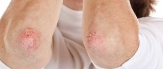
With follicular hyperkeratosis, “goose bumps” appear, that is, dense reddish pimples with a spiky shape. They are located at the base of the hair follicles. Most often, all this is observed on the side or back of the thigh, buttocks, arms, and face. In rare cases, the patient may experience itching.
Plantar hyperkeratosis is characterized by roughening of the skin on the entire sole of the foot or only in the heel area. Calluses and bleeding cracks often occur, which causes discomfort and pain.
With the seborrheic form, characteristic pigmented formations appear, which over time lose color and become colorless plaques.
The symptoms of hyperkeratosis are more pronounced in adults; in children, the pathology is often similar to dermatitis. At the initial stages of development there are no symptoms.
Stages of development
The following stages of disease development are distinguished:
- First . In some areas, the skin turns pale and becomes dry. No thickening of the stratum corneum has yet been observed.
- Second. When touching the skin, particles falling off are noticeable. Slight thickening is possible.
- Third . The skin peels off greatly and loses its smoothness, becoming rough and rough to the touch.
- Fourth. At this stage, multiple complications arise that lead to serious changes in the appearance of the epidermis. The thickening of the stratum corneum is visible to the naked eye.
Dermatological diseases
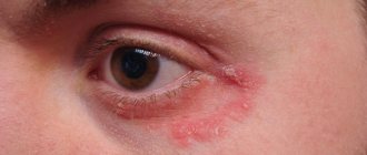
Often, dry skin on the elbows indicates the presence of a skin disease. “It could be atopic dermatitis (a chronic inflammatory skin disease). Dry skin is always characteristic of atopic, i.e., allergic conditions: dermatitis, bronchial asthma, vasomotor rhinitis, and sometimes food allergies. Psoriasis may also appear on the elbows. With this disease, rapid development of cells occurs, they do not have time to mature, the stratum corneum is not built, and the skin looks drier,” explains Skorogudaeva.
What can cause itchy skin? More details