Eyelid melanoma is an aggressive malignant tumor that develops from melanin-producing cells and is located on the upper or lower eyelid. However, this is not the only neoplasm that develops in this area, but it is the most malignant and, fortunately, the rarest. Melanoma of the eyelid is diagnosed in approximately 5-7% of all melanomas. It develops mainly in people of the older age group. The peak incidence occurs at 50-60 years of age.
- Types of eyelid tumors
- Causes of development of melanoma of the eyelid
- How is eyelid melanoma diagnosed?
- Methods for treating eyelid melanoma
- Prognosis for recovery from melanoma of the eyelid
Types of eyelid tumors
Eyelid tumors can be benign or malignant.
Benign neoplasms
Benign eyelid tumors grow slowly, superficially, and do not affect the underlying tissue. Their course is favorable and the reason for visiting a doctor, as a rule, is a cosmetic defect. Most often, benign neoplasms develop from epithelial tissue, but the source can be hair follicles and other skin elements (trichoepithelioma, Malert epithelioma, fibromas, lipomas).
The most common benign eyelid tumors are:
- Papillomas are nodular neoplasms of a dirty yellow or dirty gray color. They can be located on a leg or a wide base. The neoplasm resembles a villous mushroom or cauliflower in appearance. Sometimes it can spread widely throughout the tissue of the eyelid, affecting the tear ducts, conjunctiva of the eyelid and even the paranasal sinuses. May become malignant.
- Senile warts (senile warts) are similar in appearance to papillomatous nevus. It looks like a brown, yellow or grayish nodule, dense to the touch, does not cause pain. Compared to papilloma, they have more pronounced keratinization. Does not malignize.
- Keratoacanthosis - manifests itself as white spots covered with scales. If left untreated, it may become malignant (risk 20%).
- Trichoepithelioma - a neoplasm arises from hair follicles, has the appearance of a dense nodule, 1-3 mm in size, rarely 1 cm. It can be multiple. In some patients, the number of tumors exceeds a dozen. Mostly young people suffer. If left untreated, they can malignize into basal cell carcinoma.
- Syringoadenoma is a tumor growing from the epithelium of the sweat glands. The tumor is a multilocular neoplasm. It grows very slowly.
- Fibroma - has the appearance of a node on a wide base or on a narrow stalk. It grows from mesodermal tissue and can reach several centimeters.
Malignant tumors of the eyelids
Malignant tumors of the eyelids have infiltrative growth and tend to destroy underlying tissues and metastasize. Basal cell carcinoma progresses most favorably. It is also called a semi-malignant tumor. The most aggressive growth is in melanoma. It not only infiltrates the underlying tissues, but also gives distant metastases.
- Basalioma is a type of skin cancer. It looks like a nodule with a ridge-like surrounding. It grows slowly, germinating into the underlying tissue. In advanced cases, it can spread to the structures of the eyeball and even the orbit. Metastases are casuistically rare. Most often, basal cell carcinoma is located on the outer side of the eye, a little less often - on the lower and upper eyelids, and very rarely in the area of the inner corner. When located in the lower eyelid area, the neoplasm proceeds more favorably. The most aggressive growth is in basal cell carcinoma of the inner corner of the eye.
- Skin cancer of the eyelids. Usually this disease is represented by squamous cell skin cancer. At the initial stage, the tumor has the appearance of redness, which eventually thickens and ulcerates. The tumor is prone to infiltrative growth and metastasis.
- Meibomian gland cancer is a very rare type of cancer and has an extremely aggressive course. The tumor is localized in the upper eyelid, looks like a basal cell carcinoma, but grows quickly and metastasizes.
- Melanoma of the eyelids is the most malignant neoplasm localized in this area. The first signs are the formation of a spot with blurry contours, surrounded by an area of increased pigmentation and redness. The stain quickly spreads over the surface of the skin. Also, melanoma of the eyelid can be presented in a nodular form. In this case, it will be a node with vague contours, which is prone to bleeding. It grows rapidly, infiltrating the underlying tissue.
Decoding by color and shape
The shape and color characteristics of moles can make adjustments to the interpretation of signs.
By color
Red color is a predisposition to search for the meaning of life and reflection. Failures in relationships are associated with hot temper and impatience.
Pink moles – stability, luck, longevity.
Light shades indicate sympathy from others.
Black moles mean that their owner will have to learn a number of lessons throughout his life. Trials, blows of fate, difficult circumstances that will have to be overcome are not excluded.
The brown color of moles is interpreted as a chance to increase social status.
By shape
Let us dwell on the interpretation of the most unusual forms of moles.
The Sun is a multifaceted, interesting, cheerful personality with an ebullient temperament.
An incomplete circle is the presence of karmic debts.
The waning moon means success in activities related to finance.
The full moon is a gift of foresight, many surprises.
The imprint of lips means strong intuition, a sense of humor, the protection of higher powers, success in a profession related to raising children.
A cat is a mystery, mystery, the ability to heal people and influence their destinies.
Large moles enhance positive interpretations, while small ones weaken negative predictions.
Causes of development of melanoma of the eyelid
Why melanoma occurs in the eyelid area is not completely known. As with most cancers, this tumor develops due to mutations that have occurred in melanocytes. As a result, they gain the ability to reproduce uncontrollably and spread to neighboring tissues.
Several risk factors have been identified, the presence of which increases the likelihood of developing eyelid melanoma:
- Belonging to the white race. The risk of developing melanoma is especially high in people with light skin color.
- Age over 50-60 years.
- The presence of dysplastic nevi in the eyelid area.
- Excessive insolation.
- Sunburn.
- The presence of melanoma in close blood relatives (hereditary predisposition).
In what cases should you consult a doctor?
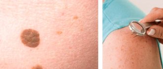
Hanging moles are easily injured, which can result in a malignant neoplasm. Therefore, when they appear, it is recommended to consult a dermatologist and have the problematic mole removed. However, there are cases when a visit to a specialist should not be postponed under any circumstances. If:
- Hanging moles are growing.
- A hanging mole hurts or itches.
- The mole suddenly changed color.
- A hanging mole becomes inflamed, swelling appears around it and it bleeds.
- A light spot appeared around the mole.
- The mole has noticeably thickened.
How is eyelid melanoma diagnosed?
Although eyelid melanoma is a superficial tumor that is easily accessible for visual inspection, it is not always possible to diagnose the disease at an early stage, since patients do not pay attention to it and do not go to see a doctor. Therefore, much attention is paid to self-examination. You can suspect a malignant process based on the following signs:
- The neoplasm has fuzzy, blurry edges.
- Asymmetrical shape.
- Uneven coloring of the tumor. In melanoma, areas of hyperpigmentation may be replaced by depigmented areas.
- The neoplasm increases in size.
- The skin pattern changes. Melanoma is characterized by a smooth surface; peeling or ulceration is less common.
- There is redness around the tumor.
- The melanoma itself may be itchy, painful, and cause a tingling or burning sensation.
As it grows, eyelid melanoma spreads to the underlying tissues, involving the conjunctiva, sclera and other structures of the eyeball in the process. In order to diagnose melanoma in time and begin treatment, it is necessary to regularly see an ophthalmologist, especially over the age of 50 years.
Methods for treating eyelid melanoma
The standard treatment for melanoma is surgical removal within healthy tissue. However, with such a complex localization, it is not always possible to carry out a full-fledged surgical intervention, since a large defect is formed that disfigures the person’s appearance. In addition, the anatomy of the eyelid is disrupted, which leads to various consequences for the eye - constant lacrimation, inflammation, eye injury, etc. Therefore, when melanoma is localized on the eyelids, other treatment methods are used, including radiation therapy and minimally invasive surgery.
Close focus radiotherapy in the treatment of melanoma of the eyelid
The essence of the technique is to irradiate the tumor from a short distance with low voltage values. Due to this, radiation is mainly absorbed by tumor cells and minimally affects other tissues. This treatment method is effective only for small, superficial tumors. Irradiation is carried out daily up to a total focal dose of 60-80 Gy.
Radiation therapy for eyelid melanoma
Radiation therapy is the exposure of a tumor to a concentrated stream of ionizing radiation. In the treatment of melanoma of the eyelid, contact radiation therapy has become widespread. For this purpose, a special plate containing radioactive material is used. In appearance it resembles a bottle cap. It is attached to the eye and left for 4-5 days, after which it is removed. As part of radiation therapy for eyelid melanoma, external irradiation can also be used, in which ionizing radiation is generated by special devices.
Excision of melanoma with a laser scalpel
A laser scalpel involves dissecting biological tissues due to a sharp increase in temperature in a small area. This leads to the fact that the irradiated area instantly burns, coagulating proteins and “sealing” the wound. A cut 2-3 mm deep appears. To completely remove the tumor, the tissue is cut in layers. The advantage of a laser scalpel is:
- Low morbidity.
- Simultaneous sealing of vessels. This not only eliminates bleeding, but also prevents the spread of melanoma cells.
- Cells are dissected in a non-contact manner, and heating the tissue leads to the death of possible microorganisms in the wound. Thanks to this, the risks of infectious complications are reduced.
The use of electric knife in the treatment of melanoma
When using an electric knife, high voltage alternating current is supplied to the tissue. Because of this, it is locally heated and charred, which leads to the formation of a cut. At the same time, coagulation of blood vessels occurs, which reduces the risks of bleeding and hematogenous spread of melanoma cells. The advantages of this removal method are similar to those of a laser scalpel, but there are a number of disadvantages:
- Trauma to tissue during removal process. This not only leads to the development of an inflammatory response in the dissection area, but also damage to the tumor being removed, which complicates its morphological and molecular genetic testing.
- There is a high risk of keloid scar formation, which in itself is not desirable, especially if it is in the eyelid area.
- Risk of permanent scar formation.
In this regard, the use of electric knife when removing melanoma is limited.
Prognosis for recovery from melanoma of the eyelid
In general, eyelid melanoma is an aggressive tumor and is diagnosed at late stages, when the prognosis for treatment is unfavorable. Five-year survival can be achieved in only half of the patients. But in the initial stages, with a melanoma thickness of less than 0.75 mm, the chances of a complete recovery are much higher.
| Read more about dermatological research at Euroonko | |
| Consultation with a dermatologist-oncologist | from 5,100 rub. |
| Skin examination using the German FotoFinder device | RUB 13,400 |
| Diagnosis of melanoma | from 5,100 rub. |
Book a consultation 24 hours a day
+7+7+78
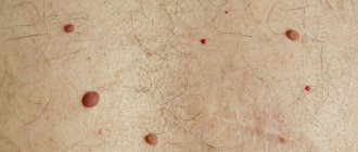
Fundamental differences between hanging moles and papillomas
The term hanging moles defines benign neoplasms that develop from skin pigment cells (melanocytes) under the influence of various causative factors, the main of which are heredity and hormonal imbalances in the human body. Papilloma is also a tumor formation, the development of which is provoked by the human papilloma virus. It is integrated into the genome of skin cells, leading to an increase in the activity of their division, which is the main mechanism for the formation of papilloma. A consultation with a dermatologist, who, if necessary, prescribes additional studies, allows you to accurately determine the nature and mechanism of development of formations on the skin.