Atheroma can occur for a variety of reasons. The radio wave method of removing atheroma is the fastest and most painless. If you need atheroma removal in St. Petersburg, feel free to contact us. Atheroma is a cyst that appears due to blockage of the sebaceous gland duct. It is completely painless, has a round shape and soft consistency. Atheroma can appear in a variety of places in the body, but today we will talk about atheroma of the ENT organs, which most often appears on the earlobe and behind the ears. If a person has a large number of such neoplasms, doctors diagnose atheromatosis. The main cause of atheroma is blockage of the sebaceous glands. These are exocrine glands, which are located in the upper layers of the skin. The sebaceous gland includes the acini, which has a large number of alveoli, as well as an excretory duct. There are sebaceous glands that do not communicate with the hair follicle; their excretory ducts are located on the surface of the skin. The walls of the alveoli are made of cells that contain a small amount of fat. These cells are regularly renewed, and dead cells are excreted during normal metabolism along with fat. If the excretory channel becomes clogged with dead particles, the sebaceous gland begins to enlarge, thereby forming atheroma. Atheromas never appear in places where there are no sebaceous glands, that is, on the palms and soles. In some cases, atheromas are congenital; this occurs due to disturbances in the functioning of the sebaceous glands during embryogenesis.
Main symptoms of atheroma
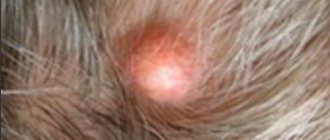
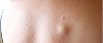
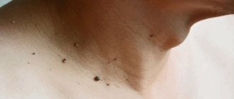
Atheroma is a rounded neoplasm that rises above the skin and is completely painless. It should feel soft to the touch or slightly compacted. Atheroma can vary in size from a few millimeters to ten centimeters. Over time, it can grow, due to the fact that the sebaceous gland continues to work and secretes secretions. So, the symptoms of atheroma:
- The appearance of a new formation on the surface of the skin.
- The texture of the tumor may be firm or elastic.
- The contours of the neoplasm should be clear.
- The subcutaneous capsule must be mobile.
- There may be a visible excretory duct in the middle.
Sometimes atheromas can burst open on their own, which is accompanied by the release of contents and an unpleasant odor. Due to the fact that the contents of the atheroma are a nutrient medium, bacteria can begin to multiply in it, which can lead to its suppuration. In this case, redness, swelling, and increased body temperature appear.
Classification
In practical dermatology, two classifications are used: according to the type of structure of capsule cells (histological) and according to the type of formation. Practitioners mainly use the second one. There are:
- Congenital (or primary) atheromas, the formation of which is caused by congenital changes in the ducts. The size of such formations is up to 0.5 cm, they are multiple and are detected soon after the birth of the child.
- Acquired (or secondary) atheromas develop in initially unchanged gland ducts in people with skin damage or impaired sebum production. The size of cysts can reach several centimeters. As a rule, they are single.
Causes of atheroma formation
Most often, atheroma appears as a result of poor personal hygiene. There may also be a hereditary predisposition to the appearance of atheromas, but this theory has not yet been proven, since scientists have not been able to find the gene that is responsible for their appearance. Atheroma of the earlobe can also occur due to an unsuccessful ear piercing, as a result of which the duct of the sebaceous gland is injured. The causes of atheroma can be very different:
- The presence of rare genetic diseases.
- Incorrect use of cosmetics.
- Damage to the skin during the removal of acne and pimples.
- Incorrectly performed facial cleansing.
- Hormonal disorders.
- Hyperhidrosis.
Possible complications
There are a number of consequences of atheroma:
- Risk of developing into phlegmon with an extensive abscess.
- Risk of relapse if opened independently.
- Risk of formation of suppuration and inflammation.
- The risk of postoperative scars that arise as a result of surgical removal of large atheroma.
- Inflammation of the scars may occur if the atheroma is removed in the clinic.
- As a result of incorrect diagnosis, a relapse may occur.
It is also worth noting that atheroma is a very rare disease; according to statistics, only 7-10% of the world's population gets sick with it. Atheroma is a benign neoplasm that never becomes malignant.
Diagnosis of atheroma
Diagnosis of atheroma is not difficult. This is usually done through a routine skin examination. Atheroma does not pose a danger to life. If there is no suppuration, then you can limit yourself to ordinary observation without undertaking any treatment. There is a high probability that the atheroma will open on its own. But if it has reached a very large size and begins to ache, then it is better to remove it. If the contents of the atheroma break out on their own, the inflammation may subside. Further prospects: atheroma may disappear completely, or it may continue to fester periodically. Despite the fact that atheromas are benign tumors, if you discover a neoplasm, it is best to consult a doctor. Let the doctor tell you what kind of tumor it is and whether treatment is required.
Who treats atheroma of the ENT organs
If atheroma appears on the ears, then you should seek help from an ENT doctor
. This specialist specializes in the diagnosis and treatment of diseases of the ear, nose and throat. The study of these organs is united within the framework of the science of otorhinolaryngology. You should contact an ENT doctor if you or your family are concerned about diseases of the upper respiratory tract, runny nose, tumors in the ear area, pain when swallowing and breathing, as well as enlarged parotid lymph nodes. All these symptoms are known to many other doctors, but only an experienced otolaryngologist can prescribe the most effective treatment for you. Timely detection and treatment of diseases of the ENT organs will save you from complications that can spread to other organs.
List of sources
- Kapustina O.G. Diagnosis and optimization of treatment of skin tumors in the outpatient practice of a dermatologist. dis. Ph.D. honey. Sci. – M., 2009. – 163 p.
- Dubensky, V.V. Skin neoplasms in the practice of a dermatovenerologist Text. / Edited by V.V. Dubensky // Tver: “Publishing house-Triad”. 2002. - 148 p.
- Bezrukov S.G., Grigorieva T.S. Histomorphological features of the structure of the membrane of facial atheroma // Tauride medical and biological bulletin. - 2013. - T. 16, No. 1, part 3 - P. 37-41.
- Kubanova A. A., Kubanov A. A., Rakhmatulina M. R., Malova I. O., Sokolovsky E. V., Apolikhina I. A., Melkumyan A. G. Federal clinical guidelines for dermatovenereology. Dermatovenereology 2015: Skin diseases. Sexually transmitted infections. M.: Business Express, 2016. 768 p.
Treatment of atheroma
An ENT doctor can offer the following treatment methods for atheromas:
- Medication.
- Surgical intervention.
If atheroma occurs behind the ear, it is recommended to immediately consult a doctor who will prescribe the necessary treatment for atheroma. The most effective way to treat this disease is surgery. And if the doctor directs you to undergo surgery, then you should not refuse; this operation is very easy to tolerate and is performed under local anesthesia. After the surgeon has completed his work, you will be able to forget about this disease forever. A good surgeon, when cutting the skin, acts as carefully as possible, without damaging the atheroma capsule. Then the capsule is removed from under the skin, and the cavity in which it was located is thoroughly disinfected with antiseptics. It will be much harder for those who waited until the inflammatory process appeared. If an atheroma tumor is detected, surgery is not performed. First, you will need to remove the inflammatory process so that the operation can be performed. Surgical intervention does not require an inpatient stay, but is performed on an outpatient basis. If the tumor is small in size, no sutures are applied. But if the atheroma has reached a large size, then there is a chance that you will have stitches, which will be processed once every 2 days. Recently, in surgery, instead of surgical intervention, laser and radio wave removal of atheroma have begun to be widely used. In this case, the contents of the atheroma are simply burned out or evaporated. As a rule, these methods are used in the early stages of atheroma development.
Clinical picture
When consulting a dermatologist, the patient undergoes a standard examination. Visually, the doctor determines the presence of a neoplasm with high mobility and absence of painful sensations during pressing. In the absence of complications, the skin over the cyst remains unchanged; sometimes an enlarged sebaceous duct is clearly visible in this area of the dermis.
If atheroma is characterized by rapid development, then small ulcerations may appear on the dermis. When an infection occurs and the subsequent development of the inflammatory process, the skin becomes red.
Atheroma without inflammation and secondary complications does not affect the life and health of the patient. Most patients seek professional help for aesthetic purposes. In other cases, surgical intervention becomes a necessary measure to stop developing inflammatory processes with the accumulation of purulent contents.
Radio wave removal of atheroma
Radio wave removal of atheroma is considered the most effective and safe method. The procedure is carried out using special equipment that converts radio wave radiation into energy. Exposure to energy destroys the contents of atheroma without causing damage to healthy tissues. Today, such removal of atheroma is a very common and popular method. This type of atheroma removal has a large number of advantages, including:
- Completely eliminating the risk of relapse.
- Removal of atheroma is carried out without suturing.
- An almost completely painless procedure.
- Excellent cosmetic effect.
- Possibility to return to work immediately after surgery.
Moreover, this method of removing atheroma makes it possible to reduce the risk of scar tissue, but this cannot be promised with the intervention of a surgeon. Radio wave removal of atheroma lasts for twenty minutes using local anesthesia. First, the tumor is processed and then exposed to radio waves through a special device. The radiation completely “burns out” the tumor and allows the atheroma to be removed along with the capsule, leaving behind a small depression. After this, the doctor will treat the damaged area of skin with iodine and apply a bandage. If a large tumor is removed, in some cases a hospital stay may be necessary.
We not only know how to remove atheroma correctly. We do it WITHOUT SCARS!
It is not enough to remove atheroma. We need to make sure that nothing reminds us of its existence.
. Especially when it comes to removing atheroma on the face or head.
Exclusive multi-stage seam processing
in “Platinental”, allows you to achieve the formation of a thin and invisible scar after surgery:
- The minimum necessary incisions in the natural folds hide the very fact of the surgeon’s intervention.
- special adhesive strips
match the edges of the wound without suturing
- application of special medical glue protects the wound from germs, eliminates the need for daily dressings and creates a favorable microclimate for fast and beautiful healing.
- When removing large atheromas, special multi-layered
self-absorbing sutures are applied.
This technique allows you to raise the seam level with healthy tissue
and form
an invisible scar
.
- sutures placed in Platinental heal very quickly and are extremely comfortable to wear: you can shower the very next day after the operation.
By following the recommendations for care after removal of atheroma, you can be sure that no one will suspect the fact of surgical intervention.
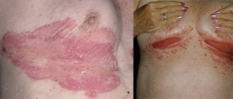
Scar after removal of atheroma on the head. Performed by surgeon: Maxim Vasiliev.
Recurrence of atheroma
During the operation, the atheroma is completely excised. Relapse can only occur if the sebaceous gland duct is poorly forgiven. The neoplasm must be removed completely, in some cases along with nearby tissues if the capsule has melted. The cause of relapse may also be the formation of a new cyst due to proximity to the postoperative scar. In some cases, relapse occurs due to incorrect diagnosis, when a lipoma or dermoid cyst is mistaken for atheroma. These tumors can also be treated surgically, but the operation is performed in a slightly different way. According to statistics, relapse of atheroma occurs in 15% of patients who have undergone surgery, and in 10% it is the result of opening an abscess cyst, when it is difficult to remove its contents due to purulent content. Such tumors must first be cured and removed after 2-3 weeks. It is recommended to remove atheromas in the winter season, when the cyst has just appeared and has not yet become inflamed. Relapse may also occur due to hyperhidrosis or a hereditary predisposition to obstruction of the sebaceous glands. In these cases, neoplasms appear not in the place where the operation was performed, but in the nearby excretory ducts of the gland.
Only radio wave removal of atheroma completely eliminates the risk of relapse. Before deciding on this procedure, try to find experienced surgeons to avoid possible unpleasant consequences due to an incorrectly performed operation. You can find out where to remove atheroma from reviews of people who have undergone this operation.
Why should atheromas be removed by a doctor?
It is believed that atheroma can be squeezed out on its own, just like a comedone. In fact, this will not solve the problem, even if the atheroma is small.
Firstly, after self-removal, a large and extremely noticeable depression remains on the skin. Secondly, your efforts were in vain: in a few weeks a new one will “grow” in the same place - or rather, the old atheroma will be filled with sebum.
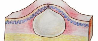
This is because atheromas have a specific structure - they are always surrounded by two capsules. The first is the atheroma’s own capsule, in which its contents are packed, like a bag. The second capsule is external. It separates the atheroma from the tissues surrounding it.
If the atheroma capsule is not removed or at least a tiny fragment of its shell remains under the skin, this 100% guarantees the appearance of a new atheroma in the same place in the future. Hence the myth about its incurability.
In fact, you can get rid of atheroma. It is important to choose the right specialist.
“Before” and on the 12th day after removal of myofibrolipoma of the forehead area. Tips&tricks on the forehead for faster resolution of swelling. Surgeon Vladislav Gladysheva.
Advantages of visiting our clinic
If you need removal of atheroma using the radio wave method in a clinic in St. Petersburg, then you can safely contact us. Our clinic employs highly qualified specialists with extensive experience who can competently diagnose and prescribe effective treatment. During their work, our ENT doctors use the most modern equipment to treat atheroma. We try to find an individual approach to each patient in order to take into account the characteristics of his body as much as possible. Our center employs highly qualified surgeons who can successfully remove atheromas without the risk of relapse. So, by contacting us, you can enjoy the following benefits:
- Consultation with a doctor with extensive experience.
- Competent diagnostics.
- Correct prescription of treatment.
- Treatment of atheroma is carried out using the most advanced technologies and modern equipment.
- Individual approach to each client and polite treatment.
- Favorable prices.
In addition, our center also employs pediatric ENT doctors , during whose appointments your children will not be afraid. You can find out more detailed information about how atheroma is removed using the radio wave method, prices and much more by contacting our employees by phone.
Diagnosis and risk factors
To correctly diagnose a tumor, it is necessary for a doctor to examine and palpate it. If the development of a malignant tumor is possible, the doctor at the clinic will prescribe additional diagnostics (a sample of the contents of the atheroma capsule is taken during surgery).
The most common inflammation of the cyst is in the following cases:
- After the onset of puberty;
- In men, inflammation of the sebaceous gland cyst can occur on any part of the body except the head;
- In women, the head is most often affected;
- In cases where acne appeared during puberty;
- After a heavy tan;
- If there were microtraumas of the skin on the body.
If atheroma appears, its formation cannot be prevented. People who have already had similar neoplasms are also at risk of developing cyst inflammation.
What symptoms do you see a surgeon for:
- Presence of hernial protrusion
- Daggering pains in the abdomen
- Bloating
- Pain in the right hypochondrium
- Bitterness in the mouth
- Nausea
- Presence of neoplasms on the skin
- Swelling and redness of the skin
- Bone fractures and bruises
- Wounds of any location
- Vomit
- Enlarged and painful lymph nodes