Etiology of the disease
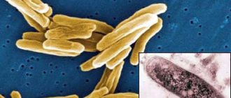
- The causative agent of skin tuberculosis is Mycobacterium tuberculosis, which was first discovered by R. Koch in 1882.
- The tuberculosis bacillus belongs to the family of radiant fungi, the genus of mycobacteria, which, in addition to it, includes the causative agents of leprosy, scleroma and more than 150 species of atypical mycobacteria.
- Mycobacteria reproduce by division and budding. This process lasts 24 hours.
- MBT exhibit significant stability in the external environment. They cannot be frozen out. They remain viable in boiling water for up to 15 minutes. They live in manure for up to 15 years, and up to 1 year in wastewater. When dried, the pathogen remains viable for 3 years.
- Mycobacteria are resistant to phagocytosis (macrophages cannot destroy the mycobacterium, although they begin to fight it - incomplete phagocytosis).
- There are human, bovine and intermediate types of MBT.
- The pathogen has the appearance of an elongated rod of a rather complex structure: a three-layer cell wall and intracellular membrane contain polysaccharides, lipoprotein complexes and proteins. Proteins are responsible for antigenic properties (tuberculin). Polysaccharides play a role in antibody detection. Lipid fractions help MBT resist acids and alkalis.
Rice. 2. Mycobacterium tuberculosis.
Ways of spreading skin tuberculosis
- Patients with tuberculosis spread the infection through sputum, urine, through fistulas and household items. The source of infection is also sick animals and food products contaminated with MBT from sick animals. During the formation of immunity, primary skin tuberculosis occurs (tuberculous chancre, lichenoid tuberculosis of the skin).
- There are a number of diseases that are associated with tuberculosis not directly, but indirectly. These are allergic vasculitis, resulting from allergic immune (“paraspecific”) inflammation in response to infection with mycobacteria (rosacea-like Lewandowski tuberculide).
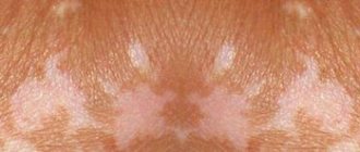
Rice. 3. The photo shows tuberculosis of the skin of the face and neck.
How does the disease progress?
The mechanism of development of such an infection includes several stages:
- entry of the pathogen into the upper respiratory tract (on the mucous membranes), from where, with a weak immune response, they enter the lungs;
- penetration into the alveoli of the lungs and further through their walls (without specific changes);
- entry into the lymph nodes and lymphatic pathways with further reproduction (here the infection can take a latent form and persist without external phenomena);
- circulation of microbacteria through the bloodstream without reproduction (about 2 weeks);
- spread of infection through tissues and organs, manifestation of a primary disease or latent infection;
- formation of immunity - formation of granulomas around microbacteria;
- infection (if the process does not progress, the granuloma becomes covered with connective tissue or resolves, the microbacterium inside it turns into the L-form);
- disease – transition of L-forms into rods.
Pathomorphology
With tuberculosis, a tubercle (tuberculum) appears around the embedded tuberculosis bacilli, the components of which are:
- inside the tubercle there are phenomena of caseous necrosis (damage) of tissues and Mycobacterium tuberculosis (a component inherent only in tuberculosis);
- surrounded by MBT cells specific for any granulomatous disease - lymphocytes, epithelioid cells and Pirogov-Langhans cells (cell proliferation);
- the outer layer (exudative component) is represented by cells macrophages, neutrophils, eosinophils (nonspecific component).
Rice. 4. Histological preparation of a tuberculous tubercle.
With tuberculous skin lesions, tubercular structures with a nonspecific inflammatory infiltrate are more common (there are few or no MBTs in the tubercle). Tuberculous granulomas are characteristic of tuberculous lupus. With allergic vasculitis that occurs in response to exposure to mycobacteria and their decay products, diffuse forms of skin tuberculosis occur. In this case, the vessels of the skin and subcutaneous tissue are affected.
Treatment
Chemotherapy, antibiotics
Bactericidal and bacteriostatic drugs are used to achieve complete recovery. In this case, the correct combination of drugs is important, taking into account the resistance of some bacteria to this type of therapy. First, treatment is aimed at suppressing the growth of bacteria and eliminating their resistance to medications. Residual infection in the cells is then eliminated. Duration of therapy is 6-12 months.
Surgery
Pulmonary resection is practiced as a radical technique. The operation is performed for bronchial stenosis, fibrous-cavernous lesions, pleural empyema, abscesses in the lungs, tuberculomas prone to progression, etc. In addition, decortication is used, that is, removal of fibrous layers, and cavernotomy - cleansing of the opened cavity.
DOTS
A treatment system consisting of several levels: bacterioscopic examination, chemotherapy, anti-tuberculosis treatment.
Sources:
- N.M. Koretskaya. Tuberculosis in children and adolescents in modern conditions // Siberian Medical Review, 2010.
- A.V. Mordyk, E.A. Tsygankova, L.V. Puzyreva, A.A. Turitsa. Tuberculosis in children of the Russian Federation at the present stage // Pediatric pharmacology, 2014, v. 11, no. 3, pp. 27-30.
- https://www.ncbi.nlm.nih.gov/pubmed/24548085 Marais BJ. Tuberculosis in children // J Paediatr Child Health. 2014 Oct;50(10):759-67. doi: 10.1111/jpc.12503. Epub 2014 Feb 19.
- V.N. Krivohizh. Modern methods of early detection of tuberculosis among children and adolescents // Health is the basis of human potential: problems and solutions, 2013, pp. 570-585.
The information in this article is provided for reference purposes and does not replace advice from a qualified professional. Don't self-medicate! At the first signs of illness, you should consult a doctor.
Primary cutaneous tuberculosis
This form of the disease is extremely rare. The disease develops during the development of primary tuberculosis. Children under 10 years of age are most often affected. Initially, a papule appears reddish-brownish in color. Next, an ulcer (tuberculosis chancre) appears in the center of the papule. Peripheral lymph nodes increase in size. Often ulcerate. Ulcers take a long time to heal. In their place, thin scars appear. With a weakened immune system, the disease returns again, disfiguring the body with scars and scars.
Types and forms of pathology
According to the period of occurrence in medicine, stages are distinguished:
- infiltration;
- decay;
- contamination;
- resorption;
- seals;
- the appearance of scars;
- calcification.
| Form | Peculiarities |
| Chronic and early | It occurs against a background of body temperature up to 40℃, cough, pain in the lungs, uneven breathing, wheezing, decreased appetite, and loss of strength. |
| Respiratory damage | Includes pathologies:
|
| Other localizations | There may be lesions:
|
Secondary tuberculosis of the skin
The disease is represented by a variety of localized and disseminated forms that appear in previously infected people. Tuberculous lupus accounts for up to 75% of all cases.
In the last 10 years, there has been a predominance of disseminated forms of cutaneous tuberculosis.
Ordinary or vulgar tuberculous lupus (Lupus vulgaris)
In the recent past, lupus was the most common form of cutaneous tuberculosis. Today, disseminated forms are more often recorded. MBT penetrate into the skin from regional lymph nodes through the lymphatic tract and hematogenously (with the bloodstream). The disease often occurs in childhood, lasts a long time, with periodic exacerbations, and spreads slowly.
Symptoms of the disease
The disease affects the skin of the nose, face, neck, red border of the lips, mucous membranes of the mouth and eyes. The skin of the extremities is rarely affected. Tuberculous tubercles merge and form lupomas. Their color is yellowish-rusty. Size – up to 0.75 mm. At first, the lupomas are located deep, and then they begin to protrude above the skin.
The shape of the lupoma is round, the consistency is soft, with significant pressure from the probe, the elements of the lupoma rupture, causing pain and bleeding. Lupomas often merge. Their surface is smooth and shiny. If you press a glass slide onto the affected area, the lupomas become the color of “apple jelly” (apple jelly symptom). With a favorable outcome, resorption begins in the center of the tubercles and replacement of the damage with thin skin in the form of tissue paper.
Rice. 5. Photo of lupoma.
Rice. 6. Symptom of “apple jelly” in tuberculous lupus
Rice. 7. Lupus vulgaris.
Rice. 8. Consequences of Lupus vulgaris.
Forms of tuberculous lupus
Tuberculous lupus of the mucous membranes
The most severe form of Lupus vulgaris. The disease affects the mucous membranes of the nose, eyes and mouth. Initially, red-yellow formations (plaques) appear on them. Their surface has a grainy appearance, reminiscent of fish eggs. Over time, the process affects the cartilage of the nose and ears. Next comes spontaneous rejection of damaged dead tissue, which ends in permanent facial disfigurement.
Rice. 9. The photo shows a lesion of the tongue due to tuberculous lupus.
Rice. 10. Damage to the oral mucosa.
Tumor form of tuberculous lupus
Tuberculous tubercles merge, forming a tumor-like formation up to 3 cm in diameter. As the process progresses, a breakdown of the underlying tissue appears, accompanied by damage to the cartilage and lymph nodes.
Rice. 11. The photo shows a tumor form of tuberculous lupus.
Flat form of tuberculous lupus
Tuberculosis foci merge, but the affected area does not protrude above the level of the skin. As the disease progresses, ulcers appear with uneven edges and a granular bottom.
Rice. 12. Flat shape of Lupus vulgaris.
Psoriatic form of tuberculous lupus
Tuberculosis foci merge. The surface of the damage is covered with many small scales.
Rice. 13. Psoriatic form of Lupus vulgaris.
Rice. 14. Psoriatic form of Lupus vulgaris.
Exfoliative (scaly) form of tuberculous lupus
Tuberculosis foci merge. The surface of the damage is covered with many large whitish scales, which adhere tightly to the underlying tissues. The appearance of the lesion resembles a butterfly.
Rice. 15. Exfoliative (scaly) form of Lupus vulgaris.
Rice. 16. Exfoliative (scaly) form of tuberculous lupus.
Rice. 17. Exfoliative (scaly) form of tuberculous lupus.
Sarcoid-like form of tuberculous lupus
Merging, tuberculosis foci form tumor-like formations of a reddish color - lupus carcinoma. The process is prone to malignancy.
Tuberculous lupus is the most common form of cutaneous tuberculosis. It is characterized by a chronic, slow progressive course and a tendency to melt tissue. The disease usually begins in childhood and lasts for years or decades, sometimes throughout life.
Clinical picture
Tuberculous lupus is most often localized on the face, especially on the nose (in 80% of cases), cheeks, upper lip, less often on the neck, torso, extremities, and often on the mucous membranes (70% of patients). Pulmonary tuberculosis is found in 5-10% of lupus patients, and tuberculosis of bones and joints occurs with the same frequency. Skin infection occurs predominantly by hematogenous or lymphogenous route.
The disease begins with the appearance of lumps - tubercles ranging in size from a pinhead to a pea, brown-reddish in color with a yellowish-brown tint, soft, doughy consistency with a smooth, slightly shiny surface. Lupoms are poured out in a group, at first they are located in isolation, then they merge; along their periphery there is always a stagnant-hyperemic zone.
The “apple jelly” phenomenon (diaskopia, obelmus). If you press a glass slide onto the lesion, yellow-brown spots appear. This symptom is explained by the squeezing out of blood from dilated blood capillaries and the transmission through the epidermis of magnifying glass with a granulomatous structure
“Probe” symptom (Pospelov’s sign). If you press a button-shaped probe on the tubercle (lupu), then it is easily immersed into the depths of the tissue. In this case, light bleeding and minor pain occur. The symptom is more pronounced with fresh lupoma. This occurs due to caseous necrosis, accompanied by the death of collagen and elastic fibers.
There are several clinical forms of tuberculous lupus: macular (Lupus planus maculosus), tubercular (Lupus planus tuberculosus), tumorous lupus (Lupus tumidus), erythematous, squamous-hyperkeratotic, verrucous and ulcerative.
- In some cases, destructive ulcerative changes involve deep underlying tissues (cartilage, bones, joints) in the process with the formation of mutilations, fibrous, keloid scars and disfigurement of the nose, ears, fingers, eyelids, limbs (Lupus mutilans).
- When the mucous membranes are affected (Tuberculosis luposa mucosae), small bluish-red tubercles the size of a millet grain are formed, which, due to their close grouping, give the affected area a peculiar granular appearance. Diagnosis is facilitated by the simultaneous presence of manifestations on the skin. When the mucous membranes of the nose are damaged, a soft, lumpy, bluish infiltrate is formed, which disintegrates to form an easily bleeding ulcer.
- Pityriasiform lupus (Lupus vulgaris pityriasiformis) is characterized by pithiarisiform scaling, and lupus psoriasiformis (Lupus vulgaris psoriasiformis) is characterized by silvery-white scales.
- There are other types of the disease: serpiginating, exfoliative, rupioid, crustose, etc.
Tuberculous lupus can be complicated by erysipelas, lymphangitis, pyoderma, and the development of skin cancer (Lupus carcinoma).
Histologically, epitheloid and giant cells surrounded by lymphocytes are detected in the dermis. Giant Langhans cells are located in the central part of the tubercle, in the peripheral zone there are lymphocytes and plasma cells in large numbers; caseous necrosis is poorly expressed. Mycobacterium tuberculosis is detected with difficulty and in small quantities.
Diagnosis and differential diagnosis
Differential diagnosis is carried out with tubercular syphilis, tuberculoid form of leprosy, tuberculoid form of cutaneous leishmaniasis, actinomycosis, discoid form of lupus erythematosus.
Colliquatic cutaneous tuberculosis (scrofuloderma)
After tuberculous lupus, this form of skin tuberculosis is in second place in terms of frequency of the disease. It got its name from the Latin scrofulae - swollen lymph nodes of the neck and colliquescere - to melt. MBT enter the skin from infected lymph nodes through the lymphatic ducts. Cracks and ulcerations appear above the area of enlarged lymph nodes. The process is localized on the lateral parts of the neck, chest and collarbones. Young women are predominantly affected.
Symptoms of the disease
At the beginning of the disease, dense, painless nodules appear, which quickly increase in size, forming nodes tightly fused to the underlying tissues. Their sizes range from 3 to 5 cm. The skin over the lymph nodes acquires a bluish tint. Over time, the node becomes soft and opens. A cold abscess forms (suppuration without any manifestations of an inflammatory reaction). Pus with blood clots and pieces of destroyed (necrotic) tissue begins to be released from the fistula tracts. The ulcer has soft edges. The bottom of the ulcer is covered with a yellowish coating. Numerous granulations are visible. As the ulcer heals, irregularly shaped scars appear, which are connected by jumpers and bridges. The top of the scars is covered with papillary processes.
Rice. 18. Scrofuloderma.
Rice. 19. Scrofuloderma.
Rice. 20. Scrofuloderma.
Combination of lichenoid and colliquative tuberculosis of the skin (scrofuloderma)
Tuberculosis (from the Latin tuberculum - tubercle, English tuberculosis) is an infectious disease caused by Mycobacterium tuberculosis, characterized by pronounced clinical heterogeneity and the development of a specific inflammatory reaction in the organs and tissues of the human body [1]. The threat of a pandemic, a high mortality rate, an increase in cases of multidrug-resistant tuberculosis and the economic damage caused by the disease make the incidence of tuberculosis one of the most important problems in global health [2, 3]. The most common active form of tuberculosis affects people of working age from 18 to 44 years (in 2014 - 62.3% of the total number of patients) [4]. According to the World Health Organization, tuberculosis has the highest morbidity and mortality rates, with 8 million new cases of tuberculosis and 3 million deaths associated with tuberculosis reported annually [5]. In the Russian Federation in the 21st century. the peak incidence was observed in 2008. However, by 2014, the decrease in the incidence of tuberculosis compared to 2008 was 30.1% (85.1 per 100,000 population) [4]. According to the literature, the mortality rate from tuberculosis in the Russian Federation in 2014 decreased by more than half due to an increase in the mortality rate of patients with co-infection with HIV and tuberculosis [4]. It should also be noted that multidrug-resistant tuberculosis is increasing worldwide [5]. The Russian Federation still ranks first in identifying cases of multidrug-resistant tuberculosis: 24.2 per 100,000 population in 2013 and 24.8 per 100,000 population in 2014 [4]. In connection with the actualization of the problem of tuberculosis in general, interest in its extrapulmonary form has increased in recent years [6, 7], including cutaneous tuberculosis (STB). However, due to less contagiousness and pathogenesis, incidence rates of extrapulmonary tuberculosis are at a significantly lower level compared to respiratory tuberculosis [2]. There is no convincing data on an increase in the number of cases of TC [6, 7]. There is no generally accepted classification of TC. According to the National Guidelines for Phthisiology 2014, cutaneous tuberculosis is divided into 2 main groups (Table 1) [1]: • true cutaneous tuberculosis (localized, granulomatous or bacterial); • skin lesions as a result of allergic immune (paraspecific) inflammation, mainly in the form of allergic vasculitis, called disseminated, hyperergic cutaneous tuberculosis and classified by Daria as “tuberculides”.
In the 2013 National Guidelines for Dermatovenereology, miliary tuberculosis and lichenoid tuberculosis are classified as disseminated forms [8]. According to the literature, TB ranks 5th among all locations of extrapulmonary tuberculosis (osseous-articular, genitourinary and digestive systems, lymph nodes) [9]. This distribution is associated with the fact that in the vast majority of cases, TB is secondary to tuberculosis of internal organs [1], which suggests the possibility of pathomorphism of tuberculosis infection and the appearance of erased and atypical, difficult to diagnose forms of skin lesions [6, 7, 10]. Thus, according to the literature, patients with undiagnosed TC have been treated for a long time for chronic pyoderma, allergic vasculitis, lupus erythematosus, perioral dermatitis, lichen planus, pruritus, eczema, Beck’s sarcoidosis, rubrophytosis, Dühring’s dermatitis herpetiformis, migratory thrombophlebitis [10] . Complicating the diagnostic search are the so-called paraspecific tissue reactions or “masks” of tuberculosis that occur in response to tuberculosis infection and are characterized by various nonspecific changes in organs and tissues. Lesions of the skin and mucous membranes are often represented by erythema nodosum and keratoconjunctivitis, rarely by hemorrhagic vasculitis and panniculitis. This phenomenon is observed mainly in primary and disseminated tuberculosis [11]. There have also been changes in the general structure of TC incidence. So, in the middle of the twentieth century. the most common were tuberculous lupus and colliquative TC; currently, on the contrary, predominantly disseminated forms of TC are recorded [3, 10]. Based on materials from the clinic of skin diseases of the First Moscow State Medical University named after. THEM. Sechenov analyzed the dynamics of clinical forms of tuberculosis over 70 years, from 1895 to 1964, and found that during this period there were 525 patients with various forms of skin tuberculosis in the clinic. Of these, 70.2% suffered from lupus vulgaris, 5.3% from scrofuloderma, 10.2% from papulonecrotic tuberculosis, 11.6% from erythema induratum, and other forms accounted for 2.7% [12]. From 1991 to 2000, the clinic recorded only 2 TB patients, one of whom was a resident of the Far East. After a long period of absence of patients with both TC and paraspecific tissue reactions for 6 months. In 2015, the clinic recorded 5 cases of TC, of which 1 case was scrofuloderma, 1 case was papulonecrotic tuberculosis associated with pulmonary tuberculosis, 2 cases were erythema induratum, 1 case was TC associated with kidney tuberculosis as a complication of immunosuppressive therapy. cyclosporine therapy; in 1 – lupus vulgaris. The patient with papulonecrotic skin tuberculosis is a resident of Dagestan, the patient with erythema induratum is a resident of Astrakhan, the other 3 patients live in Moscow. Thus, the problem of TC remains relevant and requires further study. Causative agents of skin tuberculosis: M. tuberculosis, M. bovis, in rare cases - bacilli Calmette-Guerin (BCG). TC is transmitted through the skin - primary infection, and also spreading through the lymphogenous or hematogenous route when internal organs are damaged - secondary infection. The most contagious form of TC is scrofuloderma (colliquatic TC). The term "scrofuloderma" was first introduced by Jadasson. French authors called this disease "gommes scrophuleuses" or "gommes tuberculeux". A feature of this pathology is the development of gummous nodes in the subcutaneous tissue, which undergo caseous softening and ulceration resulting in a bridge-shaped scar. There are two clinical varieties: primary, characterized by the initial formation of tuberculous granulomas in the skin, and secondary, as a result of the spread of the pathological process from regional lymph nodes [6, 7]. Primary scrofuloderma can be localized on any part of the skin, secondary - mainly in the parotid, submandibular and supraclavicular areas, on the lateral surfaces of the neck [6, 7]. The rashes are initially presented as small, dense, mobile, painless bumps the color of normal skin, which, quickly increasing in volume, turn into sharply demarcated nodes that are not fused with the surrounding tissues. Gradually, the nodes acquire a bluish tint, soften, and the skin over them loses mobility. They can remain in this state for a long time, forming “cold abscesses,” or open with the formation of deep ulcers, the release of purulent-necrotic contents, and the formation of fistulas and pockets. The elements resolve into bridge-shaped disfiguring scars [8, 9], along the periphery of which new tubercles and nodes form, as a result of which the lesion takes on a serpentine character. There is a pronounced evolutionary polymorphism [6, 7]. The pathomorphology of primary and secondary scrofuloderma is similar. The epidermis is intact, in the upper parts of the dermis the changes are nonspecific: foci of necrobiosis, infiltrate of mononuclear cells, in the lower parts of the dermis and subcutaneous fatty tissue - tuberculoid granulomas consisting of epithelioid, giant cells and lymphocytes, with caseous necrosis in the center, when stained according to Romanovsky - Giemsa detects mycobacteria [9, 13, 14]. To confirm tuberculosis lesions, the latest diagnostic methods are used, such as the Diaskin test, ultrasound examination of soft tissues, epiluminescence dermatoscopy, polymerase chain reaction, and immunohistochemical detection of Mycobacterium tuberculosis antigens [2]. Treatment of TB is carried out together with TB specialists with specific anti-tuberculosis drugs, the prognosis is favorable. A feature of this form of tuberculous skin lesions is a long-term chronic relapsing course with a tendency to spontaneous scarring of ulcers [9]. Differential diagnosis of TC is carried out with sarcoidosis, papulonecrotic angiitis, secondary and tertiary syphilis, lymphocytoma, B-cell lymphoma of the skin, leprosy, discoid lupus erythematosus, deep mycoses, leishmaniasis and other dermatoses, depending on the clinical form of TC. The prognosis is relatively favorable in the absence of resistance to anti-tuberculosis therapy [9]. Considering the rarity of the pathology, we present our own clinical observation of a case of colliquative tuberculosis of the skin.
Clinical case
Patient B., 67 years old, was in the Moscow City Scientific and Practical Center for Combating Tuberculosis with complaints of rashes on the skin of the upper body, moderate general weakness, accompanied by an increase in body temperature in the evening to 37.5 ° C, loss of appetite.
Over the past 2 years, the patient notes a weight loss of 22 kg. From the life history
it is known that before retiring she worked in a hot shop and had contact with acids and dust.
Concomitant diseases: varicose veins of the saphenous veins of both lower extremities, chronic venous insufficiency of II–III degree; bilateral retinal angiopathy, initial cataract, chronic blepharoconjunctivitis; chronic iron deficiency anemia; vascular (toxic) hypoacusia; hypertension stage II, degree I, risk of degree 2. From the medical history.
About 3 years ago, the patient first noticed the appearance of a tumor-like formation in the supraclavicular region on the left.
The local surgeon assessed the formation as an abscess and opened it. No scarring of the ulcer was observed. Subsequently, at the site of the opened nodes, the appearance of deep ulcerative defects like fistulas was noted in the axillary region on the left, on the anterior chest wall on the left. Antibacterial therapy was carried out periodically. 1.5 years after the onset of the disease, a computed tomography scan of the chest organs was performed, which revealed exudate in the left pleural cavity and destruction of the second rib on the left. She was consulted at the Moscow City Scientific and Practical Center for the Fight against Tuberculosis at the State Budgetary Healthcare Institution of the Department of Healthcare of the City of Health, and a diagnostic and treatment puncture of the left pleural cavity was performed, during which the process could not be identified, and no growth of secondary flora was obtained. An abscess of the anterior chest wall was punctured and acid-fast mycobacteria were detected using fluorescent microscopy. Objectively
.
The consciousness is clear, the physique is asthenic. Body temperature rises in the evening to 37.5° C. The skin outside the lesions is pale; visible mucous membranes are pale pink and clean. The state of the muscular system corresponds to age. Active and passive movements in the joints are fully preserved. On auscultation over the right lung, breathing is harsh, on the left it is weakened mainly in the lower parts, there is no wheezing. Percussion sound with a boxy tint in the lower sections on the right, on the left - shortening of the percussion sound in the lower sections. Respiration rate – 20 per minute. Heart sounds are clear, rhythmic, no murmurs; heart rate - 72 beats per minute. Blood pressure – 120/80 mm Hg. The abdomen is soft and painless. The liver protrudes 1 cm from under the edge of the costal arch. The spleen is not palpable. Kidneys: Pasternatsky’s sign is negative on both sides. Urination is free and painless. No focal neurological symptoms were identified. Local status.
The skin process is chronic inflammatory in nature. The rashes are multiple, localized mainly on the skin of the trunk and limbs. They are represented by brownish-pink dense lichenoid tubercles, with clear boundaries, scalloped outlines, arranged in the form of arcs or rings, leaving the central area unaffected. At the site of regressed rashes there is pigmentation with superficial atrophy, there are no scars. On the left anterior chest wall, in the area of attachment of the third rib to the sternum, a spherical node with a diameter of 2 cm, dense consistency, mobile, painless, the skin over it is not changed. In the supraclavicular region on the left, in the left axillary region and on the anterior chest wall on the left there are numerous retracted bridge-shaped scars up to 3 cm in length and deep ulcerative defects with scanty thick discharge. The peripheral groups of lymph nodes are enlarged, the nodes are not fused to the skin, and are mobile. Particularly large conglomerates are identified in the left axillary region. The nail plates of the hands and feet are not changed. There are no subjective sensations (Fig. 1). Preliminary diagnosis: a combination of lichenoid and colliquative TC. In order to verify the diagnosis, a histological examination of the affected area of the skin was carried out, as well as an examination to identify concomitant pathologies. The results of histological examination demonstrated the following changes: in the dermis there were multiple, locally confluent macrophage-giant cell granulomas without necrosis. The morphology fits into the picture of granulomatous inflammation (Fig. 2).
As a result of laboratory tests, no gross pathology was identified. An electrocardiogram and results of a study of pulmonary function were unremarkable. X-ray
. In the left pleural cavity there is a shadow of exudate up to the anterior segment of the IV rib, enveloping the entire lung; at the level of the II rib there is a thickening of the pleura of a homogeneous structure, up to 15 mm thick; local pneumosclerosis in S1-2 on the left and S4-5 on the right; There is an increase in intrathoracic lymph nodes. Computed tomography of the chest: CT signs of nodular formation? Ultrasound examination of the thyroid gland reveals diffuse changes in thyroid tissue and lymphadenopathy. A comprehensive examination made it possible to diagnose a combination of lichenoid and colliquative TC (secondary scrofuloderma). Thus, it was revealed that patient B. had primary pulmonary TB in the past, as evidenced by local pneumosclerosis in S1-2 on the left and S4-5 on the right. Such indurative areas, as a manifestation of previous pulmonary TB, are biologically active. It is known that such residual tuberculous changes, the diameter of which is more than 1 cm and the number of which exceeds 5, and the hilar localization of the process are potentially dangerous, especially in people over 60 years of age [15, 16]. This is due to the fact that typical and modified variants of mycobacteria, especially the L-form, persist in these indurative areas [17]. At the age of 67 years, patient B. experienced endogenous reactivation of tuberculosis infection in a favorable epidemiological situation; acute progressive pulmonary tuberculosis arose with damage to the lymph nodes, pleuropneumonia and lymphohematogenous generalization of the disease to the skin. In the skin, the process is characterized by a combination of lichenoid and colliquative skin tuberculosis (secondary scrofuloderma).
Ulcerative skin tuberculosis (Tuberculosis cutis ulcerosa)
The disease most often affects men who already had tuberculosis of internal organs. Tuberculosis bacilli enter the skin from the urine, feces or sputum of the patient himself, affecting areas of natural external openings: the skin around the anus, glans penis, nose, area around the mouth, mucous membrane of the tongue.
Symptoms of the disease
Merging, tuberculous tubercles form small yellowish nodules. Over time, the nodules suppurate and open with the formation of painful ulcers that make natural acts difficult.
The ulcers have soft, undermined edges. The color of the ulcers is pale red. The size of the ulcers is up to 1.5 cm, the bottom is granular. Javas often bleed. Sharply painful. As the disease progresses, tuberculous tubercles reappear at the bottom of the ulcers and around them. The tissues are destroyed, forming yellowish microabscesses, the so-called “Trel grains”, containing a large amount of MBT. The disease is difficult. Ulcers heal slowly. In their place, thin scars form, located below the skin level (atrophic scars).
Rice. 21. Ulcerative tuberculosis.
Warty skin tuberculosis (Tuberculosis cutis verrucosa)
Warty skin tuberculosis occurs among veterinarians and slaughterhouse workers who come into contact with the corpses of animals with tuberculosis. This form of the disease is rare. The skin of the back of the hand or feet is affected. The skin of patients is affected by tuberculosis from constant contact with infected sputum.
Symptoms of the disease
First, tubercles appear, the skin above which becomes bluish in color. Gradually, the tubercles merge with each other, forming dense infiltrates of a bluish color. Over time, they become covered with warty growths, dense scales and cracks. The lesion is surrounded by a bluish-red shaft, followed by a zone of shiny skin. During the healing process, the horny masses are rejected, and scars appear at the site of infiltration. The course of the disease is long-term.
Symptoms and first signs of tuberculosis in children
First of all, tuberculosis in children is manifested by such clinical signs as:
- weakness;
- stopping weight gain;
- irritability;
- fatigue during school hours;
- absent-mindedness;
- lagging behind peers in studies;
- body temperature – up to 37.5°C;
- enlarged lymph nodes;
- sweating;
- chills.Source: N.M. Koretskaya Tuberculosis in children and adolescents in modern conditions // Siberian Medical Review, 2010
Forms of tuberculosis and early signs
| Form | Symptoms |
| Tuberculosis of the bronchial glands | Cough, elevated body temperature for a long time, lethargy, fatigue, pallor, thinness. |
| Pulmonary form | Cough, shortness of breath, weakness after sleep, apathy, decreased performance, absent-mindedness, unhealthy blush, thinness, pale skin, sweating, chills, dry (and then “wet”) cough for more than 3 weeks, sputum with blood. |
| Lymph node involvement | An increase in the size of the lymph nodes, their softening, suppuration (the contents leak out), the formation of fistulas in places where pus comes out, scrofuloderma (skin lesions in the form of tumors with subsequent release of the contents). |
| Involvement of bones and joints in the process | Slow development, pain when moving, changes in gait, lameness. |
| Damage to the meninges (tuberculous meningitis) | Anxiety, lethargy, loss of appetite, bad mood, headaches, fever, vomiting, convulsions. |
| With damage to the gastrointestinal tract | Constipation or diarrhea, increased body temperature, blood in the stool, pain. Source: A.V. Mordyk, E.A. Tsygankova, L.V. Puzyreva, A.A. Turitsa Tuberculosis in children of the Russian Federation at the present stage // Pediatric pharmacology, 2014, v. 11, no. 3, pp. 27-30 |
Clinical manifestations by age of children
| Age | Symptoms |
| Infancy (up to one year) | Low mobility, apathy, weakness, attacks of suffocation or coughing, retraction of part of the chest, weight loss (including muscle mass), loss of appetite, cessation of crying, insomnia. Early detection and diagnosis are extremely important, because at this age this pathology is most dangerous. |
| 5-8 years | Decreased activity, weakness, lack of sleep and appetite, loss of body weight, cough, condensation of the chest. |
| 8-15 years | Rapid appearance of pain in the lungs against a background of apathy and weakness, active urge to cough, shortness of breath even at rest, thinning or discoloration of the skin, the appearance of cracks, wounds, hemoptysis, changes in lymph nodes in size and structure, intoxication of the lungs (at the last stage) . |
Manifestations of the chronic form:
- sleep disorders;
- liver enlargement;
- lagging behind peers in physiological development;
- dry and pale skin;
- mild euphoria.
Papulonecrotic tuberculosis of the skin (tuberculosis cutis papulo-nectrotica)
Papulonecrotic tuberculosis of the skin is more often recorded in young people. The disease is manifested by the appearance of papules, the size of which does not exceed 3 cm in diameter. The skin of the buttocks, abdomen, extensor surfaces of the limbs and chest is affected. The skin over the papules becomes pinkish-bluish in color. Over time, the papules become ulcerated. At the site of the ulcers, a grayish-white crust appears, which is replaced by a whitish scar.
Rice. 25. Tuberculosis cutis papulo-nectroticа. Multiple bluish-purple infiltrates and nodes on the legs.
Rice. 26. Papulonecrotic tuberculosis of the skin of the legs. Multiple bluish-purple infiltrates and nodes on the legs.
Rosacea-like Lewandowski tuberculosis
This form of the disease occurs as an allergic vasculitis. Erythema (redness) and telangiectasias (numerous dilated small vessels) appear on the skin of the face. Gradually the skin acquires a bluish-reddish tint. Rashes (tuberculids) are dense formations up to half a centimeter in diameter, without necrotic elements. During healing, atrophic scars form at the site of tuberculides.
Rice. 27. The photo shows Lewandowski’s rosacea-like tuberculosis.
Lichenoid tuberculosis of the skin (Tuberculosis cutis lichenoides seu lichen scrofulosorum)
Lichenoid tuberculosis of the skin often develops in weakened children, less often in adults with tuberculosis. The disease is manifested by the appearance of tubercles, which are covered with light gray scales. The tubercles are located symmetrically, most often on the skin of the abdomen and limbs. After healing, pigmentation or small scars remain at the site of the rash.
Rice. 28. The photo shows lichenoid tuberculosis of the skin.
Indurative tuberculosis of the skin (indurated erythema)
This form of the disease manifests itself in 2 varieties - Bazin's erythema nodosum and Hutchinson's ulcerative erythema. The disease often affects patients with tuberculosis of internal organs.
Erythema nodosum of Bazin
The disease is more often registered in women aged 16 to 40 years. Dense nodes or flat, extensive infiltrates that appear on the skin of the legs, thighs, buttocks and upper extremities are bluish-red in color. The size of the seals is 3 – 8 cm. Seals and infiltrates are often located symmetrically, in the deep layers of the skin and subcutaneous fatty tissue. With regression of tuberculous elements, ring-shaped atrophic areas and pigmentation remain.
Hutchinson's ulcerative erythema
Sometimes the lesions merge, forming an extensive lesion with ulcerations in the center and dirty gray granulations. This form is named after the English dermatologist who first described it - Hutchinson's ulcerative erythema. Without treatment it lasts for a long time, sometimes even years. After healing, sunken pigmented scars remain.
Rice. 29. The photo shows indurative tuberculosis of the skin (indurated erythema of Bazin).
Rice. 30. Ulcerative erythema of Hutchinson.
Diagnosis of skin tuberculosis
Rice. 31. View of Mycobacterium tuberculosis in the light of a fluorescent microscope.
The diagnostic process consists of the following components:
- Bacteriological diagnosis (detection of MBT in discharge from ulcers and lymph node punctures).
- Diagnostic biopsy followed by histological examination, which allows to identify the morphological components of the tuberculous tubercle.
- Detection of tuberculosis of internal organs.
- Detection of scars on the skin as a result of a previous tuberculosis lesion.
- Tuberculin diagnostics.
- Trial treatment.
Diagnostic biopsy together with bacteriological examination is the most significant method in diagnosing tuberculosis.
Dermatologists are prescribed to detect skin tuberculosis. Knowledge of risk groups, which include patients in this category, early symptoms of the disease and diagnostic methods helps doctors to detect the disease in a timely manner.
Identification of the pathogen in the early stages of the disease will allow the patient to be successfully cured.
Pulmonary tuberculosis
Which doctors should I contact?
Tuberculosis is treated by phthisiatricians, but with the first complaints they most often turn to a general practitioner, pediatrician or pulmonologist, who, after an examination and diagnostic measures, will recommend a consultation with a phthisiatrician.
Treatment of pulmonary tuberculosis
The success of treatment largely depends on early diagnosis of the disease. Tuberculosis therapy is long-term and ranges from 4 months to several years.
Stopping medication on your own, skipping or reducing the dosage can cause resistance, that is, resistance of mycobacteria to antibiotics. During treatment, you must stop smoking and drinking alcohol.
Tuberculosis therapy involves a combination of several antibacterial drugs. Basically, all medications are taken in tablet form, but at the onset of the disease or in severe cases they require intravenous administration.
Tuberculosis treatment is carried out both at home and in a specialized hospital. Hospitalization is indicated for the initial detection of tuberculosis, for the treatment of severe forms of tuberculosis and for surgical treatment of pulmonary tuberculosis.
During treatment for tuberculosis, it is very important to eat well, getting the required amount of calories, vitamins and nutrients from food.
The calorie content of food should be 10% higher than with a normal lifestyle. Dairy products, lean meat, fish, and nuts are rich in protein, which is necessary to strengthen the immune system. Be sure to include vegetables, herbs, and whole grains in your menu. You should avoid fast food, fatty, smoked foods, sugar and sweet carbonated drinks.
Complications
Nonspecific complications characteristic of many pulmonary diseases include hemoptysis, pulmonary hemorrhage, and spontaneous pneumothorax. Spontaneous pneumothorax is the entry of air into the pleural cavity due to damage to lung tissue. The presence of air in the pleural cavity compresses the lungs and interferes with the normal breathing process. Cases have been described where the formation of bronchopleural fistulas leads to pneumothorax.
Rare but dangerous complications include: atelectasis (collapse of part of the lung, which ceases to participate in gas exchange and the breathing process), lung abscess (formation of an abscess in the lung tissue), amyloidosis of internal organs (as a result of a long-term inflammatory process, protein metabolism is disrupted in organs amyloid protein begins to be deposited, as a result of which their normal functioning is disrupted), chronic pulmonary failure.
Specific complications characteristic of pulmonary tuberculosis include: tuberculosis of the bronchi, trachea, larynx, root of the tongue; tuberculous pleurisy; tuberculous empyema is an accumulation of pus in the pleural cavity.
Prevention of pulmonary tuberculosis
Specific prevention includes vaccination.
Vaccination is carried out for newborn children in the maternity hospital on the 3rd–7th day in the absence of contraindications.
The vaccine is administered intradermally, after which a local tuberculosis process is formed, which is not dangerous to general health. Subsequently, the body develops specific immunity against mycobacteria. This means that a vaccinated child with good post-vaccination immunity, when encountering mycobacteria, either does not become infected or suffers a mild infection.
Immunity acquired after BCG vaccination lasts an average of 5 years. Repeated vaccinations are carried out at 7 and 14 years of age.
Prevention of tuberculosis in adults includes annual medical examination with fluorography of the lungs.
Sources:
- Karachunsky M.A. Differential diagnosis of pulmonary tuberculosis // Pulmonology and allergology. – T.1. – 2005. P. 6–9.
- Federal clinical guidelines for the diagnosis and treatment of respiratory tuberculosis. Russian Society of Phthisiatricians. – 2014. – P. 43.
IMPORTANT!
The information in this section cannot be used for self-diagnosis and self-treatment. In case of pain or other exacerbation of the disease, diagnostic tests should be prescribed only by the attending physician. To make a diagnosis and properly prescribe treatment, you should contact your doctor.