Causes of appearance How pinguecula manifests itself Diagnostics Treatment Advantages of treatment at MGC Cost of treatment Video of surgery to remove pinguecula Video of our doctor Complications of the disease
A pinguecula is a benign round-shaped formation located on the conjunctiva of the eye. The pinguecula usually has a characteristic appearance: small in size (no larger than a match head), yellowish in color, slightly protruding above the surface of the conjunctiva, most often found on the nasal side, but can also be located outside the cornea. The disease can be unilateral or bilateral.
Treatment
Pinguecula has a long chronic course and a favorable prognosis, is not prone to degeneration and in most cases does not require any treatment. If you experience symptoms of eye irritation or dryness, you should make an appointment with your doctor to determine the exact cause of these complaints. Based on the results of the examination, if necessary, the doctor may prescribe medications to moisturize the eyes (“artificial tears”), anti-inflammatory eye drops if there are signs of an inflammatory reaction.
If the pinguecula is inflamed, patients using contact lenses should temporarily stop wearing them so as not to injure the mucous membrane of the eyes.
We do not recommend that patients use traditional methods of treating pinguecula (blueberries, aloe, honey, etc.), because they are not effective and often cause allergic reactions, aggravating the condition.
If the pinguecula causes physical or psychological discomfort to the patient, it can be removed surgically or using a laser. The operation is performed on an outpatient basis and does not require special preparation.
What is a pinguecula of the eye?
A pinguecula is an elastic, compacted yellowish formation that appears in the nasal part of the conjunctival membrane of the eye (in the area of contact with the cornea). It is a mistake to associate this pathology with various types of tumors, since only in rare, especially advanced cases, such a change in the conjunctival membrane can become malignant.
Pinguecula is classified as a benign disease, usually affecting both eyes but not affecting visual acuity.
The worldwide prevalence of this eye pathology is extremely high. Doctors note that with pinguecula of the eye, the causes and treatment may vary. As a rule, the disease manifests itself in the form of a whitish or yellowish growth. If the neoplasm occurs on the protein membrane, it is called a pinguecula, but when it is localized on the cornea, forming a wing-like structure, it is called a pterygium. In fact, pinguecula represents only a slight change in eye tissue due to the increased content of fats and proteins in the body, so often the growth does not affect the cornea and does not cause pain - the disease is asymptomatic. But it cannot be said that the pathology is entirely harmless - it brings aesthetic inconvenience, since it is noticeable to others, and also often causes chronic irritation of the organs of vision.
In general, the disease is not dangerous. It rarely causes dysfunction of the visual apparatus and even less often develops into a malignant tumor, but sometimes it is necessary to treat the pinguecula. In the article we will consider in what cases and what therapy doctors prescribe.
Information about the described pathology first appeared in 1550 BC. It is interesting that from those ancient times, records of the Egyptians have been preserved, who described the pinguecula as “specific fatty deposits in the eye.” Modern doctors believe that the disease can develop due to genetic predisposition or occur in an isolated form. Pathology is diagnosed equally in women and men. People living in countries with hot and dry climates are at particular risk.
Advantages of treatment at MGK
All patients of the Moscow Eye Clinic are guaranteed to receive attentive attention from medical staff and a choice of the most appropriate treatment methods.
The clinic is equipped with the most modern medical equipment, which ensures the highest level of diagnosis of any eye pathologies, even at the initial stage of the disease, and the possibility of making an erroneous diagnosis is excluded.
The Moscow Eye Clinic employs leading domestic specialists who have sufficient experience in treating eye diseases. Our specialists love their work and always strive to achieve the best treatment results.
Pinguecula removal in the clinic is performed on an outpatient basis, on a one-day basis. The operation is painless, performed under local drip anesthesia, and has a short recovery period (several hours). After removal of the formation, our specialists will give recommendations to prevent the reappearance of pingueculae.
All questions you are interested in can be asked by phone in Moscow and the MGK hotline number (the call is free) or online, using the appropriate form on the website.
Author:
Yakovleva Yulia Valerievna 5/5 (1 rating)
Honey. portal:
Symptoms
As a rule, the pterygoid fold grows slowly and, even when already noticeable, may not cause noticeable discomfort for a long time. The growth usually begins in the corner of the palpebral fissure, most often from the side of the nose, but sometimes from both corners at the same time, and spreads in a triangular wedge (or “wing”) towards the pupil. The leading, apical part of the triangle is called the head, the place of transition into the main body of the fold is called the neck of the pterygium.
Serious problems begin from the moment the head grows into the cornea and gradually blocks the pupil, grossly disrupting the natural optics of the eye. Visual acuity decreases, and would decrease even if absolute transparency was maintained (and this is not the case: the transparency of the cornea under the fold and around the ingrown head decreases, hyperemia and neovascularization in the pterygium itself are also possible) - due to inevitable astigmatism, i.e. the inability of the eye to focus an image at one point, let alone accurately focus on the retinal macula.
In addition, the pterygium tends to periodically become inflamed, which manifests itself with approximately the same symptoms of conjunctivitis as described at the beginning of the article. In this case, pain and irritation, a feeling of constant dryness and a foreign body, and lacrimation (as a reflex attempt of the visual system to compensate for the deficiency of the tear surface film) dominate.
Based on the geometric extent and area of overlap, five degrees of pterygium are distinguished: in the first, initial, the head only approaches or reaches the limbus, without overlapping the cornea and without affecting visual acuity; in the fifth, most severe degree, the pupil is completely blocked and vision deteriorates to 0.1 or lower.
Main types of therapy:
When the film has grown to the optical zone of the cornea and interferes with vision, surgical intervention is indicated. Among the indications for surgery:
- Development of astigmatism;
- Significant loss of vision;
- Restriction of eyeball movement;
- Frequent relapses of the disease.
But it is not at all necessary to wait for the connective tissue to spread and grow into the central zone of the cornea - the pterygoid hymen can be removed earlier at the request of the patient for cosmetic purposes. This will only prevent the formation of scars on the cornea, simplify the operation and improve the prognosis. Treatment is carried out on an outpatient basis, under local anesthesia and usually takes no more than 20 minutes. The patient goes home for recovery on the same day.
Causes
Reasons contributing to the development of growths:
- advanced age;
- weather conditions (wind);
- influence of ultraviolet rays;
- infrared radiation;
- climate conditions;
- poor state of the environmental situation (pollution by smog, dust).
Various growths on the patient's eyes can cause the eyes to become swollen, itchy, or red.
Diagnostics
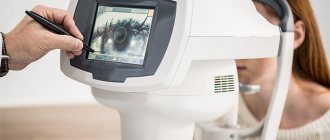
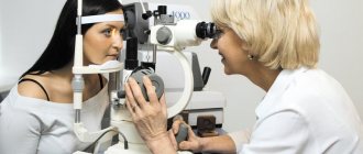
Pterygium usually does not cause diagnostic difficulties, since everything, as they say, is too obvious: the pathology is localized in the most convenient way for ophthalmoscopy. There are certain morphological criteria for the differential diagnosis of stationary (stalled) and progressive pterygium; For these purposes, a slit lamp is used.
Fig. 3 Diagnosis of pterygium - slit-lamp examination (biomicroscopy)
Malignant growth
The most common types are:
- Basalioma is the most popular neoplasm of the eyelids, destroying tissue locally, most common in patients over fifty years of age, usually located in the outer corner of the eye and involves a nodule, progressing very slowly, growing into surrounding tissues.
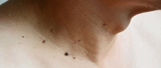
- Progressive nevus is a pigmented neoplasm of the eyelids, it can be congenital or develop during sexual maturation, characterized by a change in color, the appearance of a halo, and peeling. Both classical surgery and laser surgery are effectively used as a method of treatment.
- Melanoma of the eyelid is the most malignant neoplasm of the eyelids, most often it is a speck with uneven contours, and less often it is a node prone to bleeding. The color of melanoma can vary from yellowish to almost black. The choice of excision method and subsequent treatment depends on the volume and nature of the melanoma.
Rehabilitation measures
It is noteworthy that after laser surgery, the patient may experience re-formation of the growth after some time. Quite quickly, the pinguecula can form again, and sometimes it can be much larger than before the operation. To prevent this, for the first time (about a month) after the operation, you should wear a bandage so that irritating factors do not affect the operated eye. A person may experience redness of the organs of vision, which, however, disappears quite quickly. Also, in the summer, the patient must wear glasses that protect the eyes from UV rays.
Pinguecula is a fairly common diagnosis, which many doctors, however, consider not serious. With effective and proper treatment, you can quickly deal with this growth without worrying about eye health and visual acuity.
Preparation for surgery for pterygium and rehabilitation period
It is possible to prescribe anti-inflammatory, antiallergic drops or glucocorticosteroids (GCS) to reduce redness, swelling and pain.
Be prepared that it may take several weeks for you to fully recover from surgery. At first, the cornea will react sensitively to any external influences, and the image may be unclear - this is normal. To reduce the frequency of relapses, metabolites, corticosteroids and anti-VEGF drugs are used, which prevent the formation of new vessels, prevent the progression of the disease, relieve swelling and improve vision.
How is pterygium diagnosed and treated?
Treatment for a film on the eye can be conservative. In the early stages, it is aimed at relieving symptoms and improving the patient’s well-being. The decision to undergo surgical intervention is made if the pterygium progresses and causes pain. The treatment plan is drawn up by an ophthalmologist after identifying the suspected causes and diagnosis.
To characterize the patient’s condition, the specialist will first perform viziometry, a routine vision test. To assess the condition of the anterior segment of the eye (cornea, anterior chamber, lens, conjunctiva and eyelids), biomicroscopy using a slit lamp will be required. This diagnostic method will allow you to assess the stage and degree of growth of pterygium. As auxiliary methods to clarify the diagnosis, the eyes are examined using an ophthalmoscope and a refractometer to examine the fundus in detail. To predict further growth or possible relapses, a study of lacrimal function, keratotopography and angiography are performed.
By the way, pterygium is very similar to pinguecula - a harmless yellowish-white formation on the conjunctiva. It occurs as a result of aging or exposure to sunlight and usually does not require treatment. It happens that the pinguecula becomes inflamed, in which case moisturizing drops help. Both pterygium and pinguencula can cause discomfort when wearing contact lenses because they impede lens movement. Pinguenicula is also usually sensitive to external mechanical stress and ultraviolet radiation.