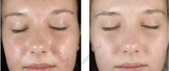
The eye area is subject to age-related changes, like other parts of the body. Fine wrinkles form around the eyes, eyelids droop, and bags form under the eyes.
Often this zone is the first to age; external changes immediately attract attention.
Why is the area around the eyes tender and sensitive?
There is practically no subcutaneous fat layer, no powerful muscles. Therefore, the first signs of withering appear in this part of the face.
Canthopexy will restore youth to the eyes and refresh the look.
Features of eye anatomy
The area of the organ of vision has structural features: the skin in this area is thin. It contains a small amount of collagen fibers. Therefore, here, before wrinkles form in other areas, skin tone is lost.
Under the influence of ultraviolet radiation, the skin quickly loses fluid.
Under the skin is the periorbiatle muscle. Due to it, the eyelids open and close. When stressed, skin creases appear in the corners of the eyes; at a young age they quickly return to normal; with aging, they remain and wrinkles and “crow’s feet” form.
Under the muscle there are plates of cartilage, the muscle fiber is attached to them by tendons and ligaments. The ends of the tendons of both eyelids are attached to the edge of the cartilage. They connect with each other, forming the corners of the eyes - external and internal.
The cartilage is located on the periosteum. The tendons of the outer corner are longer and stretched than the inner one. During life, they stretch even more, the outer canthus begins to descend, sometimes this is a congenital feature.
Canthoplasty (canthopexy) - correction of eye shape
Plastic surgery does not stand still, so every day people all over the world have a chance to become more perfect.
Recently, the procedure of canthopexy and canthoplasty has become very popular. Canthopexy and canthoplasty allow you to change the shape of the eyes, tighten the corner of the eyelids, thereby achieving an aesthetic appearance of the entire face. The procedure received its name due to the fact that any operation performed in one way or another affects the canthal tendon in the eyelid area, which is responsible for support.
This procedure is most popular among people who have severe ptosis (sagging eyelids) and too narrow/wide eye shape. Canthoplasty is also suitable for those who have drooping eyelids due to age-related changes. Canthopexy helps to “rejuvenate” the look, since the procedure tightens the lower eyelid and also removes ectropion (a case where the eyelid is slightly inverted).
Preparation
If canthopexy is required, preparations should be made before surgery. They first visit an ophthalmologist, he diagnoses the pathology of the organ of vision, determines contraindications and indications for surgical intervention. Be sure to visit a therapist, he will determine the general condition and identify diseases of the internal organs. Before the operation, laboratory and instrumental studies are performed.
General blood test - determine the indicators of formed elements, ESR. It is used to judge the presence or absence of inflammation, anemia, and disorders in the circulatory system.
A general urine test allows you to assess the condition and functioning of the kidneys; since surgery is ahead, they should be normal. Protein, red blood cells, white blood cells, mucus, epithelium, and the presence of ketone bodies are determined.
Blood biochemistry – evaluates the condition of internal organs: liver, pancreas, gall bladder, kidneys. Indicators are determined: liver enzymes, creatinine, bilirubin, c-reactive protein, albumin.
A coagulogram is done to determine bleeding time and blood clotting, this is important during surgery.
Perform an analysis for infections: HIV, syphilis, hepatitis.
From instrumental studies, an ECG is performed to assess the functioning of the heart, since surgery and anesthesia are to be performed.
During the week before surgery, it is not recommended to drink alcoholic beverages or smoke.
The surgery is performed in the plastic surgery department. Beforehand, on a computer, using a special program, the patient is shown what he will look like after the operation.
What is contopexy
Every woman wants her eyes to be alluring and captivate any man. In youth, it’s easy to maintain the beauty of your eyes: properly moisturize this delicate area, which is prone to the earliest appearance of signs of aging. But the skin around the eyes itself is thin and contains a small amount of collagen. Tonic, cream, mask - with the help of this complex the area around the eyes will be in normal balance until a certain point. With age, irreversible changes occur in the skin and soft tissues of the upper and lower eyelids, as a result of which blepharoplasty surgery becomes the only radical way to rejuvenate the area around the eyes. Today, there are many types of eyelid surgery, and now we will focus on one of them. We will talk about canthopexy.
Execution technique
There are two options for the operation: canthopexy of the outer eyelid - lateral. It is performed quickly, under local anesthesia. If the inner corner of the eye is affected, this is medial canthopexy.
For additional blepharoplasty or canthoplasty, general anesthesia is used.
The technique is determined by the doctor. A small incision about 1 cm long is made above the upper eyelid. This allows access to the tendon that is connected to the canthus. The doctor stretches the tendon and attaches it to the periosteum with special absorbable sutures.
There are situations when tendons are partially removed. Then stitches are placed for a week. The operation lasts about two hours.
The scar after canthopexy is not noticeable; it is made on a fold of skin. After surgery, the patient is allowed to go home; long-term hospitalization is not necessary.
What is the difference between classic blepharoplasty and canthoplasty?
In fact, patients often hear about the blepharoplasty procedure, but it is worth understanding that the difference between these operations is quite significant.
Blepharoplasty is effective in cases of slight sagging eyelids, bags under the eyes, crow's feet and shallow wrinkles. Canthoplasty is a more serious procedure. It implies a different method of performing the operation.
Due to age-related loss of support, our eyelids gradually begin to droop. It is in such cases that canthopexy (canthoplasty) can restore the necessary support to the eyelid, get rid of a “tired” face, and even restore facial asymmetry in case of facial nerve paralysis. Canthopexy (canthoplasty) also helps with thyrotoxicosis (thyroid disease), when patients experience unpleasant consequences in the form of exophthalmos (protrusion of the eyeballs).
In the case of the same ectropion (inverted eyelid), blepharoplasty is useless. Moreover, this procedure can even worsen the problem.
However, at the moment it is possible to carry out both procedures simultaneously, which will make the result longer and more effective.
Rehabilitation
Any operation is stressful for the body, so a period of recovery is required. After surgery, swelling, hematomas, bruises remain, and the conjunctiva is irritated.
You may experience burning, pain, increased photosensitivity, and lacrimation. The doctor prescribes painkillers and eye drops for this period to reduce irritation of the conjunctiva. The discomfort will disappear after 7 days.
After the operation, a feeling of tension in the eyelids is bothersome for 1–2 months, which then goes away without a trace.
To fully restore the body, you need to rest. Avoid eye strain, limit the time spent at the computer and watching TV. It is better to postpone reading. Avoid bright light for 14 days; use sunglasses in sunny weather; in addition, they protect against sand and dust getting into the eyes, and, consequently, infection.
You cannot go to the bathhouse or gym for 10 days. It is not recommended to use decorative cosmetics on the eye area or wear contact lenses. Excessive physical activity and heavy lifting are prohibited.
Indications for canthopexy
- Reduced elasticity and “stretching” of the lower eyelid, i.e. its horizontal elongation due to stretching of the canthal ligaments (mainly lateral);
- decreased tone of the orbicularis oculi muscle and its displacement below the edge of the cartilaginous (tarsal) plate;
- weakening or separation of the orbicularis oculi muscle, which is responsible for contraction of the lower eyelid with age or due to injury;
- Age-related depression of the eyes due to atrophy of the retrobulbar (behind-the-eye) fatty tissue (age-related enophthalmos).
Figure 1. The diagram shows that the lateral canthal ligament consists of an upper and lower part. Just below there is a thin ligament called the inferior retinaculum (retinaculum inferior).
In addition to external signs of lower eyelid weakness, you can perform a pinch test to determine its laxity. It is necessary to take the fold of skin of the lower eyelid in its central part, pull it down and release it. Normally, the eyelid immediately returns to its original position. If the lower eyelid can be pulled back by more than 6 mm and it slowly, “reluctantly” returns to its original position, this indicates weakness of the lower eyelid.
In most people, the most prominent point of the cornea and the zygomatic eminence are at the same level - this is called the “neutral vector”. If the zygomatic eminence protrudes forward in relation to the eye - “positive wind”.
In Figure 2 “Neutral vector” - red dotted line - the eyeball and the zygomatic eminence are on the same line.
In Figure 3, the black dotted line depicts a “positive vector” - the cheekbone protrudes forward in relation to the most protruding point of the cornea of the eyeball. The “neutral vector” is marked with a red dotted line.
“Neutral” and “positive vectors” indicate good development of the skeleton and soft tissues of the face, providing good support and support to the lower eyelid.
When measuring with a ruler the distance from the outer orbital edge to the most protruding point of the cornea, the result will be 15-17 mm. Ophthalmologists call this position of the eyeball normophthalmos. If the value is less than 15 mm, the eye is located inside relative to the inferior orbital margin, i.e. in the enophthalmos position.
In Figure 4, the black dotted line shows a “negative vector” - in profile, the eyeball protrudes forward in relation to the cheekbone, the eyes seem bulging. The “neutral vector” is marked with a red dotted line.
The “vector” is “negative” if the orbital edge is flat, smoothed and the eye protrudes forward relative to it, in which case the lower eyelid has no support and, in turn, does not provide sufficient support to the eyeball.
The “negative vector”, as a rule, is caused by underdevelopment of the facial skeleton and a deficiency of soft tissues in the midface. In such people, under the influence of gravity, “weakness” of the lower eyelids, ptosis (drooping, sagging) of the lower eyelids and cheeks appears quite quickly, compared to patients who have more developed bones of the facial skull.
In the presence of a “negative vector”, the eyeball is moved forward in relation to the cheekbone by more than 17 mm, this is called exophthalmos. The latter can be congenital (with the so-called “shallow orbit”, characteristic mainly of the Negroid race), or also appear as a result of age-related changes, endocrine disorders or volumetric processes in the brain skull (in such cases, it is most often one-sided).
Performing classic lower blepharoplasty when weakness of the lower eyelid is detected, especially in combination with the presence of a “negative vector,” is dangerous, since even minor removal of eyelid skin can lead to eversion - ectropion. In such cases, eyelid surgery is complemented by canthopexy or endoscopic midface lift.
Canthopexy technique
Rice. 2. The changes in the lower eyelid that occur with age are schematically depicted. In addition to excess skin, a fine network of wrinkles and “hernial sacs” of varying degrees of severity, a decrease in the tone and elasticity of the lower eyelid is noteworthy. The “weakness” of the lower eyelid manifests itself as follows: it does not fit tightly to the eye, moves forward a little, sags, and seems to be “pulled” down. The outer corner of the eye drops below the inner one (normally, in young people, the outer corner of the eye is at the same level or 2 - 3 mm higher than the inner one). As a result, the eyes appear bulging, rounded, and a white stripe of sclera is visible between the cornea and the edge of the lower eyelid. When viewed directly, the slightly expanded costal edges of the lower eyelids are visible. Progression of lower eyelid laxity can lead to ectropion.
Rice. 3. The dotted line shows preoperative markings.
The markings for canthopexy are performed in the operating room and are similar to those for lower blepharoplasty. The line is drawn 1-2 mm away from the ciliary edge of the lower eyelid. The main difference is a small 0.7-10 mm incision, which will be located in the sub-eyebrow area.
Rice. 4. Making an incision with a scalpel along the lines of preoperative markings.
Rice. 5. Fixation with an absorbable thread of the orbicularis muscle of the lower eyelid to the periosteum of the upper orbital edge (subeyebrow region).
Rice. 6. Application of intradermal sutures.
Even if eyelid correction is performed under general anesthesia, the tissues of the lower eyelid are first infiltrated (flooded) with saline solution with the addition of an anesthetic and vasoconstrictor drugs. Then the skin is dissected with a scalpel along the lines of preoperative markings. In most cases, the plastic surgeon begins with a standard lower blepharoplasty: peeling away the skin, highlighting and removing the hernia bags.
After this, through an additional incision in the sub-eyebrow area, the periosteum is sutured with an absorbable thread, which is passed under the canthal ligament, the orbicularis oculi muscle is captured in the area of the outer corner, and then the thread is drawn in the opposite direction under the canthal ligament and finally fixed to the periosteum with a U-shaped suture. An internal suture placed in a similar manner ( canthopexy)
) will firmly hold the lower eyelid in the desired position.
The eyelid correction ends with excision of excess skin and application of an internal suture with a non-absorbable thread so that only two nodules are exposed to the skin: at the beginning and at the end of the incision. The stitches are removed on day 5. Postoperative scars will quickly fade and become invisible.
Rice. 7. The figure schematically shows a supporting canthopexy.
Supportive canthopexy differs in that it involves only the canthal ligaments, which are sutured together and fixed to the periosteum of the orbit above the lateral ligament of the eyelids.
The eyes are the area of our face that receives the most attention. When meeting for the first time, the first visual contact occurs using the eyes. The eyes most quickly display age or a hint of it. You can convey many feelings and emotions through your eyes: disappointment, joy, hope, regret.
If they say: “The eyes glow”, then they mean a cheerful, radiant look. In order for this look to remain as long as possible, you need to approach this problem competently. The canthopexy operation is absolutely safe and is performed in a short time, and the effect will last from 5 to 10 years.
Photos before and after blepharoplasty of Asian eyelids
The following photos illustrate the incredible transformation of one of my patients, not only externally, but also internally. After surgery for Asian eyelid surgery, the girl became more open, sociable and self-confident. So:
Photos before and 2 months after Asian eyelid surgery
Below is the result of Europeanization of the Asian eyelids of the same girl after 8 months
The work of plastic surgeon S. N. Zharkova, Moscow
Look how noticeably the girl’s appearance has changed thanks to the operation of Europeanization of the Asian century.
See more photos of the results of operations to Europeanize Asian eyelids here
When should you give up?
Reviews from men about blepharoplasty confirm its safety and low morbidity. We must not forget that with certain concomitant pathologies, surgical intervention can be harmful.
You should refrain from the procedure if you have:
- diabetes mellitus in the stage of decompensation;
- increased intraocular pressure;
- low secretory capacity of the lacrimal glands;
- diseases of the heart and blood vessels in the acute period;
- high risk of bleeding.
Before the procedure, the plastic surgeon prescribes a certain range of laboratory tests. This helps to identify and exclude contraindications for blepharoplasty.
Most of them can be eliminated.
This means that consultation with a specialist or a course of treatment may allow surgical intervention. But the final decision remains with the plastic surgeon.
Operation to Europeanize the eyelids
Preparing for surgery
Eyelid surgery can be performed under either general or local anesthesia. You can also find the required scope of examinations on my website by following the link.
Progress of the operation
The operation to Europeanize the eyes is one of my favorite operations. I do it this way:
Marking For me, as a specialist, marking during Europeanization of eyelids sometimes takes more time than the operation itself. This is a very important stage. And there can be no mistakes, as, indeed, at each stage.- Making the cut I perform the cut only with a scalpel!!! There is no more precise tool. I carry out internal manipulations using a laser or radio knife. An incision in the skin of the upper eyelid is made where we want to see the fold of the upper eyelid in the future.
- Removal of excess fat tissue, often I also remove sub-eyebrow fat. The more fat is removed, the deeper the fold is. Almost all fat must be removed, otherwise you will not get a good result.
- Resection of excess skin As a rule, the excess is very small. It is necessary to take into account that the skin will be needed in order to deepen the fold. Excessive skin resection may also lead to failure.
- Excision of part of the orbicularis oculi muscle is absolutely always performed. Since in Asian eyelids the orbicularis muscle is overstretched and there is quite a lot of it.
- To form a fold, the skin must be fixed with interrupted sutures to the aponeurosis of the muscle. I do this with absorbable and non-absorbable threads, that is, 50/50. This gives a soft, beautiful scar and a guarantee of no relapse.
Video from the operating room during eyelid Europeanization surgery
On video: the progress of the operation to Europeanize Asian eyes. The operation was performed under local anesthesia. Plastic surgeon Zharkova S. N.
Complete recovery takes about two months, after which the final result of the plastic surgery can be assessed.
The duration of the operation is 1.5-2 hours. The patient spends several more hours in the hospital after the operation.
Patient reviews of Asian eyelid blepharoplasty
This review was left by my patient on one of the plastic surgery portals, so I’ll just quote:
July 07, 2022, 11:41:26, Agnes, Moscow
Operation: Blepharoplasty of Asian eyelids
Since childhood, I dreamed of having blepharoplasty and getting rid of the Asian eyelid. For a very long time I could not dare to have an operation, and when the decision was made, I was faced with a problem - I could not find a doctor who could be trusted with such a serious matter. Over the course of a month, I had consultations with about 10 doctors and could not find my surgeon. And when I got an appointment with Svetlana Nikolaevna, I immediately realized that I would have the operation with her. And she was not mistaken: she is actually a competent doctor and surgeon who knows her business. Before the operation, I received detailed consultation and answers to all my questions, the operation itself was painless, and an hour and a half later, when the pressure bandage was removed from me, I immediately saw how my eyes and face as a whole had changed. And one more thing - after the operation I prepared for pain (according to the stories of my friend and mother), I bought a whole bunch of painkillers, but I never felt any discomfort, I don’t know whether it was Svetlana Nikolaevna’s merit or I was just lucky, but I was incredibly happy. To summarize, I would like to thank my surgeon, Svetlana Nikolaevna Zharkova. If someone, like me, is looking for a surgeon in Moscow who can be entrusted with a serious operation, then be sure to go for a consultation with Svetlana Nikolaevna.
You can read more reviews from my patients in the Reviews section.
How is the operation performed?
Like any surgical treatment, blepharoplasty has a preparatory period.
A few days before the procedure, the patient is advised to refrain from drinking alcohol. It is worth balancing your daily diet. Eliminate heavy, high-calorie foods. Pay attention to sufficient fluid intake.
At the preoperative appointment, the doctor uses a marker to mark problem areas and future incision lines.
The type of anesthesia depends on the extent of surgery. For a short, low-volume procedure, local anesthesia is used. General anesthesia is used for long and complex operations.
The technique of plastic surgery depends on the anatomical features of the patient’s facial structure.
The surgeon uses an electrocoagulator to stop bleeding from vessels of various sizes. During the operation, soft tissue is excised and the skin is tightened. The final stage is the application of an aseptic dressing.
Complications
The risk of negative reactions after blepharoplasty is minimal, but possible. Most often this is due to the physiological characteristics of the patient.
The appearance of bruises is possible in the case of a developed network of capillaries at the incision sites.
Edema appears due to intense microcirculation and restoration of damaged tissue.
Tear production is a natural process of the body. Tear contains antiseptic substances that stimulate healing and reduce inflammation.
These phenomena are temporary and pass without a trace.
The formation of scars after high-quality plastic surgery occurs extremely rarely. Careful selection of a doctor and clinic and careful adherence to recommendations help to avoid complications.
A separate issue: the divergence of the edges. This can happen if you put stress on your eyes and facial muscles early. To correct this condition, an emergency visit to a plastic surgeon is necessary.