Otitis externa is an inflammation of the outer ear, which includes the pinna and external auditory canal (EA).
Author:
- Filatova Evgenia Vladimirovna
otolaryngologist, rhinosurgeon
3.80 (Votes: 10)
- Causes of a boil in the external auditory canal
- Causes of development of bullous otitis media
- Treatment
Otitis externa is an inflammation of the outer ear, which includes the pinna and external auditory canal (EA). Like most inflammatory diseases of the ENT organs, otitis externa can be acute or chronic.
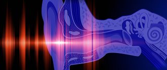
Features of the structure of the outer ear
The auricle and ear canal are covered with skin that extends to the eardrum. There are two sections in the ESP: cartilaginous and bone; at the border of these sections, a physiological narrowing is determined - the isthmus, which is why the three-dimensional model of the external auditory canal resembles an hourglass. Under the skin of the auricle and the cartilaginous part of the external auditory canal there is elastic cartilage, due to which this part of the auditory canal is mobile during chewing and articulation. In the bony section, under the skin, there is periosteum and bone. The skin of the external auditory canal differs from any other skin in that its thickness contains cerumenous glands that produce earwax (cerumen). The function of cerumen is to protect the skin from constant moisture and microorganisms, and the presence of a cerumen film is an important factor in the normal functioning of the outer ear. The dimensions of the external auditory canal may vary markedly depending on individual characteristics. Often in the area of the isthmus there are osteophytes - bony protrusions that reduce the lumen of the ESP, and sometimes completely block it. Anterior, posterior and inferior to the auricle in the subcutaneous tissue there are lymph nodes that can become inflamed with otitis externa.
Acute external otitis is an inflammation of the external ear that occurs within 1 month.
Forms of external otitis:
- diffuse external otitis;
- local external otitis or NSP boil;
- acute bullous (hemorrhagic) external otitis;
- myringitis;
- dermatitis of the auricle;
- erysipelas of the auricle;
- malignant (necrotizing) external otitis;
- chondroperichondritis of the auricle.
The most common is acute diffuse external otitis, in which bacterial inflammation of the skin of the entire ear canal occurs, often spreading to the eardrum or the skin of the auricle.
Inflammatory diseases of the external ear
IN
Inflammatory diseases of the external ear are common diseases that occur in all age groups and are characterized by a variety of clinical manifestations, requiring a clear physician orientation in matters of diagnosis and treatment. It is very important to be able to use clinical data to differentiate otitis externa from acute otitis media and mastoiditis, since a diagnostic error or inadequate treatment can cause serious complications, including intracranial ones, that threaten the patient’s life.
Etiology and clinic
In recent years, there has been a tendency towards an increase in the frequency of external otitis, which is due, in particular, to unfavorable environmental factors, irrational use of drugs, primarily antibiotics. One of the leading factors in the pathogenesis of external otitis is trauma to the epidermis of the external auditory canal, in particular after severe removal of wax or discharge from the ear with hard objects, as a result of maceration when water gets into the ear, with chronic purulent otitis media. Speaking about the need for proper, gentle removal of sulfur, one should take into account its bactericidal and fungicidal properties, mechanical protection of the skin of the external auditory canal from unfavorable exogenous factors.
Inflammation of the outer ear can be caused by various microflora: Staphylococcus aureus, Streptococcus pyogenes, Pseudomonas aeruginosa, Enterobacter, fungi of the genus Candida, Aspergillus, Penicillium,
viruses, pathogens of syphilis and tuberculosis.
Pseudomonas aeruginosa is highly resistant to most antibacterial drugs. When identifying this gram-negative aerobe, one must certainly take into account the possibility of nosocomial infection during diagnostic and therapeutic manipulations in the ear canal.
Otomycoses
One of the current problems is the progressive increase in external otitis of fungal etiology (otomycosis), especially in childhood. The increase in the incidence of otomycosis in children is due to dysbacteriosis and various factors that weaken the resistance of the child’s body. The occurrence of otomycosis in both adults and children can be caused by immune, hormonal, metabolic disorders, allergies, long-term treatment with antibacterial, hormonal drugs, cytostatics, and radiation therapy of neoplasms. The occurrence of mycotic lesions of the outer ear may be preceded by long-term local use of glucocorticoid drugs for otorrhea caused by a purulent-inflammatory process in the middle ear.
The development of otomycosis is possible when working in dusty conditions, in pressure complexes with high pressure and humidity.
Characteristic symptoms of otomycosis are intense, almost constant itching, noise in the ear, and discharge from the ear. The nature of the discharge depends on the type of pathogen: caseous masses of white or grayish color are characteristic of Candida albicans
, black -
Aspergillus niger
, yellowish -
Aspergillus flavus
, greenish -
Penicillium
. Pain in the ear is absent or mildly expressed, but quite often patients complain of local headache (Kunelskaya V.Ya., 1989, Evdoshchenko E.A., 1985, Apostolidi K.G., 1996).
An increase in the proportion of mycotic infection in ear pathology has led to an increase in atypical clinical manifestations and complicated forms of the disease.
In 1960, A.K. granulating external otitis as an independent nosological form
, the occurrence of which is caused by the penetration of the
Monilia
. The disease lasts a long time, with the formation of granulations on the eardrum and the walls of the ear canal.
A rare form of inflammatory lesions of the outer ear is malignant or necrotizing otitis externa.
, the frequency of which is less than 1% of all dermatoses of the external ear. It occurs mainly in older people suffering from diabetes. The severity of the disease is due to sequestration of the bony part of the external auditory canal, the possibility of developing intracranial complications and damage to VII, IX–XI pairs of cranial nerves. Mortality in complicated necrotizing otitis remains high, ranging from 23 to 67% (Gukovich V.A., 1988).
Limited and diffuse otitis externa
Infectious external otitis may occur in the form of a boil in the external auditory canal
(limited external otitis – otitis externa circumscripta) or in the form of “spread” inflammation (
diffuse external otitis
– otitis externa diffusa).
The main complaints of patients are pain and itching in the ear. The itching is minor and is observed only at the beginning. Patients are primarily concerned about intense pain in the ear, reaching a maximum on the 3rd–4th day of the disease, intensifying with movements in the temporomandibular joint (chewing, opening the mouth), with possible irradiation to the temporal or maxillary region.
Inflammation of the outer ear should be differentiated from inflammation of the middle ear (Table 1).
In cases where the boil is localized on the posterior wall of the ear canal, it is often necessary to make a differential diagnosis with mastoiditis, a possible complication of acute otitis media, in which the process spreads to the cellular system of the mastoid process. The main clinical differences, in addition to those listed above, are presented in Table 2. In doubtful cases, correct diagnosis is facilitated by the analysis of X-ray symptoms. In the projections of Schüller, Mayer, Stenvers, with mastoiditis, loss of air supply and disruption of the structure and integrity of the bone base of the mastoid cells are determined, while with a boil of the external auditory canal, no radiological changes in the temporal bone are observed.
When hemolytic streptococcus penetrates into damaged skin of the auricle, erysipelas
. The main symptoms of erysipelas are a characteristic sharp, shiny-tinged, hyperemia of the auricle, involving the earlobe and clearly demarcated from unaffected areas; pain, infiltration and increase in the volume of the auricle. Hyperthermia and chills are noted.
Neuroviral infection with severe course - ear form of herpes zoster
– Herpes zoster oticus, a distinctive feature of which is an acute rash of pink spots, and soon vesicles with transparent contents, located along the sensory nerves. There are sharp neuralgic pain in the ear and the corresponding half of the head, itching, tingling, dizziness, sensorineural hearing loss and damage to the facial and trigeminal nerves. An increase in body temperature and general malaise are often observed. The blisters begin to dry out on days 6–8, and the crusts disappear by the end of the third week of the disease. Diagnosis is especially difficult in the first days of the disease, when there are still no characteristic herpetic rashes, and neuralgic pain can simulate various dental and neurological diseases.
A very common, painful disease is microbial eczema of the outer ear.
- a secondary erythematous-vesicular disease that develops against the background of chronic streptococcal or fungal skin lesions, in which the process may involve both the auricle and external auditory canal, and the eardrum. Eczema occurs in cases of mechanical, chemical or thermal irritation of the skin, in the absence of proper treatment and hygiene with prolonged suppuration from the ear. The disease does not have a single etiology and can be explained from the point of view of two complementary theories - neurogenic and allergic. Severe mental trauma, damage to peripheral nerves, various diseases of internal organs, hormonal disorders, diabetes mellitus, and rickets contribute to the development of eczema. Clinically, the disease has features of both a microbial process and eczema, that is, it is manifested by the formation of small vesicles in the area of the infectious focus, the presence of crusts, erosions, cracks, and purulent discharge. The earliest and most persistent symptom is intense itching, especially pronounced during exacerbations of the process.
Treatment
Thus, inflammatory diseases of the outer ear are varied in etiology, pathogenesis, clinical picture and severity. The structure of the causative agents of external otitis is heterogeneous and includes bacterial gram-positive and gram-negative microbial flora, fungi and viruses. This determines the need for a differentiated approach to the treatment of each individual patient, based on detailed clinical and laboratory research.
Difficulties in treatment are largely due to the increasing virulence of pathogens and their resistance to widely used antibiotics and antiseptics, which is due to the unreasonably wide, often uncontrolled use of the latter in clinical practice.
Antibiotics
Drug therapy for inflammatory diseases of the outer ear involves the rational use of antibiotics, based both on a detailed assessment of the patient-microorganism-drug interaction system, and on the results of identifying the microorganism that caused the disease during bacteriological examination. An indispensable condition for prescribing an adequate antibacterial agent is to establish the sensitivity of the flora in vitro
using discs impregnated with antibiotics.
Unfortunately, in some cases, for example, when isolating mixed flora, the reaction of bacteria to an antibacterial agent in vivo
may be altered (Belousov Yu.B., Moiseev V.S., Lepakhin V.K.).
In recent years, fluoroquinolones and oral cephalosporins have been shown to be highly effective
, having a wide spectrum of action, having the ability to easily penetrate tissues, creating a high concentration in them. However, one should take into account the resistance of Pseudomonas aeruginosa to all group I cephalosporins and most group II and IV cephalosporins, as well as the side effect of fluoroquinolones, which consists in their penetration into the growth layer of the bone, which precludes the use of fluoroquinolones in childhood.
Macrolide antibiotics have been used in the treatment of bacterial infections of the upper respiratory tract and ear for more than 30 years.
, of which erythromycin is the best known.
The use of erythromycin is limited by its poor absorption when administered orally, low bioavailability, relatively narrow spectrum of antibacterial action and the likelihood of developing side effects from the gastrointestinal tract. Among the macrolide antibiotics synthesized recently is roxithromycin, which has improved physicochemical, biological and pharmacokinetic properties. The spectrum of antibacterial activity of roxithromycin includes streptococci, some types of staphylococci, and atypical microorganisms ( Mycoplasma, Legionella, Chlamydia
). The drug penetrates well into tissues, fluids and cells of the body, creating high intracellular concentrations. It is quickly absorbed in the gastrointestinal tract and is resistant to acidic environments. For adults, the drug is prescribed either at a dose of 150 mg twice a day or 300 mg once a day, for children - at a rate of 5 mg/kg twice a day.
Enzyme preparations
Among the means of antibacterial therapy, there is a large group of drugs that are alternative to antibiotics. Their use becomes especially relevant when isolating antibiotic-resistant strains, when it is impossible to carry out antibiotic therapy due to intolerance by the patient. It is advisable to use enzymes, in particular lysozyme
and
trypsin
. Lysozyme and trypsin are non-toxic and do not cause allergic reactions. The combined use of these enzymes (Veremeenko K.N. et al., 1974, Brofman A.V., 1980) promotes the healing of damaged tissues and inhibits the growth of pathogenic microorganisms, and provides an immunomodulatory effect.
The disadvantages of enzyme preparations are their instability under conditions of unstable pH and temperature, pyrogenic properties, and therefore the use of enzymes immobilized on a cellulose carrier is promising (Davidenko T.I. et al., 1993, Apostolidi K.G., 1996).
The use of multienzyme turundas containing immobilized trypsin and lysozyme, as well as purified trypsin powder immobilized on a cellulose carrier, has a number of advantages in the treatment of external otitis of bacterial etiology (staphylococci, Pseudomonas aeruginosa, Proteus, Klebsiella, Escherichia coli), otomycosis (fungi of the genus Candida, Aspergillus
), granulosa external otitis.
The treatment method consists of either daily or every other day injection of turunda with immobilized enzymes into the external auditory canal
after mandatory thorough evacuation of pathological discharge from it. Treatment for 10–14 days allows for recovery. A persistent clinical effect is confirmed by the results of a microbiological study. The advantage of enzyme therapy is the preservation of the normal flora of the external auditory canal, which significantly reduces the risk of colonization by pathogenic microorganisms and prevents relapse of the disease.
Treatment of otomycosis
A serious problem is the treatment of otomycosis, especially in children. The most effective can be considered a complex of therapeutic measures, including both a direct effect on the pathological focus in the outer ear
, and
restoration of intestinal microbiocenosis
. Intestinal dysbiosis is a determining factor in maintaining disturbances in the microbiocenosis of the external ear, and therefore, in case of otomycosis, the elimination of dysbiotic conditions of the intestine is justified (Naumova I.V., 1999).
Modern antifungal drugs
broad spectrum of action have significantly higher therapeutic activity and bioavailability in comparison with traditionally used nystatin.
against fungi of the genus Candida
.
Triazole compounds ( ketoconazole, fluconazole
) are highly effective, inhibiting the synthesis of sterols in fungal cells and blocking a number of cytochrome-dependent enzymes.
For topical use, antifungal drugs may be recommended, prescribed, according to the mycogram, on the turundas once a day for 3–4 weeks.
of eubiotics for 1–3 months
– preparations of bifidobacteria, E. coli, lactobacilli.
It should be taken into account that with otomycosis in children, all types of physiotherapeutic effects on the ear, the use of hormonal drugs, as well as penicillin and tetracycline antibiotics are contraindicated.
For indolent, difficult-to-treat processes, along with antibiotics and antihistamines, the use of immunomodulators and adaptogens is indicated.
Eleutherococcus extract is used orally, 20–30 drops per day for a month, it is well tolerated. For the purpose of antiviral and immunomodulatory effects, it is advisable to use interferon inducers of general and local action and recombinant a-interferon.
Laser therapy
At the Ear, Nose and Throat Clinic of the MMA named after I.M. Sechenov has accumulated significant experience in the use of therapeutic lasers in the treatment of inflammatory diseases of the external ear. The variety of laser systems currently available allows the doctor to choose the type of laser radiation depending on the task at hand. Thus, helium-neon laser
(red spectrum range, wavelength 0.63 µm).
To influence the deep layers of soft tissues of the external auditory canal, it is advisable to use low-energy laser radiation in the near-infrared region of the spectrum - infrared lasers
with a wavelength of 0.8–1.2 µm. The light guide is inserted into the fibrocartilaginous part of the auditory canal or, if it is sharply narrowed, it is installed in the tragus area. The ability of laser radiation to provide anti-inflammatory, analgesic, immunocorrective, hyposensitizing effects allows one to achieve a lasting clinical effect during a course of therapy consisting of 7-10 daily sessions lasting 3-5 minutes (Ovchinnikov Yu.M. et al., 1996).
Ozone therapy
In recent years, ozone has been used to treat infectious and inflammatory diseases of the outer ear (Velikanov A.K. et al., 1994, 1995, Ovchinnikov Yu.M., Sinkov E.V., 1999). The use of ozone in medicine is based on its anti-inflammatory, bactericidal, antiviral, fungicidal, immunocorrective, and analgesic effects. It is characteristic that ozone is equally active against gram-positive and gram-negative microflora, fungi and has a neutralizing effect on microbial toxins.
Both gaseous ozone and ozonated solutions are used to wash the ear, in particular, an ozonated isotonic solution of sodium chloride, obtained by bubbling a solution of 0.9% NaCl with ozone until an ozone concentration of 5 mg/l is achieved. A necessary condition for ozone therapy is the elimination of excessive ozone accumulation in the room and the use of an insulator sealed around the auricle during treatment sessions. The course of ozone therapy is 6–10 sessions with the duration of one session being 5 minutes. Ozone is used in complex treatment along with the prescription of drug therapy, which can significantly reduce the time of treatment of patients and, accordingly, reduce economic costs. The use of ozone increases the effectiveness of antibacterial drugs. The therapeutic effects of ozone are of particular relevance for patients with external otitis, suffering from allergic diseases for which antibiotic therapy is impossible.
Phototherapy
A serious problem is the treatment and prevention of external otitis under conditions of exposure to extreme factors on the human body, such as high blood pressure, antiorthostatic hypokinetic conditions, deep-sea diving, and space flights. The impact of these factors leads to a decrease in the body's defenses, activation of pathogenic and opportunistic microflora and, as a consequence, to the development of acute inflammatory diseases, including external otitis (Viktorov A.N., 1987, Ovchinnikov Yu.M., Yarin Yu M., 1997).
One of the promising but insufficiently studied treatment methods is phototherapy with polychromatic light of a wide optical range.
, which has antibacterial and fungicidal activity. Research conducted at the ENT Clinic of the MMA named after I.M. Sechenov proved the effectiveness of light in the optical range, similar to the sun, as a preventive and therapeutic agent for external otitis caused by Staphylococcus aureus, Enterobacter, Pseudomonas aeruginosa, and fungi (Ovchinnikov Yu.M., Yarin Yu.M., 1997, 1999). It has been established that even a single exposure to the solar spectrum is detrimental to bacterial microflora. For fungicidal therapeutic effects, it is necessary to carry out at least three sessions, since a single effect on the fungal flora can enhance its growth. Phototherapy has no effect on spores of pathogenic fungi. The fungicidal effect is manifested in the initial phase, the phase of accelerated uneven growth and the development phase of the fungal colony. Carrying out complex therapy using a xenon irradiator made it possible to reduce the time for relief of diffuse bacterial external otitis by 4–6 days. To ensure a lasting positive result for otomycosis, it is necessary to carry out a second course of treatment after 7 days, including 3 light therapy sessions.
Carrying out courses of preventive effects on the external auditory canals for persons exposed to extreme factors during work allowed to normalize the indicators of microbial contamination of the skin of the auditory canals and the qualitative composition of microbial associations, increasing colonization resistance.
Acute diffuse otitis media
Provoking factors: excessive toilet of the ears, water getting into the ear, microtrauma of the skin of the NSP, factors of decreased immunity (hypothermia, excessive tanning, ARVI, chronic diseases). Pathogens: Staphylococcus aureus, Pseudomonas aeruginosa.
Symptoms
This is severe pain in the ear, ear congestion, liquid discharge from the ear canal in a small amount, there is also an increase in the behind-the-ear or parotid lymph nodes, inflammation of the skin of the auricle, there may be an increase in temperature, and symptoms of intoxication. On examination, hyperemia and swelling of the skin of the NSP are noted, the skin is uneven, with overlays of transparent yellow or purulent discharge, and desquamated epidermis is visible. The lumen of the ESP is often sharply narrowed and the eardrum may not be visible. If it is visible, it is usually also uneven and moderately hyperemic. Enlargement and tenderness of regional lymph nodes, thickening, tenderness and hyperemia of the auricle are often detected. Hearing, while maintaining the lumen of the lumen, is slightly reduced or normal.
Diagnostics
To make a diagnosis, in addition to examination, it is necessary to exclude middle ear disease, for which tuning fork tests (a hearing test using a set of tuning forks), tympano- and impedance measurements (measurement of the mobility of the eardrum), and a study of hearing thresholds (audiometry) are performed. These studies are carried out to exclude diseases of the middle ear. Laboratory diagnostics includes a general blood test, blood sugar, microflora culture from the diseased ear to prescribe antibacterial therapy.
Treatment
The main direction of treatment is antimicrobial and anti-inflammatory therapy. Antibiotics and anti-inflammatory drugs (non-steroidal anti-inflammatory drugs, corticosteroids) are prescribed locally or systemically. For severe pain, there is a good effect from behind-the-ear blockades with corticosteroids and local anesthetics. A gentle ear toilet and rinsing is carried out to free the ear from discharge and epidermal masses. Additionally, you can use physical treatment methods: quartz tube or laser therapy.
Etiology and pathogenesis
Predisposing factors include:
- injury to the ear canal with sharp objects (matches, hairpins);
- malnutrition;
- avitaminosis.
The main cause of chronic furunculosis, which occurs with frequent relapses, is considered to be a violation of carbohydrate metabolism. The penetration of pathogenic microorganisms into the hair follicle leads to acute inflammation, which results in the formation of one or more boils located on the lower, front and/or back walls.
Limited (local) otitis externa or boil of the external auditory canal
A furuncle is an inflammation of the hair follicle that spreads to the subcutaneous tissue. Bristly hairs grow at the entrance to the NSP and around it, and inflammation develops in their follicles.
Causes of a boil in the external auditory canal
The most common pathogens are streptococci. Provoking factors and symptoms are similar to those of diffuse otitis media.
Diagnostics
Upon examination, a narrowing of the ESP is visible due to infiltration in the area of one of its walls. The boil goes through three stages in its development:
- Infiltration stage: at this stage, a painful infiltrate of the NSP skin is formed, local hyperemia and thickening of the skin about 0.5-1.5 cm in size appear.
- The abscess formation stage is the appearance of a purulent cavity and a necrotic core in the area of infiltration. At this stage, the presence of an infiltrate is also determined, in the center of which a yellow-green purulent core is visible, usually with a hair in the center; when pressing on the infiltrate, fluctuation (the presence of fluid in it) can be determined.
- The resolution stage is the release of pus from the cavity with gradual recovery.
Treatment
Treatment varies and depends on the stage. At the infiltration stage, systemic and local antibacterial and anti-inflammatory therapy is prescribed. In the stage of abscess formation, it is worth performing surgical treatment of the boil - opening and draining, cleaning the cavity from pus, dressings are performed within several days. Antibacterial and anti-inflammatory drugs are also prescribed. During the resolution stage, it is sufficient to use only local antibacterial drugs and ear toilet.
2. Reasons
Abscess suppuration in the area of the outer ear is an infectious pathology. In the vast majority of cases, the pathogen is bacterial cultures, most often pyogenic (pyogenic) staphylococci.
The most significant risk factors include:
- incorrect treatment and insufficient sanitary care of the ear canal for purulent otitis;
- sharing linen (towels, pillows, etc.) with a family member suffering from pyoderma in one form or another;
- scratching, using objects not intended for this purpose (and non-sterile), such as matches, hairpins, etc., to clean the ear canal;
- metabolic disorders, in particular carbohydrate metabolism (for example, with diabetes mellitus);
- weakened immune system;
- hypovitaminosis (especially A, B and C);
- ototrauma.
Visit our Otolaryngology (ENT) page
Acute bullous or hemorrhagic otitis externa
This is a form of inflammation in which blisters (bullas) containing bloody fluid appear on the skin and on the outer surface of the eardrum. If such a bubble is opened, then bloody discharge flows out of the ear.
Causes of development of bullous otitis media
Most often, bullous otitis occurs against the background of acute respiratory viral infection or influenza, but a bacterial infection can also be the cause.
Treatment
An ear canal toilet is required. Antibacterial and anti-inflammatory drugs are prescribed locally, and drugs that strengthen the vascular wall and reduce vascular permeability should also be prescribed systemically. Opening bullae is not recommended.
4.Treatment
The basis of etiopathogenetic therapy is the prescription of antibiotics and strengthening the immune system. Alcohol turunds, bactericidal ointments, UV irradiation, UHF are used locally. In case of rapid painful development, the abscess is opened surgically, observing all antiseptic precautions; The rod is removed, rubber drainage is used according to indications, then treatment is carried out according to the above scheme. The prognosis is favorable in case of timely adequate intervention, sanitation of existing foci of chronic infection (teeth, tonsils, etc.) and effective correction of endocrine and metabolic processes, if necessary.
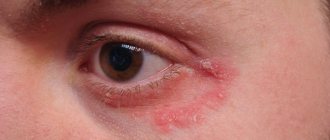
Dermatitis of the ear
It can be separated into a separate form, since inflammation of the skin of the auricle often occurs, without involving the NSP. The causes of the disease are a bacterial infection plus a factor of sensitization of the body to bacterial toxins. With this type of inflammation, redness and thickening of the skin occurs, with the formation of small serous blisters, similar to those in eczema. When the bubbles open, yellow crusts form in their place. Diagnosis is made based on examination, and consultation with a dermatologist may be required. Treatment is also antibacterial and anti-inflammatory, and antihistamines may be prescribed.
Erysipelas of the auricle
Acute erysipelas is an infectious disease and is caused by streptococcus pyogenes; later, other flora may join. With such inflammation there are symptoms of skin damage and a general inflammatory syndrome - weakness, fever, chills. Factors provoking the disease: decreased immunity, abrasions and burns of the skin, swimming in polluted waters, contact with a carrier of pyogenic streptococcus or a sick person. Depending on the depth of damage to the skin, three forms are distinguished:
- Erythematous, when pain occurs, redness of the skin (it has a “varnished” appearance) slightly rises above the level of healthy skin and has a clear border with it.
- The bullous form, more severe, is characterized by the same skin lesions as in the erythematous form, but with the formation of large blisters above the surface of the skin with transparent or cloudy contents.
- Necrotic form. With this form, deep damage to the skin occurs, right down to the reticular and papillary layers, and deep ulcers with purulent plaques form. Due to the addition of various microflora, it is more severe than other forms. Typically, patients with this form require surgical debridement of areas of necrosis.
Erysipelas
This is a serious disease that often causes complications, and its treatment should take place in an infectious diseases hospital. If erysipelas is suspected, consultation with an infectious disease specialist is required. Isolated erysipelas of the auricle is rare and is usually combined with lesions of the skin of the face or neck.
Treatment
Massive antibacterial and anti-inflammatory therapy, detoxification therapy, and antistreptococcal gamma globulin are prescribed.
Malignant otitis externa (necrotizing otitis externa)
This form of external otitis is distinguished as a separate one, but in fact it is a negative course of ordinary diffuse external otitis. During the course of the disease, inflammation spreads to the structures of the temporal bone, damage to the cranial nerves and the development of intracranial complications may occur. The name of the disease is not associated with the development of a malignant tumor, it simply reflects the course of the disease. Fortunately, this disease is rare; it affects patients with severe immune disorders, such as AIDS, conditions after radiation and chemotherapy, decompensated diabetes mellitus, and patients receiving immunosuppressive and cytostatic therapy. The leading pathogen is Pseudomonas aeruginosa, but any infection is polymicrobial. During the process of inflammation, necrosis of the skin of the ear canal occurs and inflammation passes to the periosteum and bone, followed by its destruction. Complaints in this disease include severe pain, discharge from the ear of a purulent-inflammatory nature, with a putrid odor, and hearing loss quickly occurs. Later, symptoms of damage to the cranial nerves appear: difficulty swallowing, choking when eating, numbness of the face, paralysis of the facial muscles. When the process spreads to the middle and inner ear, systemic vertigo may develop. When inflammation spreads into the cranial cavity, cerebral and meningeal symptoms develop. On examination, one notices pronounced inflammation of the skin of the NSP, skin with foci of necrosis, a large amount of purulent-necrotic discharge, the eardrum and the structures of the middle ear are often indefinable. Symptoms of damage to the cranial nerves are determined - facial asymmetry, ptosis of the lower eyelid on the affected side, asymmetry of the soft palate, impaired mobility of the epiglottis. In diagnosis, culture for bacteriological examination and computed tomography of the temporal bones are important. Treatment is always carried out in a hospital setting. Massive antibacterial and detoxification therapy is carried out. Infusions of human gamma globulin, antipseudomonas serum or hyperimmune human plasma are prescribed. Surgical treatment is also performed - necrotomy (removal of necrotic areas of skin and bone), and subsequently, upon recovery, plastic surgery of the defect is performed using one’s own tissues.
3. Symptoms and diagnosis
The furuncle of the external auditory canal in terms of dynamics does not differ from abscess processes of other localization: the infiltration stage is followed by violent suppuration, then the abscess opens (spontaneously or during medical intervention - but in no case should this be done independently!), and after passive or forced evacuation of purulent-necrotic masses results in fairly rapid healing and scarring. At the stage of infiltration, pain is expressed; the pain can be very severe, throbbing or shooting in nature. As a rule, body temperature is elevated, sometimes to high values. Any mechanical impact, including chewing, swallowing or talking, sharply increases pain. Severe swelling and a significant size of the boil can lead to significant obstruction of the ear canal and, accordingly, to transient hearing loss on the affected side. When the peripheral endings of the vagus nerve are involved in the process, coughing and vomiting are possible.
The diagnosis usually does not cause any difficulties for an experienced otolaryngologist. In case of repeated or multiple boils, measures are taken to laboratory identification of the infectious agent; as necessary, an endocrinologist, infectious disease specialist and other specialized specialists are involved in the diagnostic and treatment process.
About our clinic Chistye Prudy metro station Medintercom page!
Chondroperichondritis of the auricle
This disease is an inflammation of the perichondrium and cartilage of the auricle caused by bacterial flora. Chondroperichondritis can be caused by trauma to the auricle with damage to the skin, suppuration of an auricular hematoma, deep burns of the auricle, a boil or suppurating atheroma of the ear, and erysipelas. There are infiltrative and purulent forms. A few days after the described traumatic factor, infiltration and hyperemia of the skin of the auricle develops, accompanied by pain, with or without fever. An important diagnostic criterion is that the earlobe remains unaffected, since there is no cartilage in its thickness. Treatment: antibacterial and anti-inflammatory therapy. If there are purulent cavities, an opening with removal of pus and gentle necrotomy of the destroyed cartilage is performed. The disease is difficult to treat and can last for weeks or months. Often the consequence of chondroperichondritis is cicatricial deformation of the auricle; sometimes there may be complete destruction of the cartilage.