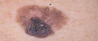
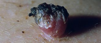
Description
Papillomas on the surface of a mole are an ambiguous phenomenon that requires careful study and diagnostic measures.
A nevus (mole) is a benign formation, which, as an independent manifestation, is harmless to the human body. Its structure is made up of special cells - melanocytes, arranged in layers.
Inside the cavity there are follicles, sweat and sebaceous glands. However, the basal epithelial layer is absent. It is from this that the formation of papillomas occurs.
What does papilloma look like?
Papilloma is a benign neoplasm that appears on the body when infected with papillomavirus.
This neoplasm is benign in nature, but differs in the nature of its origin. The development of papillomas occurs when infected with human papillomavirus.
The growth may be light pink or flesh-colored. The shape resembles a nipple on a stalk, rising above the surface of the skin. Has a predisposition to increase in length.
Consists of a loose, soft structure and choroid plexuses. The diameter can reach from 2 to 15 millimeters. As a result of injury, its rapid growth is noted.
When exposed to certain factors, various changes can occur in the body, against the background of which a condition is observed when a papilloma grows from a mole.
If such a phenomenon occurs, you need to worry. Separated from each other, these neoplasms do not pose a danger to human health. But when a papilloma begins to grow from a mole, it may indicate cellular dysplasia.
Reasons for the appearance of formations on a mole
The human body is covered with benign growths that do not pose a health hazard. A mole with growths is an alarming signal that requires the attention of a dermatologist or oncologist. Any growth, white or pink, is considered abnormal.
One of the unpleasant properties of moles is the ability to degenerate into malignant ones; a striking symptom is the appearance of a process on the pigmented area under the influence of various factors. Causes:
- The influence of ultraviolet rays. Excessive sun tanning increases the production of melanocytes, with a large accumulation, which break through into new growths on the skin. The shoots grow on the face, back, and in areas most exposed to sunlight. The rays affect the increase in the size of the nevus, change the color to a darker side, or provoke burnout, acquiring a light shade. The risk of degeneration increases with age for sunburn received in early childhood.
- Genetic disorders, inherited, are expressed by the inability to adapt to the negative influence of external factors.
- Hormonal disorders during pregnancy, puberty, menopause.
- Injury to growths. Providing poor-quality care is fraught with the development of an inflammatory process with the release of purulent fluid.
- A bite of an insect.
- The presence of a large number of moles and freckles on the body. Growths may appear on formations with uneven edges.
All factors are triggers in the development of melanoma.
Causes
The main provoking factor in the formation of a neoplasm on a nevus is the penetration and activation of the human papillomavirus.
Infection is possible in several ways:
- injury (infection enters the body through scratches, cuts, microscopic cracks);
- unprotected sexual contact with a person who is a carrier of an active virus;
- use of general hygiene products (washcloth, soap, towel, etc.);
- transmission of a viral infection from mother to newborn during passage through the birth canal.
In addition, papilloma can occur as a result of its damage, especially if the nevus is hanging or convex.
What is papilloma
Papilloma (wart) is a benign formation caused by papillomavirus (HPV).
Growths can appear on various parts of the body, both on the skin and on the mucous membranes, including the surfaces of internal organs.
Favorite places for warts to be localized are the groin and axillary areas, feet, and neck area.
Some types of the virus are highly oncogenic, causing cancer formation.
The tendency to malignancy is more typical for hanging and large papillomas.
Infection with the human papillomavirus can occur not only through sexual contact, but also through household contact.
Papillomas can vary in shape and size, and sometimes remain completely invisible.
HPV is able to penetrate the body through the skin and mucous membranes.
The pathogen immediately begins to reproduce, causing organ damage.
Signs of infection do not appear immediately, so the patient may find out that he has an infection months or even years later.
Symptoms
Papilloma developing from a mole is quite difficult to notice immediately. First of all, an alarming signal is a change in the shape of a mole, as well as its growth.
What causes papillomas?
Experts identify a wide variety of causes for the appearance of papillomas.
In addition, the following signs will indicate transformation:
- blurred boundaries;
- pain during touch;
- change in shade;
- bleeding;
- characteristic discharge from a mole.
It is worth noting that papillomatosis is distinguished by its rapid development, despite the fact that the first symptoms of manifestation may go unnoticed.
Moles and papillomas: what must be removed and when
Experts do not recommend removing moles if they behave calmly and do not cause discomfort to their owner.
If any complications arise from the nevus, and doctors strongly recommend removing it, then it is advisable to do this as soon as possible.
It is strictly not recommended to do this yourself at home.
It is necessary to get rid of unwanted moles only in a medical facility.
Before removing the tumor, the specialist will conduct the necessary examination, after which he will suggest one of the methods for removing the tumor.
Doctors are well versed in which nevus removal methods are the most effective and safe.
As for papillomas, you will also need to consult a doctor.
After an examination and a series of diagnostic tests, the doctor will prescribe removal of growths using the most gentle method and antiviral therapy.
Treatment of papillomas and warts comes down to their removal; the difference is that for each type of tumor, the doctor selects the method that will be most optimal for each specific case.
If it is necessary to send biomaterial for diagnostics, radio wave therapy or surgery will most likely be used.
In order to remove tumors, the following techniques are used:
- Cryodestruction . The technique is based on the use of low temperatures. Liquid nitrogen is used to freeze papillomas, which provokes necrosis of pathological tissues.
- Chemical removal method . Medications containing high concentrations of substances that cause chemical burns are used. The formations dry out and subsequently fall off.
- Electrocoagulation. High frequency current is used for removal. The tumors are burned out.
- Laser therapy. This technique is the most preferable, since after removal of papillomas there are no traces left on the skin. After the procedure, the recovery period is short.
- The radio wave technique is based on the use of radio waves, which promotes excision of the growth.
- Surgical method of removal . A scalpel is used to cut out the affected tissue, after which the material is sent to the laboratory for histological analysis. The technique is indicated for suspected malignancy of the process.
For small papillomas, conservative therapy is carried out using antiviral drugs.
Diagnostics
If a papilloma formation is detected on a mole, you must seek help from a specialist. First of all, the doctor will collect the necessary information regarding the patient’s medical history, the time when the growth of an additional tumor was noticed, and what symptoms accompany the process.
After this, a visual inspection of the affected area is carried out, which allows you to determine the size and color of the growth.
If this data is insufficient, an additional diagnostic examination is carried out, including several methods:
- biopsy - necessary to take a fragment of the affected tissue;
- histology – a histological examination of a previously taken sample of material is carried out to determine the nature of the neoplasm and the mole itself on which it appeared, which makes it possible to determine the degree of malignancy of the pathological process;
- PCR diagnostics to detect urogenital infection.
Also, if necessary, a laboratory blood test can be performed, which will also show some deviations from the norm in the presence of the disease.
What to do if a growth on a mole falls off
A growth on a hanging mole may fall off due to poor blood circulation. If the appendage has fallen off, you need to see a doctor to have the tissue tested.
To avoid unpleasant complications, it is recommended to regularly examine the formations covering the body.
If alarming symptoms appear, you should contact the clinic. Signs include:
- the appearance of a wart or any type of growth on a mole;
- change in size, shape;
- color change. Dangerous shades are considered: black, red and white;
- itching, burning, redness, peeling;
- inflammatory process with the formation of ulcers that secrete pus and an unpleasant odor;
- solid build-up structure;
- blister formation, weeping surface, discharge of clear liquid.
If such signs are detected, it is recommended to seek help from a doctor who will help you understand the causes of the deviations. Diagnostics involves histological examination of biomaterial to determine malignancy. Additional tests may be needed to determine the presence of metastases and their growth. The ability of metastases to spread through the circulatory and lymphatic systems causes rapid damage to the lungs, liver, and brain.
We recommend reading
- Causes and dangerous consequences of mole removal
- Is it possible to peel off the crust after removing a mole yourself?
- Are watery moles dangerous?
Ignoring the symptoms leads to serious consequences - the development of melanoma, a dangerous type of cancer. Oncology of this type causes rapid progression.
The disease is fatal; timely treatment in the early stages is important.
Once a growth appears, treatment is possible according to the following options:
Regular observation if no malignant signs are detected, using a photographic method that allows you to monitor changes in color, size, structure.
Removing a growth on a mole using modern medicine methods. Cryodestruction – freezing of the formation using liquid nitrogen. Liquid nitrogen causes tissue necrosis, manifested by the appearance of a bubble. Then a crust appears that does not require intervention. It should fall off on its own, leaving a pink spot. Disadvantage: it is impossible to control the depth of exposure of liquid nitrogen to the growth; incomplete removal is possible, requiring a repeat procedure.
Laser therapy – removal using a laser that does not cause pain or bleeding. Elimination of small moles, without scars. But the biomaterial cannot be sent for research.
Radio wave method - allows you to excise the growth. Removal if the formation is small. Provides the possibility of studying biomaterial.
Removal of the tumor along with a portion of the skin. For the treatment method, surgical intervention is a suitable option. During the operation, the tumor and part of the skin are removed to exclude the possible development of oncology. In addition to surgery, additional treatment of chemotherapy and radiation will be required.
Timely treatment and compliance with all prescriptions increase the chances of recovery, otherwise it can result in death.
Treatment methods
The tactics of therapeutic measures will depend on the results of the examination.
Self-medication is dangerous with complications!
Attention
Despite the fact that our articles are based on trusted sources and have been tested by practicing doctors, the same symptoms can be signs of different diseases, and the disease may not proceed according to the textbook.
Pros of seeing a doctor:
- Only a specialist will prescribe suitable medications.
- Recovery will be easier and faster.
- The doctor will monitor the course of the disease and help avoid complications.
find a doctor
Do not try to treat yourself - consult a specialist.
You just have to remember that treatment should be carried out in a complex manner. It is mandatory to prescribe a course of antiviral drugs, as well as medications that help increase and strengthen the patient’s immune system.
Why can our articles be trusted?
We make health information clear, accessible and relevant.
- All articles are checked by practicing doctors.
- We take scientific literature and the latest research as a basis.
- We publish detailed articles that answer all questions.
To remove papilloma on a mole, doctors can use several methods.
Traditional surgery
This surgical technique is used if the papilloma in diameter exceeds more than one centimeter, or if the development of an oncological process is suspected.
The procedure consists of complete excision of the tumor along with the nevus on which it formed, as well as uninjured areas of the skin. This effect reduces the risk of relapse, that is, the reappearance of tumors.
Laser therapy
Refers to low-traumatic techniques. It involves influencing the growth using a laser beam. This tactic is used when formations appear that do not exceed five millimeters in diameter.
Cryodestruction
The procedure is carried out using liquid nitrogen. Under the influence of low temperatures, the papilloma freezes and will be removed. This technique is not recommended for getting rid of papillomas on moles localized in the facial area, as it has a number of side effects.
These include swelling of the treated area, death of hair follicles, and increased pigmentation.
Electrocoagulation
High frequency waves are used. Using the procedure, you can remove the formation, as well as immediately take a sample of pathological tissue for further cytological and histological examination.
Radiotherapy
Removal of moles with papilloma is carried out under the influence of a radio wave scalpel. It can be used in the formation of multiple papillomas, as well as if they are located on the genitals.
Could the appendage be a cancerous tumor?
A malignant tumor that appears due to the activity of melanocytes can be recognized by its appearance:
- superficial location in the form of a process on the skin or mole;
- knotty - the most dangerous and aggressive. It looks like a nodule and can appear on a pigmented area of the skin;
- subungual melanoma - affects the lower extremities, in the area of the nail plate of the big toes, soles.
Any growth on the surface of a mole may turn out to be a cancerous tumor that requires immediate treatment.
Why is it dangerous?
The danger of this condition, in which there is a growth of papilloma inside a mole, lies primarily in the malignancy of the pathological process, that is, certain types of moles, under the influence of certain factors, can degenerate into a malignant tumor.
During the process of degeneration, dark-colored inclusions appear in the structure of the nevus, which, as they progress, begin to grow and outwardly resemble a wart.
In addition, when the papilloma enlarges, it is quite easy to injure it, which can cause bleeding. Against this background, an infection can penetrate into the resulting wound and spread through the bloodstream throughout the body. This condition, in turn, can lead to the development of sepsis or an abscess.
How quickly does malignancy occur?
Malignancy is the process of transforming healthy cells into cancer cells. Daughter cells acquire malignant features. Pathology occurs at different speeds depending on the individual characteristics of the patient’s body.
Stages of development
Malignancy occurs in several stages.
- Initiation External and internal harmful factors provoke cell mutation. The appearance of oncogenes causes rapid growth and reproduction of affected cells. In a healthy body, such cells die, but due to initiation they continue to divide. However, this stage is not yet the beginning of the development of oncology.
- Promotion At the second stage, oncogenes are activated, provoking the proliferation of diseased cells.
- Evasion of diseased cells from differentiation Differentiation is changes in the functions, size, and shape of a cell that allow it to perform its genetic functions. Poorly differentiated cells eventually form an altered area of tissue, which later becomes the basis of the tumor. Such a mole already leads to skin cancer.
- Tumor progression Uncontrolled proliferation of altered cells leads to the germination of a diseased area of tissue, which begins to destroy the walls of blood vessels. The affected cells spread throughout the body, ending up in the lymph nodes and distant organs. There the cells settle and the formation of metastases begins.
Prevention
To avoid possible risks, you need to be careful about your skin. First of all, this applies to people who have fair skin, blue eyes and a lot of freckles on their body. It is this category that is more susceptible to cancer processes.
To avoid the growth of moles and the formation of papillomas on them, it is not recommended to spend a lot of time under the sun. Equally important is proper nutrition and healthy lifestyle.
Moles and papilloma individually are not dangerous to human health. However, if a papilloma growth is formed from the nevus itself, then in this case you need to be extremely careful, and it is better to consult a doctor, since such a condition may signal a possible process of malignancy.
Could it be a wart or other formations?
There are two types of age spots: congenital and acquired.
Acquired moles may develop warts or papilloma. There are characteristic features that distinguish nevus from other growths.
The following is considered normal:
- the presence of a light brown or dark brown tint;
- round or oval shape with clearly defined edges;
- size up to 10 millimeters.
Description of a benign formation that does not pose a health hazard.


