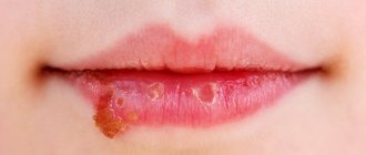
The terms "dermatitis" and "eczema" are synonymous. The term eczema is usually applied to the dermatitis seen in atopic individuals. The term "dermatitis" means inflammation of the skin. It can be acute with weeping, formation of crusts and vesicles, subacute or chronic with dryness, peeling and cracks.
Skin rashes are almost always itchy and are usually complicated by secondary infection.
Dermatitis can be exogenous (eg, contact, irritant, infectious or photodermatitis) or endogenous (eg, atopic, seborrheic, discoid).
Most often, the diagnosis is made on the basis of a detailed history and morphological studies of skin rashes, taking into account the characteristics of their distribution.
Atopic dermatitis
Atopic dermatitis begins in childhood, between 2 and 6 months of age, and affects about 2% of the population, but is now experiencing an increase in prevalence. A family history of atopy is present in 70% of patients. Hay fever (hay fever) and/or asthma may develop as your child gets older. More than 90% of children recover by age 12, but it is very difficult to predict this in an individual child.
Atopic dermatitis in children affects the face, neck, and torso. Later, the flexor surfaces are involved in the process: popliteal, elbow, wrist and ankle joints, and as a result of repeated scratching, lichenification of the skin can occur. Hand dermatitis usually appears later in life. Secondary infection is common and often leads to worsening of the dermatitis. Pruritus can be debilitating and cause great distress for patients and families. Some patients, especially infants and young children, have food allergies, most often to eggs, fish and dairy products. With atopic dermatitis, many skin disorders can be identified. Immunological abnormalities include a tendency to increase immunoglobulin E, a predisposition to anaphylactic reactions, and a decrease in local cell-mediated immunity. This leads to increased sensitivity to viral infections.
Make an appointment now!
Leave your contact details or call and our manager will contact you to make an instant appointment
FUNGAL DISEASES OF THE FOOT
The first place among fungal diseases is occupied by mycosis of the feet. The disease is widespread. The incidence of mycosis of the feet in the general population ranges from 5 to 20% and reaches 50% among patients with immunodeficiency, endocrine disorders, and somatic diseases. Mycosis is often observed in patients suffering from various dermatoses. With mycosis of the feet, in 40–50% of cases, the nail plates are affected. According to foreign researchers, onychomycosis occurs in 2.6-35% of people aged 55 years and older, and according to researchers from the UK and the USA, in 4.7% of people in this age group. Onychomycosis in children is rarely recorded, its prevalence ranges from 0.2 to 0.44%, according to scientists from Belgium, Canada and the USA, and from 0.6 to 1.5%, according to domestic researchers.
Mycosis of the feet is a collective name commonly used to refer mainly to two diseases caused by Trichophyton rubrum (T. rubrum) and Trichophyton mentagrophytes var. interdigitale (T. interdigitale).
The main causative agents of onychomycosis are dermatomycetes (from 60 to 95%): of these, T. rubrum is in first place, causing damage to the toenails, hands and any part of the skin; on the second - T. interdigitale, affecting the nails on the first and fifth toes and the skin of the third and fourth interdigital folds, the lateral surfaces of the toes, the upper third of the sole and the arch of the foot. Nail damage can be caused by T. violaceum, T. tonsurans, T. schoenleinii, T. mentagrophytes var. gypseum, T. verrucosum, rarely by fungi of the genus Microsporum. Other causative agents of onychomycosis include Epidermophyton flocossum, yeast-like and mold fungi. Of the mold fungi, Scopulariopsis brevicaulis is the most common, causing predominantly damage to the nails on the first toes of older people, then various species of Aspergillus, Penicillium, Cephalosporium, Alternaria, Acremonium, Fusarium, Scutalidium, etc. At the same time, reports of damage to nails began to appear more and more often , caused by non-dermatomycetes and having an unusual clinical picture.
Infection with mycosis of the feet and hands can occur through direct contact with a patient in a family or group, or through shoes, clothing, household items (bathroom rugs, washcloths, manicure accessories, etc.). The causative agents of the disease are found in large quantities in pieces of affected nail plates or scales from lesions on the skin. More often, people who constantly use a bathhouse, shower, or swimming pool are susceptible to fungal disease. When walking barefoot, a patient with mycosis of the feet leaves fungus-infected scales on the floor, in the foot basin, which easily stick to the skin that is damp after washing, especially in the area of the interdigital folds. The penetration of fungi into the epidermis is facilitated by a violation of the integrity of the skin, caused by sweating or dry skin, diaper rash, abrasion, minor trauma, poor drying of interdigital folds after water procedures, etc.
Infection with fungi is not equivalent to a disease, since when they come into contact with human skin, they do not always cause clinically significant changes. Some people experience low-symptomatic lesions or mycocarriage. Infection is facilitated by injuries to the nails, fractures of the bones of the feet, hands, and impaired blood supply to the extremities. Persons with concomitant pathologies - endocrine, especially diabetes mellitus, severe somatic, immune and other disorders - are more at risk of getting sick.
The low incidence of onychomycosis in children is explained by: rapid growth of nails; smaller nail surface area available for fungal penetration; less frequent nail injuries; low prevalence of mycosis of the smooth skin of the feet in this age group.
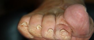
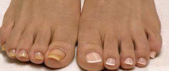
Clinical manifestations of onychomycosis are varied and depend on the type of pathogen. The nails on the feet are most often affected (up to 80%), less often on the hands, and simultaneous lesions on the feet and hands are also found. It should be noted that the appearance of nails in some cases, both with onychomycosis and with diseases of a non-fungal nature, may be the same, and therefore diagnostic errors are possible.
With onychomycosis caused by T. rubrum, nail damage is often multiple. The disease on the toes begins with the appearance of yellow spots or longitudinal stripes in the area of the lateral edges of the plates. On the hands, they appear in the center of the plate and have a lighter color - whitish or grayish, the nails become dull. Depending on the nature of the changes in the plate, the following forms of damage are distinguished: normotrophic, hypertrophic, atrophic and onycholysis-type damage. In the normotrophic form, the normal configuration of the affected nails remains for a long time; they become dull, acquire a yellowish color at the distal edge, with thickening in the corners of the plates due to subungual hyperkeratosis. In the hypertrophic form, the nail plate is thickened, and initially a yellowish color is noted; gradually the nails become deformed, transverse striations appear, a dull, dirty gray color appears, and loosening appears at the free edge. In some patients, the nails acquire a beak-like shape or change like onychogryphosis, most often on the first toes. In the atrophic form, the plates are significantly destroyed, deformed and look as if corroded at the distal edge, the nail bed is partially exposed, covered with a layer of crumbling loose horny masses, the nails become dull, yellowish and grayish in color. When affected by onycholysis, the plates become thinner, separate from the nail bed, lose their shine, become dull, dirty gray or yellowish in color. However, the matrix area retains normal coloration. One patient may experience combined nail damage.
Nail damage with onychomycosis caused by T. rubrum in children has some features: the configuration of the nail may not be changed, but its surface is rough or exfoliated; nail damage is more common at the distal edge, less often at the lateral edge in the form of slight thickening or onycholysis; change in the type of onychodystrophy (nails are lumpy, gray); the color of the nails may not change; subungual hyperkeratosis is rarely observed. Damage to smooth skin on the feet may be absent or characterized by fine-plate peeling on the inner surface of the terminal phalanges, most often the third or fourth; sometimes superficial, less often deep cracks are observed, mainly in the third and fourth interdigital folds.
With onychomycosis caused by T. interdigitale, a normotrophic form of the lesion is more often observed; spots or stripes of bright yellow color appear in the thickness of the plate, in the center of it; sometimes thickening of the nail is observed at the free edge, deformation of the plate, which looks as if eaten away.
Nail damage caused by anthropophilic trichophytons (T. violaceum, T. tonsuraus, T. schoenleinii) is usually observed with simultaneous damage to the scalp and smooth skin. With superficial trichophytosis, the disease most often begins with damage to the nails of the hands, and later the toenails are also involved in the process. Gray spots or stripes appear at the distal edge and on the lateral parts of the nail, the plate becomes thickened, with grooves, and crumbles. With favus, a yellowish spot forms in the thickness of the nail, which, gradually increasing, covers the entire plate. The configuration of the nail does not change for a long time, then the plate thickens, becomes deformed and begins to crumble. Usually the nails of the hands and toenails are affected in patients who have suffered from favus for a long time.
Nail damage caused by zoophilic trichophytons and microsporums is extremely rare. There are isolated reports in the literature, including a description of two children with microsporic onychomycosis observed by the author. The clinical picture resembled onychodystrophy, the disease began with leukonychia, then changes in the configuration of the nail appeared, later destruction of the plate from the distal or proximal edge was noted, it did not grow to the bed, the color was normal or yellowish.
With onychomycosis caused by yeast-like fungi of the genus Candida, the lesion begins from the posterior or lateral ridges, mainly on the fingers. The ridges become thickened, swollen, hyperemic, silvery scales are visible along the edges, the eponychion disappears, pain occurs on palpation, and sometimes a drop of pus separates. The nail plates are usually uneven; transverse grooves appear on them, running parallel to the posterior ridge; sometimes the plate is destroyed in the proximal area. However, these changes in the nail are caused by trophic disorders in the area of the roller. With candidiasis, the nail plate becomes thinner, more often at the lateral edges, less often in the distal part, does not grow to the bed, and is yellowish in color. There may be damage to the nail without changing the ridge.
Onychomycosis caused by mold fungi, as a rule, develops secondary to onychodystrophy of various etiologies; the damage is superficial. The color of the nail plate varies depending on the type of pathogen: it can be yellow, green, blue, brown or black.
Depending on the location of the lesion, it is necessary to distinguish between distal, lateral, distal-lateral, proximal, total and superficial onychomycosis. Moreover, the lateral lesion can spread along the edge of the plate by 1/3, 2/3, as well as to the matrix and deeper. This is extremely important to consider when prescribing treatment.
Diagnosis of onychomycosis is based on clinical manifestations and detection of the fungus during microscopic examination of pathological material. The type of fungus is determined by sowing on Sabouraud's nutrient medium.
Onychomycosis should be differentiated primarily from onychodystrophy of unknown etiology, with frequent changes in the nails due to dermatoses (eczema, psoriasis, lichen planus). Changes in nails due to skin diseases can precede skin rashes and even be isolated for a long time.
Onychodystrophy or trophic changes in nails can develop as a result of the direct influence of various factors: contact with washing powder, cleaning products, occupational hazards, etc., and also arise as a result of pathology of internal organs. The disease is characterized by clinical polymorphism.
With eczema on the fingers or toes, the nails become uneven due to the formation of transverse grooves, they soften, and peel off at the distal edge. As a rule, the nail fold is thickened, the eponychion may be absent, as with candidiasis, but the inflammatory phenomena are insignificant.
With psoriasis, peeling of the nails from the bed at the distal edge (onycholysis) is often observed; Thickening of the plates may be observed due to subungual hyperkeratosis, in some patients - thimble-shaped wear of the plates with peeling in the pits; sometimes the nails are destroyed and acquire a yellowish color, but the most characteristic sign is the compaction of the skin ridge at the altered distal edge of the plate, which is never observed with onychodystrophy.
With lichen planus, a deep crack is often observed in the center of the nail, but the change can also be in the form of longitudinal ridges, cracks, with pronounced subungual hyperkeratosis; the plates break off at the distal edge. Due to splitting and brittleness of the nail, partial or complete loss sometimes occurs. When choosing a treatment method for a patient with onychomycosis, it is necessary to take into account the type and shape of the lesion, the prevalence of the process, the rate of nail growth, general condition, safety, method of application, as well as material costs.
If single nails are affected from the distal or lateral edges by 1/3-1/2 of the plate, it can be cured only with the help of external antifungal agents and cleanings. According to the first method, the following is used externally until healthy nails grow back: nizoral cream twice a day, terbinafine cream (fungoterbin, mycoterbine, terbifin, exifin) once or twice a day, mycozoral cream twice a day, canizon cream or solution twice a day , mifungar cream once a day.
Cleaning is carried out using keratolytic agents: 20% urea patch (ureaplast) or mycospor ointment in a nail treatment kit. They are performed until healthy nails grow completely at intervals of two to four weeks. After removing the infected areas of the nails, apply one of the above medications to the cleaned nail bed twice a day.
The most effective preparation for external treatment of nails is mycospor ointment in the set. Treatment is carried out in two stages. At the first stage, the nail plates are removed using an ointment containing bifonazole and urea. The ointment is applied to the affected nails using a dispenser and left for a day under a waterproof plaster. Before applying the ointment again, remove the bandage, make a soap and soda bath, and remove the affected areas of the nail with a special file. The procedures are repeated until the plate is completely removed. On average, painless layer-by-layer nail removal occurs in two weeks (from four to 28 days). The advantage of this method is that the nail bed is treated already at the first stage. In the future, it is recommended to rub 1% mycospor cream once a day in the evening every day for four weeks or longer, depending on the degree of damage to the nail, at the same time the cream is applied to the skin of the feet (hands). The duration of treatment depends on the clinical form and area of the lesion. The tolerability of mycospore ointment is satisfactory, so this technique can be used in patients suffering from onychomycosis and eczema of the feet (hands).
According to the second method, you can use 5% loteryl varnish or 8% batrafen varnish externally for six to eight months when nails are affected on the hands, and 9 to 12 months on the feet. Loceryl varnish is applied to nails once a week for at least six months. Batrafen varnish is used: every other day during the first month, twice a week during the second month, once a week for the third month and beyond, until healthy nails grow back.
Patients with multiple lesions of nails and skin, with a distal-lateral type of lesion and involvement of the matrix in the process, proximal, total type, as well as in the absence of effect from local therapy, are treated with systemic antimycotics. It should be noted that the therapeutic effect can be achieved with monotherapy with highly effective systemic antimycotics, however, as our own observations and the experience of other researchers show, in case of a hyperkeratotic form, a total lesion, the affected nails and horny layers in the bed area should be removed using keratolytic agents, which allows to reduce the duration and increase the effectiveness of treatment.
Currently, there are five antifungal drugs for oral use (griseofulvin, ketoconazole, terbinafine, itraconazole, fluconazole), of which three can be considered highly effective: terbinafine, itraconazole and fluconazole.
Terbinafine (lamisil, exifin, terbizil, onychon) is an antifungal drug with fungicidal action, belongs to the class of allylamines, available in tablets of 250 and 125 mg. The daily dose for adults is 250 mg, for children it is calculated depending on the child’s body weight: for body weight up to 20 kg, 62.5 mg/day is prescribed, from 20 to 40 kg - 125 mg/day, over 40 kg - 250 mg/day. days The drug is used for onychomycosis caused by dermatophytes at a dose of 250 mg once and at a dose of 125 mg twice a day, daily. The duration of treatment is six weeks for fingernails and 12 weeks for feet. The treatment period can be more than six months for onychomycosis on the first toes, hypertrophic form, slow growth of nails.
Itraconazole (orungal, irunin) is an antifungal drug belonging to the azole class, a broad-spectrum drug, available in 100 mg capsules, it is used in patients with onychomycosis caused by dermatophytes, yeasts and molds. Itraconazole is most effective when prescribed using the pulse therapy method, i.e. it must be prescribed 400 mg in two doses (two capsules in the morning and evening) for seven days, then after a three-week break the course is repeated. Duration of therapy: three to four courses.
Fluconazole (Diflucan, Mycoflucan, Mycosist, Forkan, Flucostat, Mycosist, Mycomax) is a drug belonging to the class of triazole antifungal agents, available in capsules of 50, 100, 150 and 200 mg, and has a wide spectrum of action. It is prescribed 150 mg once a week, on a fixed day, until healthy nails grow back. The drug is effective for onychomycosis of the hands, onychomycosis of the feet (hands) in children, onychomycosis of the feet without damage to the matrix, single lesions of the nails in patients under the age of 40 years.
Ketoconazole (Nizoral) is an antifungal drug from the azole class, effective against dermatomycetes and yeast fungi. For onychomycosis, it is taken 200 mg/day (in the first week 400 mg/day) after meals every day until healthy nails grow back.
Griseofulvin is an antifungal antibiotic with fungistatic activity against dermatomycetes, in tablets of 0.125 g. Griseofulvin for children is prescribed in a daily dose of 16 mg per 1 kg of body weight; adults weighing up to 50 kg - five tablets, then for every 10 kg over 50 add one tablet. The daily dose is no more than eight tablets (1 g). The antibiotic is used in three doses with a teaspoon of vegetable oil every day for the first month, every other day for the second month, then twice a week until healthy nails grow back.
Treatment of mycosis of the smooth skin of the feet and other localizations is carried out with antifungal agents for external use. Various antifungal agents are used, depending on the severity of inflammation, peeling and keratosis (nizoral cream, mycozoral ointment, mifungar cream, canizon cream and solution, mycozon ointment, batrafen ointment and solution, exoderil cream and solution, terbinafine cream (Lamisil, exifin, fungoterbin, terbifin, terbizil, etc.)).
Regardless of the method of treatment for mycosis of the feet (hands) and onychomycosis, shoes (gloves) must be disinfected once a month, until healthy nails grow back, using a 1% solution of chlorhexidine bigluconate or a 25% formaldehyde solution as disinfectants.
Zh. V. Stepanova, Doctor of Medical Sciences, Professor of the Central Scientific Research Institute of Medical Sciences, Moscow
Note!
- The incidence of mycosis of the feet in the general population ranges from 5 to 20% and reaches 50% among patients with immunodeficiency, endocrine disorders, and somatic diseases.
- Onychomycosis occurs in 2.6-35% of people aged 55 years and older.
- The main causative agents of onychomycosis are dermatomycetes.
- Infection with mycosis of the feet and hands occurs through shoes, clothing, and household items (bathroom rugs, washcloths, manicure accessories).
- Clinical manifestations of onychomycosis depend on the type of pathogen.
- The appearance of nails in some cases, both with onychomycosis and with diseases of a non-fungal nature, may be the same, and therefore diagnostic errors are possible.
- Depending on the nature of the changes in the plate, the following forms of damage are distinguished: normotrophic, hypertrophic, atrophic and onycholysis-type damage.
- Diagnosis of onychomycosis is based on clinical manifestations and detection of the fungus during microscopic examination of pathological material.
- The most effective preparation for external treatment of nails is mycospor ointment in the set.
Discoid dermatitis
Discoid dermatitis is characterized by the appearance of round or oval spots in symmetrical areas, often on the extensor surfaces, usually in adult patients. Exogenous causes must be excluded.
Dyshidrosis is a variant of eczema in which recurrent vesicles or bullae affect the palms, fingers, toes, or both. The disease is characterized by remissions and exacerbations, which are sometimes provoked by fever, emotional stress, and active fungal infection.
Types of arthrosis development
For treatment of arthrosis of the foot joints to be successful, it is necessary to make a correct diagnosis. Each stage of disease development requires a different set of therapeutic measures. Doctors distinguish the following stages of pathology development:
- First. At this stage, noticeable fatigue of the lower extremities appears, as well as “pulling” pain after long walks or excessive physical exertion. At this stage, the patient may not even suspect that he is developing a disease, because he attributes the symptoms to banal fatigue. But if you do not visit a doctor, the disease will begin to gain momentum;
- Second. All the symptoms of the first stage begin to appear more actively. The pain becomes longer and more severe. Calluses may appear on the heels due to improper foot position, the knuckles become thicker;
- Third. The joints undergo serious changes, so the person begins to limp severely. Joint mobility decreases. X-rays show that the gaps between the joints are significantly reduced or disappear completely.
To make a diagnosis, the doctor must conduct an examination and diagnosis. It includes:
- taking an anamnesis, when the doctor asks the patient about all his complaints;
- X-rays and other examinations that will make it possible to accurately determine the condition of the joints, cartilage and bones, and also identify possible changes in their structure;
- examinations that will allow you to study the condition of muscle tissue;
- tests to find out about the state of the body and the nature of inflammation in it;
- measuring the size of the foot so that you can track the dynamics of treatment for arthrosis of the toe or any other joint.
Irritative dermatitis
In irritative dermatitis, the rash may be caused by physical or chemical irritation and damage to the skin, in which case it is not usually associated with an allergy. This type of damage can be caused by soap, detergents, food products, and building materials.
The necessary examination and adequate modern treatment for each form of dermatitis can be determined by highly qualified specialists of our center - the Three I Immunology Clinic - allergists and dermatologist. Come visit us at Novosibirsk, st. Galushchaka, 2, we will help solve your problems!
What is gout on women's legs?
Under the influence of provoking factors in women, the natural processes of uric acid secretion and entry into the body are disrupted, and the level of urea in the blood increases significantly. This is due to insufficient production of estrogen, which in the female body begins during menopause. Therefore, gout most often affects women over 50 years of age with acute deficiency of the sex hormone and pathologies of the endocrine system. The main symptoms are:
- Formation of tophi (accumulation of urates in articular tissues);
- Nephrolithiasis (accumulation of urea in the kidneys with subsequent formation of stones);
- Gouty (urate) nephropathy.
Frequently asked questions from our patients on the Internet about trophic ulcers
How to treat non-healing wounds on the legs?
To effectively treat non-healing leg wounds, it is necessary to understand how such wounds appeared. Long-term non-healing wounds on the legs can form as a result of a number of completely different pathologies, and each requires its own specific treatment.
How to treat non-healing wounds at home?
Before you begin to treat long-term non-healing wounds at home, you need to find out the cause of such wounds. Most often this is:
- Diabetes.
- Obliterating atherosclerosis.
- Chronic venous insufficiency.
If you want to cure long-term non-healing wounds, then in addition to the local use of ointments, it is necessary to deal with the underlying pathology that caused the problem.
How to treat a deep wound?
How to treat a deep wound should be asked to the doctor during an in-person consultation. Treating such wounds on your own, using the Internet, can be fraught with progression of the process and the development of a life-threatening condition.
My father developed sores on his legs six months ago, how can I get rid of them?
If “sores” on your legs appear and do not go away for a long time, as a rule, only a doctor will help you get rid of them. Long-term non-healing wounds are often a sign of a serious pathology that requires specific treatment.
Why is a trophic ulcer dangerous? - Wikipedia warns!
The main point in the treatment of trophic ulcers is to identify the underlying disease that caused the ulcerative skin defect. This approach will ensure effective treatment of ulcers and prevention of relapses. The danger of trophic ulcers lies in the fact that, in the absence of proper treatment, damaged cells are capable of malignant degeneration and the appearance of squamous cell carcinoma, and in rare cases, skin sarcoma. Therefore, even if the trophic ulcer has gone away, treatment after surgery should continue.
Trophic ulcer - treatment. Reviews from our patients.
Patient's review of laser treatment for trophic ulcers in our center.
Victor Petrovich, 65 years old.
I am a pensioner with a whole cartload of all sorts of ailments. My legs had been hurting and swollen for a long time, but I, like many, put off going to the doctor until later. I took some pills on the advice of a neighbor nurse, although I didn’t notice any particular improvement. In general, I lived to the point where I had a huge ulcer on my leg. God bless your doctors, I already thought that I would be left without a leg. They gave me laser treatment. I didn’t particularly delve into the nuances of this case; the doctors guaranteed a good result. Everything went without pain, literally after a few days my trophic ulcer began to disappear before my eyes, and after two weeks only a small scar remained. Of course, various medications were prescribed, then I went to a sanatorium and forgot about this terrible disease. Thanks to all the doctors of the Medical Center for Phlebology, especially Dr. Malakhov Alexey Mikhailovich.
A patient's review of the treatment of trophic ulcers using the EVLO method in our center.
Komissarova L.I.
Our patient with surgeon-phlebologist A.M. Malakhov. after treatment
My leg ached constantly, there was a buzzing sensation in it at the end of the day, and then a trophic ulcer formed, which became larger and larger. I didn’t dare to have surgery using the classical method, because I was afraid after reading reviews online and all sorts of complications. Well, my daughter is in the know, she found me an experienced doctor in Moscow, Alexey Mikhailovich Malakhov. Before the operation, and this is endovasal laser coagulation, at the medical center in Moscow, where I decided to have it, they did an ultrasound, donated blood and that’s it. After the operation, I was forced to get up from the table and walk for about an hour. After 3 weeks I saw my doctor. I didn’t worry about scars, the main thing was that the ulcer healed faster. It's been 3 months since the laser procedure on my leg. Everything is fine, the ulcer has become very small, but I hope it will heal completely soon.
I'm very glad that everything went well. Thanks for all. Komissarova Lyudmila Ivanovna, December 7, 2016
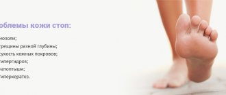
Prevention
Prevention of skin diseases of the feet:
- foot care: keep feet clean and dry;
- After water procedures, dry the skin between your fingers;
- choose loose, breathable shoes with good air circulation inside;
- use socks made from natural fabrics. Change them twice a day;
- dry your shoes for at least 24 hours before next use;
- individual replacement shoes;
- Even at home there should be personal items.
Trophic ulcer - treatment without surgery
If the cause of the ulcer is not venous pathology, then conservative measures are sufficient. A portable laser device is a great help in home treatment of the initial stages of ulcerative skin lesions up to 50 cm2 in size. A laser injected into a vein helps reduce blood viscosity, significantly reduces the risk of blood clots, and increases blood flow in problem areas. In addition, the laser beam effectively reduces pain and trophic ulcers. Laser treatment (reviews below) in combination with conservative treatment allows you to avoid surgical intervention.
Laser therapy is carried out in courses of 10-15 procedures. With the correct individual selection of treatment parameters, this treatment method does not cause complications, patients note good tolerability of the procedures, and the trophic ulcer is quickly eliminated. Laser treatment, the price of which is not too high, is very popular.
Symptoms
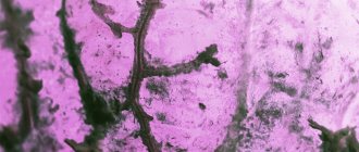
Conventionally, the following forms of mycosis of the feet are distinguished, which are presented in the photo:
- Squamous: unilateral peeling and mild itching in the arch of the foot. This form often goes unrecognized.
- Intertriginous: the folds between the toes are affected, then the process moves to the back of the foot. Weeping cracks appear, accompanied by itching. A bacterial infection may accompany the development of erysipelas.
- Dyshidrotic: on the arch of the foot, vesicles with light and then cloudy contents form, merging with each other. They open with the formation of painful erosions. This type is accompanied by itching and pain.
Mycosis of the skin of the foot in the acute period can be accompanied by fever, poor health, headache, and enlarged inguinal lymph nodes. When infected with trichophyton, the nail of the 1st or 5th finger is affected, and subsequently the process spreads to all nail plates. First, yellow spots appear at the free edge of the nail, then it thickens, loosens and crumbles. Therefore, to treat foot fungus, you need to use complex action products, for example, Clotrimazole lotion for skin and nails.