True eczema
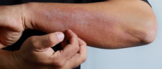
This variant of the disease is characterized by frequent exacerbations and is manifested by inflammatory reactions in symmetrical areas of the skin. The affected areas become red and swollen, and weeping surfaces with a large number of small blisters filled with serous contents are found. After the vesicles break through, small erosions form. Their surface resembles “serous wells” due to opalescent droplets of exudate.
As the disease progresses, the number of newly formed blisters decreases, erosions become covered with crusts, which subsequently transform into a scaly, scaly surface.
The main clinical manifestation of true eczema is a pronounced polymorphism of the elements of the rash. On the affected areas of the skin, rashes are detected at various stages of their resolution.
The basis for diagnosing this disease is the typical clinical picture. Most patients have a history of allergic conditions or recent emotional distress.
Eczema is a polyetiological skin disease with a predominance of an allergic component, in which various rashes with severe itching appear. This disease is prone to a chronic relapsing course, quite often complicated by the addition of a bacterial infection and mental disorders (anxiety-phobic, depressive).
The direct cause of the development of eczema has not been established; both internal and external factors play a role in its development. Triggering factors of endogenous origin include: hormonal imbalances, hereditary predisposition, pathology of the immune system in the form of dysglobulinemia (increased number of B-lymphocytes and class E and G antibodies and decreased M), formation of aggressive antigen-antibody complexes, disturbances in nervous regulation and many other. External influences include various chemical and physical influences, taking medications. The main mechanism for the development of eczema is considered to be a hereditarily determined pathology of immune reactions. Concomitant diseases of the gastrointestinal tract and biliary system can aggravate the course of the disease. The disease can occur in both acute and chronic forms. Depending on the external manifestations and triggering factors, true eczema, microbial, seborrheic, childhood and professional are distinguished.
The acute form begins with the appearance of swelling and redness of the skin, which is accompanied by rashes in the form of blisters and weeping erosions (sometimes plaques and pustules are noted). In their place, crusts and excoriations (scratching) gradually appear. New elements can be formed over a long period of time, so the external clinical picture may differ in polymorphism. When the process subsides, at the site of the rash there remains an area of thickening and redness of the skin with scaly peeling and traces of scratching. The chronic stage is caused by thickening of the skin with an enhanced pattern, areas of hyper- or hypopigmentation. Patients complain of severe itching and burning at all stages of the disease, but especially during the period of active rashes.
True eczema (idiopathic, which means without a proven cause) is characterized by the appearance of foci of rashes with unclear boundaries, localized on the face or extremities, which alternate with normal skin. After the acute stage it usually becomes chronic.
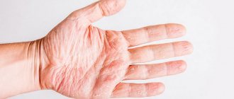
Dyshidrotic eczema is localized on the lateral surfaces of the fingers, on the soles and palms of the blisters, which are accompanied by itching and, after opening, leave a weeping surface and crusts. Horny eczema is quite common - it is characterized by hyperkeratosis with peeling of the palms and soles. Unlike true eczema, microbial eczema is characterized by the formation of purulent crusts and clear contours, which is not observed with true eczema. Typically, such foci are localized near the wound surface and are called “paratraumatic”. Seborrheic eczema usually develops in the scalp area, producing pinkish lesions with lamellar peeling and an “oily” appearance. The itching is pronounced. Occupational eczema occurs with prolonged contact with certain industrial allergens (tars, antibiotics, heavy metals, adhesives, acids, alkalis). The lesions correspond to classic manifestations, but are often located on open areas of the skin that come into contact with the allergen (hands, face, neck, legs). Other manifestations of increased reactivity of the body are often observed - allergic rhinitis and conjunctivitis, bronchospasm. Usually characterized by a chronic course, which does not resolve even after eliminating the allergen.
A dermato-venereologist is involved in the diagnosis and treatment of eczema.
To make a correct diagnosis, in addition to examining the affected areas, collecting complaints and anamnesis, it is necessary to carry out laboratory tests: general blood and urine tests, biochemical blood tests, and more specific ones - determination of the total amount of immunoglobulins E and those specific to certain allergens. According to indications, a histological examination of the affected areas of the skin is sometimes performed. To confirm occupational eczema, skin tests are performed with the suspected irritant.
Treatment includes eliminating the suspected irritant, following a hypoallergenic diet, and using antihistamines (desloratadine, cetirizine, levocetirizine). In the acute stage, more active 1st generation antihistamines are prescribed - chloropyramine, quifenadine, clemastine and others). In case of severe inflammation, injections of glucocorticosteroids are prescribed - betamethasone, dexamethasone. In most cases, severe itching can be stopped by prescribing the anxiolytic Hydroxyzine. In case of severe general condition, detoxification therapy is carried out (against the background of microbial eczema). Fucorcin, chlorhexidine, and glucocorticoids are used externally until the inflammation subsides. After this, preparations of ichthyol and naphthalan are used. If a bacterial infection or microbial eczema occurs, antibiotics are additionally prescribed.
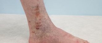
Microbial eczema
With this type of disease, the lesions are not symmetrical and are most often found in the lower extremities. The occurrence of microbial eczema is mainly associated with poor personal hygiene and hyperhidrosis.
Crusts and weeping characteristic of the pathology are found on all affected skin areas, on the periphery of which single papules filled with purulent contents are found.
Microbial eczema, in turn, is divided into mycotic, traumatic and varicose, depending on the underlying cause of the disease.
The diagnosis is established on the basis of characteristic complaints, typical clinical manifestations and the patient’s history of fungal infections, injuries and varicose veins. Also, if necessary, bacteriological studies are carried out to determine the sensitivity of the pathogen and the degree of its sensitivity to various drugs.
Basic principles of pharmacotherapy for eczema
Eczema is a common atopic disease that occurs with itching, disturbances in sleep and quality of life [31]. Eczema (from the Greek ekzeo - boil) is an acute or chronic recurrent allergic skin disease, formed under the influence of exogenous and endogenous trigger factors and characterized by the appearance of a polymorphic rash, an acute inflammatory reaction caused by serous inflammation of the skin, and severe itching [21]. Eczema accounts for 30–40% of all skin pathologies. The disease appears at any age, often occurs acutely, less often there are chronic forms. The acute form is associated with erythema and edema. Lichenification and peeling are the main features of the chronic form. This reflects morphological changes in the skin resulting from a complex interaction between genetic and environmental factors, leading to immunological and biochemical changes resulting in inflammatory lesions and pruritus [21, 30, 36]. About 1/3 of all cases of eczema are microbial eczema. In recent years, microbial eczema has acquired a tendency toward a more severe course with frequent, prolonged relapses, a significant spread of the pathological process, and is characterized by resistance to conventional treatment methods [7]. Eczema develops as a result of the complex influence of exogenous and endogenous factors, among which genetic predisposition, the state of the central and peripheral nervous system, the presence of sensitization to various allergens, pathology of internal organs, dysfunction of the gastrointestinal tract, endocrine system, and foci of chronic infection are of great importance. cholecystitis, adnexitis, ENT pathology), etc. [8, 11]. According to modern concepts, sensitization to microbial antigens against the background of changes in the immune system plays an important role in the mechanism of development of microbial eczema. Microbial allergens have quite pronounced antigenic activity, which leads to sensitization of the body. Normally, the microbial flora of the skin is non-pathogenic and participates in bacterial protection of the skin by suppressing pathogenic strains with non-pathogenic ones. Skin microflora includes resident (Staphylococcus (S.) epidermidis, S. aureus, Micrococcus spp., Sarcina spp., coryneform bacteria, Propionibacterium spp.) and transient, represented by opportunistic and pathogenic microorganisms [2]. A significant portion of microorganisms die under the influence of the bactericidal environment of the skin (acid reaction, fatty acids of sebum, lysozyme) and do not penetrate the intact skin barrier [7]. Due to disruption of the protective function of the skin and changes in the quantity and quality of skin lipids, patients with eczema may develop a bacterial infection [27, 30]. Thus, the leading role in the occurrence of the disease against the background of a genetically determined hereditary predisposition is played by a change in the microbial landscape - the predominance of staphylococcal and streptococcal flora, as well as fungi of the genus Candida [20]. According to domestic studies, when scrapings from skin lesions in patients with eczema, S. aureus is sown in 80% of cases, S. haemolyticus in 14%, and non-lipophilic yeast of the genus Candida in 40.7% [5]. In areas of exudation and weeping, the number of microorganisms can reach 1 × 10⁷ per 1 cm2. Toxins secreted by S. aureus (enterotoxins A and B, toxic shock syndrome toxin) are superantigens that simultaneously stimulate several parts of the immune response [20]. In 57% of patients, antibodies to staphylococcal enterotoxins A, B, C and toxic shock syndrome toxin were detected in the blood serum. It has been shown that enterotoxin B has the most reactogenic properties, and a relationship has been established between the severity of skin damage and sensitization to this toxin [5, 9, 13]. They bind to the b-chain of the T-cell receptor and the class II major histocompatibility complex molecule and activate an entire family of T-lymphocyte clones, which leads to increased production of pro-inflammatory cytokines and enhances the allergic inflammatory response. Bacterial colonization of the skin by S. aureus and its release of enterotoxins is one of the reasons for the formation of resistance to steroid therapy [13, 20, 29, 33, 34]. A double-blind, multicenter, randomized controlled trial demonstrated a correlation between the severity of eczema and S. aureus colonization and found bacterial colonization to be an important factor in the exacerbation of skin lesions [25]. Thus, microbial sensitization is a triggering, maintaining and aggravating factor in patients with eczema [3]. Treatment of eczema is carried out comprehensively, taking into account the form and stage of the disease, the severity of the process, endogenous and exogenous factors underlying the development of the disease [7, 12, 18, 23, 24]. Therapy for eczema begins with limiting contact with identified and potential allergens and irritants. For all forms of eczema, general hyposensitizing therapy is prescribed, including antihistamines. The main goals of treatment are to prevent disease progression, reduce itching, resolve rashes, and prevent relapses [21]. External therapy occupies a special place in the complex treatment of microbial eczema. Local treatment is determined by the clinical picture of eczema. External therapy is carried out over a long period of time and in stages, and different dosage forms are used at different stages. The principle of local therapy remains unchanged: “on wet - wet, on dry - dry” and “do not irritate the irritated”. Traditionally, lotions and wet-dry dressings are used. In this regard, during the weeping stage, lotions, wet-dry dressings made from antiseptic and astringent solutions, and aerosols with corticosteroids are used to suppress signs of acute inflammation - hyperemia, swelling, exudation, itching and pain [7, 11]. For profuse weeping, which usually occurs with true eczema (acute or exacerbation of subacute and chronic), lotions or wet-dry dressings made from antiseptic solutions are prescribed, and for mild but persistent weeping, astringent solutions are prescribed. Antiseptic solutions include solutions of boric acid (Sol. ac. borici 1%, 2% and 3%), rivanol (Sol. rivanoli 1:1000–1:4000), resorcinol (Sol. resorcini 1–2%), furacilin (Sol . furacilini 1:5000), potassium permanganate (Sol. Kalii hypermanganici 1:5000). The latter solution also has deodorizing properties. The resorcinol solution is prescribed in a dark bottle, since resorcinol quickly decomposes in the light. The group of astringent solutions consists of lead water (Aq. plumbi), drilling fluid (Liq. Burovi; Liq. aluminii acetici 1–2 tablespoons per 1 glass of water), lapis solution (Sol. Argenti nitrici 0.1–0.25% ), goulard water (Ac. borici 6.0; Sp. aethylici 4.0; Aq. plumbi 200.0). Lapis solution, like resorcinol solution, should be prescribed in a dark bottle. When choosing a solution for a lotion or a wet-dry dressing, the following factors must be taken into account. For complications caused by impetiginization of the process, antiseptic solutions are indicated, primarily rivanol and furatsilin. In cases accompanied by severe itching, Goulard's water has a good effect. For weeping forms of microbial eczema and dyshidrotic eczema, solutions of stronger concentrations are indicated: Sol. ac. borici 3%; Sol. resorcini 3%; Sol. rivanoli 1:1000, etc. For lotions for elderly and weakened patients, solutions of weak concentrations are used, since strong ones can cause burning and pain in them. Sometimes such patients do not tolerate weak concentrations well. In such cases, lotions from various herbal infusions are used: sage, chamomile, string, tea, etc. As for astringents, they are usually prescribed after antiseptic solutions, when the symptoms of inflammation have largely subsided, however, although not abundant, the persistent wetting. The application of lotions from strong solutions of astringents to extensive foci of weeping with pronounced inflammation can lead to an exacerbation and spread of the process, which is explained by the formation of a film that prevents the outflow of serous fluid and, therefore, promotes its absorption (autosensitization). Particularly dangerous in this regard are oak bark infusion and tannin solution. When the weeping subsides, creams and sequential ointments containing corticosteroid hormones are prescribed. For chronic eczema without exacerbations, treatment begins with creams or ointments. Creams and ointments containing both corticosteroid hormones and salicylic acid are applied to foci of chronic eczema, covered with dry crusts and abundant scales, as well as to foci of dyshidrotic eczema. Salicylic acid promotes the falling off of crusts and scales and deeper penetration of corticosteroid hormones into the tissues of the lesions [15]. The effectiveness of lotions and wet-dry dressings in the acute stage of eczema does not raise any doubts, however, patients are not always ready to make solutions at home and methodically carry out procedures using lotions several times a day [11]. Most classic solutions, pastes, and ointments have a strong and unpleasant odor, stain and stain linen and clothing, which limits their use, especially in outpatient practice. The use of solutions requires the use of bandages that interfere with the movement of patients, limiting their daily and professional activities. The effect of lotions, pastes, and ointments develops relatively slowly and requires hospitalization of patients and long-term step-by-step treatment [1]. The modern patient expects from external treatment not only rapid relief of inflammatory and subjective symptoms of the disease, shortening the duration of the disease, but also a convenient and pleasant therapeutic effect that does not limit his social and professional activity, and, as a result, a significant improvement in the quality of life. Therefore, in modern clinical practice, the requirements for drugs used in the treatment of chronic dermatoses have increased significantly. The drug should have an active anti-inflammatory effect, be well tolerated, have a high safety profile, have a prolonged effect that does not require repeated use, be aesthetically attractive and not limit the usual lifestyle [4, 19]. In many ways, topical corticosteroid drugs meet these criteria. External glucocorticosteroids, which first appeared in the therapeutic arsenal of dermatologists in the middle of the last century, are currently the basic therapy for inflammatory dermatoses. To date, there is no therapeutic alternative to them in terms of the speed of onset and severity (activity) of the anti-inflammatory effect. Topical corticosteroids have high anti-inflammatory, antiallergic, immunosuppressive activity, as well as vasoconstrictor and antimitotic effects [22, 28]. These drugs, having a powerful pathogenetic effect, can quickly reduce inflammatory changes in the skin, and in a short time reduce or eliminate the subjective symptoms of dermatoses (itching, burning). Improving skin status undoubtedly has a positive effect on the psycho-emotional state of patients, restoration of ability to work and daily activity, which significantly improves their quality of life. The success of therapy is largely determined by the correct choice of steroid, taking into account its activity and method of application, depending on the nature, stage, and localization of dermatosis [4]. According to the European classification of potential activity, steroids are distinguished into weak (class 1), medium (class 2), strong (class 3), and very strong (class 4). In acute inflammation, characterized by edema, hyperemia, and weeping, it is advisable to use moderate, strong, and less often very strong topical corticosteroids (evidence levels A, B). As the severity of inflammation decreases, moderate and weak corticosteroids are used (levels of evidence A, B) [16]. Of great importance is the correct choice of the dosage form of the drug, which determines the absorption activity of the steroid and the rationality of the external therapy performed (Table 1). One of the most common mistakes when prescribing topical corticosteroids is the choice of drug without taking into account the topographic and morphofunctional characteristics of the skin, as well as the absorption activity of external agents in various areas of the skin. The absorption of external medications directly depends on the structure (thickness) of the epidermis. Comparative studies have revealed significant differences in the absorption of the same drug applied to different areas of the skin. Thus, one of the criteria for choosing a topical steroid is the localization of the source of inflammation. On areas of thin skin (face, genitals, flexor surfaces), it is preferable to apply steroids of moderate potency. It should be borne in mind that it is extremely undesirable to apply drugs containing strong steroids to the above areas of the skin, which are areas of increased sensitivity to glucocorticosteroids, since this can lead to a high risk of developing both local side effects (atrophy, telangiectasia, etc.) , and systemic, as well as activation of infectious agents [4]. Taking into account the important role of infectious agents in the pathogenesis of eczema, in particular microbial eczema, the doctor is faced with the task of not only relieving inflammation and subjective symptoms in the shortest possible time, but also suppressing sensitization to bacterial and fungal antigens, normalizing the number and composition of skin microflora [7] . Infectious agents can not only contribute to the development of the disease, but also complicate its course. In this regard, in terms of external therapy, drugs with antimicrobial and antifungal properties must be added to topical glucocorticosteroids. Several drugs can be used sequentially, but external use of combined agents, which have anti-inflammatory, antibacterial and antifungal effects, is more preferable [20]. As for the antimicrobial component, it should be noted that traditionally prescribed combined external glucocorticosteroids contain active ingredients such as gentamicin, tetracycline, clotrimazole, natamycin, to which many pathogens of bacterial and fungal infections are insensitive. Currently, the ineffectiveness of traditional treatment regimens for dermatoses of combined etiology is primarily associated with the development of resistance of pathogens to prescribed drugs [6]. Thus, according to a mycological study of the sensitivity of nosocomial strains of S. aureus to antibiotics used in clinical practice for a long time, conducted in Russia, resistance to gentamicin was revealed in 31% of the isolated strains and to tetracycline in 37%. Thus, in more than 1/3 of patients, the use of these drugs as antimicrobial therapy does not provide the expected therapeutic effect [14, 17]. In another study [32], in 87% of patients with chronic eczema and atopic dermatitis, MRSA (methicillin-resistant S. aureus) strains were detected by culture from the lesions. The risk group for MRSA contamination included patients with a long course of dermatosis, elderly patients burdened with somatic diseases (diabetes mellitus, chronic infections of the lungs and ENT organs), as well as frequent and long-term hospital stays. At the same time, in women the activity of bacterial contamination of the skin with MRSA was higher, but the infection was more severe in men [5]. According to microbiological studies [10], resistance to gentamicin of MRSA strains is 56%. When choosing a combined glucocorticosteroid, you should focus primarily on the anti-infective effectiveness of its action. Irrational choice of drug leads to a further increase in the resistance of microorganisms, lack of effect from therapy and worsening of the skin process [5]. Considering that in 90% of patients the main bacterial agent is S. aureus [25], and in most cases MRSA strains are isolated that are also resistant to gentamicin, it is necessary to use a drug that has sufficient antibacterial activity against such pathogens. Fusidic acid is a strong antibacterial agent and inhibits almost all strains of Staphylococcus aureus, including S. aureus, S. epidermidis and methicillin-resistant strains, streptococci and other pathogens. The drug acts as an inhibitor of protein synthesis in the bacterial cell and has a bacteriostatic or bactericidal effect depending on the size of the inoculum. Topical application of fusidic acid is safe and effective in the treatment of bacterial skin infections [17, 26, 30]. The development of resistance generally remains low or short-lived and can be minimized by limiting therapy to no more than 14 days [35]. There is no cross resistance, like cross -sensitivity, to fusic acid, because this is the only antibiotic in its group. Therapeutic effectiveness in the local use of fusic acid is due to the unique ability to penetrate deep into the skin and accumulate in a dosage, several times higher than the minimum therapeutic one. Fusidic acid was successfully used together with corticosteroids (betamethasone and hydrocortisone). Hydrocortisone acetate - soft corticosteroid is preferable to use it with a mild degree of damage. Betamethasone belongs to a group of strong glucocorticosteroids (group III), which allows you to prescribe it with severe inflammation [17]. These drugs are especially effective in the treatment of microbial eczema, because their effect is aimed at the treatment of both inflammation and infection. The use of antibiotics and corticosteroids both separately and in fixed combinations gives a good therapeutic effect for eczema and also reduces the colonization of golden staphylococcus. The early beginning of combined topical therapy is necessary for patients with medium and severe eczema and reduces the need to use antibiotics at the later stages of the disease [25]. The appointment of one drug with a wide therapeutic range, replacing several drugs, allows excluding polypraggmasy, avoiding side effects and undesirable interactions between drugs, reducing costs and increasing patient compliance [4]. An integrated approach to the treatment of eczema will reduce the severity of inflammation, accompanied by itching and exudation, causing significant suffering, and thereby have a beneficial effect on the quality of life of patients.
Literature 1. Akovbyan V.A. Composite preparations for external treatment: the advantages are obvious // Clinical dermatology and venereology. 2003. No. 4. P. 50–53. 2. Arzumanyan V.G., Zaitseva E.V., Kabaeva T.I., Temper R.M. Assessment of staphylococcal and non-lipophilic yeast microflora of the skin in patients with skin pathology using the contact method of sowing // Bulletin of Dermatol. and venerol. 2004. 3. Bakulev A.L., Kravchenya S.S., Murashkin N.N. and others. Combined topical therapy for patients with microbial eczema // Dermatology. 2012. No. 1. 4. Belousova T.A. Modern principles of external therapy of inflammatory dermatoses // RMJ. 2008. No. 8. pp. 547–551. 5. Belousova T.A., Goryachkina M.A., Katranova D.G. Features of skin microbiocenosis in patients with allergic dermatoses: the problem of choosing external therapy // Clinical dermatology and venereology. 2013. No. 3. pp. 107–112. 6. Belousova T.A., Goryachkina M.V. Algorithm for external therapy of dermatoses of combined etiology // Pharmacotherapy in dermatovenerology. 2011. No. 5. pp. 146–152. 7. Klemenova I.A., Shebashova N.V., Lisina L.N. Microbial eczema: the use of combined topical drugs // Dermatology. 2011. No. 3. 8. Kubanova A.A., Skripkin Yu.K., Akimov V.G., Znamenskaya L.F. Eczema // Clinical dermatovenerology / ed. Yu.K. Skripkin and Yu.S. Butova. T. 2. M. GEOTAR-Media, 2009. pp. 106–117. 9. Mokronosova M.A., Maksimova A.E., Baturo A.P. and others. The influence of various methods of external therapy on the colonization of the skin by Staphylococcus aureus and the course of atopic dermatitis // Ros. allergol. magazine. 2004. No. 1. P. 58–61. 10. Murashkin N.N., Gluzmin M.I., Bakulev A.L. The role of methicillin-resistant strains of Staphylococcus aureus in the pathogenesis of severe forms of atopic dermatitis in children // Bulletin of Dermatology and Venereology. 2012. No. 1. P. 68–76. 11. Olisova O.Yu. External treatment of dyshidrotic eczema // Clinical dermatology and venereology. 2009. No. 6. pp. 93–95. 12. Orkin V.F., Olekhnovich R.M. Microbial eczema (clinic, pathogenesis, treatment) // Journal of dermatology and cosmetology. 2002. No. 2. pp. 24–26. 13. Pampura A.N., Smirnova M.O. Modern approaches to the treatment of atopic dermatitis in children // Attending physician. 2008. No. 5. P. 57–61. 14. Potekaev N.S., Sherina T.F. On the issue of the association of dermatoses and mycoses of the skin // Ros. Journal of Skin and Venereal Diseases. 2004. No. 6. P. 55–57. 15. Potekaev N.S. Eczema: remarks on modern ideas // Clinical dermatology and venereology. 2009. No. 1. P. 67–73. 16. Kubanova A.A., Kisina V.I., Blatun L.A., Vavilov A.M. and others. Rational pharmacotherapy of skin diseases and sexually transmitted infections: Hand. for medical practitioners / general ed. A.A. Kubanova, V.I. Kisina. M.: Litterra, 2005. 882 p. (Rational pharmacotherapy: ser. handbook for practicing physicians; vol. 8). 17. Skripkin Yu.K., Dvornikov A.S., Kruglova L.S., Skripkina P.A. Modern view on pathogenetic therapy of atopic dermatitis // Bulletin of Dermatology and Venereology. 2006. No. 4. 18. Skripkin Yu.K., Mashkilleyson A.A., Sharapova G.L. Skin and venereal diseases. M.: Medicine, 1997. 19. Modern external therapy of dermatoses / ed. N.G. Short. Tver: Provincial Medicine, 2001. 528 p. 20. Sukharev A.V., Gutka V.O., Patrushev A.V. and others. Combined external therapy of dermatitis complicated by secondary infection // Dermatology. 2012. No. 3–4. 21. Federal clinical recommendations of the Russian Society of Dermatovenerologists and Cosmetologists for the management of patients. M., 2013. 22. Churyukanov V.V., Belousova T.A., Goryachkina M.V. Topical glucocorticosteroids in dermatology: an idea of the mechanism of action, the relationship between effectiveness and safety // Klin. derm. and Venus. 2004. No. 3. P. 106–110. 23. Yusupova L.A., Khafizyanova R.Kh. Treatment of patients with eczema // Ros. Journal of Skin and Venereal Diseases. 2005. No. 6. pp. 20–23. 24. Yutskovsky A.D. Eczema etiologically associated with fungal infection (immune mechanisms of development, diagnosis, prognosis, features of corrective therapy, clinical examination and rehabilitation of patients): Abstract. dis. ...cand. honey. Sci. M., 1990. 25. Gong JQ, Lin L., Lin T. et al. Skin colonization by Staphylococcus aureus in patients with eczema and atopic dermatitis and relevant combined topical therapy: a double-blind multicentre randomized controlled trial // Br. J. Dermatol. 2006. Vol. 155(4). P. 680–687. 26. Guo-xing Z., Wei L., Huai-qiu H. et al. 2% Fusidic Acid Cream in the Treatment of Bacterial Infected Skin Diseases: A Multicenter Clinical Observation on Efficacy and Safety // Chin. J. Dermatovenerol. 2007. Vol. 5. . Available from: https:// en.cnki.com.cn/Article_en/CJFDTOTAL-ZBFX200705013.htm. 27. Arslanagic N., Arslanagic R. Atopic dermatitis and staphylococcus aureus // Med. Arh. 2004. Vol. 58. P. 363–365. 28. Brazzini. B., Pimpinelli N. New and established topical corticosteroids in dermatology // Am. J. Dermatol. 2002. Vol. 3. P. 47–58. 29. Bunikowski R., Mielke ME, Skarabis H. et al. Prevalence and role of serum IgE antibodies to the Staphylococcus aureus derived superantigens SEA and SEB in children with atopic dermatitis // J. Aller. Clin. Immunol. 2000. Vol.105 (4). P. 814–819. 30. Dasiga Venkata, Subrahmanya Pratap, Mariam Philip et al. Evaluation of Efficacy, Safety, and Tolerability of Fixed Dose Combination (FDC) of Halometasone 0.05% and Fusidic Acid 2% Topical Cream Versus FDC of Betamethasone Valerate 0.12% and Neomycin Sulphate 0.5% Topical Cream in the Treatment of Infected Eczematous Dermatosis in Indian Subjects : A Randomized Open-Label Comparative Phase III Multi-Centric Trial. 31. Hon KL, Wang SS, Lee KK et al. Combined antibiotic/corticosteroid cream in the empirical treatment of moderate to severe eczema: friend or foe? // J. Drugs Dermatol. 2012. Vol. 11 (7). P. 861–864. 32. Jayasekera A, Jennings L, Holden CR et al. Methi cillin-resistant Staphylococcus aureus in skin disease affects mainly elderly patients with eczema and leg ulcers who have associated chronic disease // Acta Dermatol. Venereol. 2008. Vol. 88 (2) P.156–158. 33. Leung D. Infection in atopic dermatitis // Curr. Opin. Pediatr. 2003. Vol. 15. P. 399–404. 34. Leung DY, Harbeck R, Bina P et al. Presence of IgE antibodies to staphylococcal exotoxins on the skin of patients with atopic dermatitis. Evidence for a new group of allergens // J. Clin. Inves. 1993. Vol. 92. P. 1374–1380. 35. Schöfer H., Simonsen L. Fusidic acid in dermatology: an updated review // Eur. J. Dermatol. 2010.Vol. 20(1). P. 6–15. 36. Thappa DM Eczema. Textbook of dermatology, venereology and leprology. 3rd ed. India: Elsevier (Publishers), 2009. P.131–145.
Seborrheic eczema
With this type of pathology, lesions occur mainly in the scalp area. Microbial eczema can also damage the skin on the face, around the navel, behind the ears and in natural folds.
Inflamed, hyperemic and dry areas are found in the scalp area. When they are combed, the epidermis peels off according to the pityriasis type. It is important to note that the affected skin is clearly demarcated from healthy skin.
Only in rare situations is seborrheic eczema accompanied by exudative manifestations and the formation of crusts, which, when removed, reveal a moist, eroded surface.
The diagnosis is made based on the typical clinical picture and patient complaints. In some cases, histological examination is performed.
How to deal with eczema?
Eczema is one of the common skin pathologies. What are its reasons? Is it transmitted from person to person? How is it treated?
These and other questions are answered by the dermatologist at Clinic Expert Tula, Candidate of Medical Sciences Vladislav Leonidovich Sheinkman.
– Vladislav Leonidovich, let’s start with a definition: what is eczema?
– This is a chronic recurrent skin disease with acute inflammatory symptoms caused by serous inflammation of the epidermis and dermis.
– What are the causes of eczema?
– There is a hereditary predisposition, a relationship with the nervous system - stress can also lead to disease. Post-traumatic microbial eczema occurs around the wound surface.
Is there a stress vaccine? Read our article
– What types of eczema are there?
– True (or, otherwise, idiopathic) eczema most often turns into chronic. Patients are bothered by severe itching, blisters appear on the skin, then they burst, and the weeping stage begins. There are many foci of inflammation on the body, they involve more and more new areas of the skin.
I have seborrheic eczema. It is localized on the scalp, face, arms, and neck. There is no weeping stage, but itching is present.
Dyshidrotic eczema affects the arms and legs; In this case, bubbles appear; they, of course, bother the patient, but may not open. There are painful sensations when moving the hands and feet.
Microbial eczema occurs at the site of fractures, injuries, or some microbial foci. Not only characteristic blisters appear, but also pustules.
There is also occupational eczema, which affects people who are forced to constantly come into contact with harmful substances at work.
“A doctor of this specialty is engaged in establishing the connection between existing pathologies or disabilities and the characteristics of work activity.” Quote from the material “Educational program on medical specialties. Let’s go to an appointment with an occupational pathologist.”
Horny, or tylotic, eczema affects the palms of the hands and soles of the feet.
Childhood eczema - it begins around 3-6 months of a child’s life, often has a hereditary nature, and is preceded by diathesis. Locations are the cheeks, forehead, then lesions can appear on the shoulders, hips, and legs. Eczema can spread to the head and ears.
– Is eczema contagious?
- No.
– Can eczema go away on its own without treatment?
– Since this is a chronic skin disease, it may seem to subside for a while, but sooner or later a period of exacerbation will return. Unfortunately, many people self-medicate - they use some kind of ointments, tinctures. This is something that you absolutely cannot do if you have eczema. You need to be treated by a dermatologist - only he can make the correct diagnosis and prescribe adequate treatment. And self-medication often leads to serious complications.
– How is eczema treated?
– A patient of any age must follow a hypoallergenic diet - that is, foods that can serve as disease-provoking factors should be excluded from the diet. For adults, these are alcoholic drinks, compotes, juices, lemonade, products with dyes, preservatives, sweets, chocolate, honey, jam, berries, imported fruits.
Patients should try to avoid stress. They are recommended to examine the gastrointestinal tract - problems with the liver and stomach are often detected. Do not come into contact with chemically active substances (including synthetic and chlorine-containing detergents).
Treatment consists of prescribing desensitizing, antihistamine drugs; antibiotics can be used for microbial eczema.
You can read more about antibiotics in our articles here and here
Various lotions are used in treatment - for example, with a solution of Miramistin or Rivanol, pastes, creams. Hormone-containing drugs are used exclusively under the supervision of a doctor.
– Is it possible to swim in the sea if you have eczema?
– There is such a myth: if a patient with eczema swims in the sea - especially in the Dead Sea - he will be cured immediately. Nothing like this! During an exacerbation, it is generally not advisable to swim in the sea: exposed inflammatory foci react to the salt in sea water, and the itching and rashes intensify. If you go to the sea in the remission stage - when there are no rashes, this has a beneficial effect on the prevention of all skin diseases in general.
– Is it possible to prevent the development of eczema? And if so, how?
– Prevention is possible. It is necessary to normalize the daily routine, eat rationally, and, if possible, control the functioning of the nervous system.
You can make an appointment with a dermatovenerologist here
ATTENTION: the service is not available in all cities
Interviewed by Igor Chichinov
The editors recommend:
When the sun is your enemy. What is solar dermatitis?
What is demodicosis and how to cure it forever?
Nerves under control! How to maintain mental health?
For reference:
Sheinkman Vladislav Leonidovich
Graduate of the pediatric faculty of the Smolensk State Medical Institute in 1994.
From 1994 to 1995, he completed an internship, and from 1996 to 1998, a clinical residency in the specialty “Dermatovenereology”.
Has an academic degree of Candidate of Medical Sciences.
Currently working at Clinic Expert in Tula as a dermatovenerologist. Provides reception at the address: st. Boldina, 74.
Occupational eczema
The pathogenesis of occupational eczema involves systematic exposure of the skin to irritating substances and other harmful production factors (chemicals, dust, too dry or humid air, etc.), increased capillary permeability and disturbances in the ANS. This combination leads to sensitization of the body and the development of a pathological condition.
Clinical symptoms are similar to those of true eczema: symmetry of lesions and polymorphism of rashes.
When making a diagnosis, it is important to carefully question the patient about the presence of harmful production factors and their influence on the intensification and weakening of the symptoms of the disease. Many patients note that during periods of no exposure to harmful substances, for example during the annual vacation, the skin looks absolutely healthy and rashes stop bothering them.