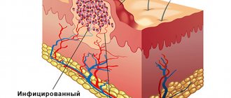
A mole is a collection of melanocytes that forms on the skin. Since melanocytes contain pigment, they are responsible for the color of moles from light brown to black. Nevi are not only congenital. They can appear throughout a person's life. Convex ones are formed if melanocytes are located deep in the skin layers. When the cells are located on the surface of the epidermis, flat moles appear.
Sometimes nevi can degenerate into a malignant neoplasm - melanoma. In such a case, there is a need for its urgent removal. In order to promptly notice any pathological changes, moles of large sizes and unusual shapes should be constantly monitored. There are several indicators that a nevus does not pose a threat to human health:
- The size of the neoplasm is no more than 5 mm;
- Uniform color of the mole;
- Smooth surface and contours of the nevus;
- The presence of hairs growing from the body of the formation.
Survey
In order to identify pathological changes in the formation, an optical device is used - a dermatoscope. By applying the device to an area of skin, the doctor can examine in detail the structure of the formation and understand whether it poses a danger to the patient . Based on this, the specialist determines which method of removing the formation is suitable for the patient.
People often come to Melanoma Unit to eliminate cosmetic defects associated with moles, warts and papillomas. For this purpose, an individual method of removing formations is selected, with maximum cosmetic effect.
Our doctors - oncosurgeons, dermatologists - remove moles, papillomas, warts using surgical, radio wave, and laser methods.
All procedures are virtually painless, safe and can be performed in one clinic visit.
Possible complications after surgical excision of nevi
After surgical removal of a mole, the following side effects may occur:
- Itching, pain, discomfort at the surgical site;
- Allergic reaction to a drug for local anesthesia;
- Prolonged bleeding;
- The appearance of keloid scars;
- Swelling in the area of intervention.
An increase in body temperature may indicate the presence of an inflammatory process, so in this case you should consult a doctor.
Surgical removal of moles is one of the effective methods of getting rid of nevi, which helps prevent the possible development of cancer pathologies. Our clinic employs qualified and experienced doctors who will help you efficiently and quickly get rid of not only nevi, but also other neoplasms.
Attention!
This article is posted for informational purposes only and under no circumstances constitutes scientific material or medical advice and should not serve as a substitute for an in-person consultation with a professional physician.
For diagnostics, diagnosis and treatment, contact qualified doctors! Number of reads: 4920 Date of publication: 08/06/2018
Dermatologists - search service and appointment with dermatologists in Moscow
Removal methods
- Surgical method
Used for cancer-suspicious formations. Such formations require the intervention of a scalpel, as there is a risk that malignant cells may remain in the skin. The formation is excised with a slight indentation from the skin, after which the doctor applies a cosmetic suture. The minor operation is performed under local anesthesia, is painless and takes no more than 30 minutes. There is no need to stay in the clinic after the operation.
Sutures are removed 10 days after surgery. Complete healing of the wound takes 3-4 weeks.
- Radio wave method (electrocoagulation)
It is used to eliminate cosmetic defects caused by moles, papillomas and other benign skin formations.
Not suitable for removing large skin lesions.
Using a high-frequency electrical discharge, the doctor removes the formation on the skin.
Advantages of the method:
- Virtually no bleeding
- Faster healing compared to surgical method
- More pronounced cosmetic effect
- Painless method
Flaws:
- Applicable only when confirming the good quality of the formation
- A small scar may still be present
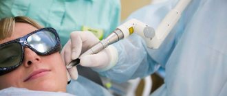
- Laser method
The doctor uses special equipment equipped with a powerful light beam that destroys tumor cells without affecting the surrounding skin tissue.
This method is used only for confirmed benign formations. Advantages of the method:
- Painless and bloodless procedure
- Pronounced cosmetic effect, does not leave scars on the skin
- Fast healing
- Removal in one visit
Flaws:
- Applicable only when confirming the good quality of the formation
Advantages and disadvantages of surgical removal of nevi
Despite the fact that surgical removal of nevi is the oldest method among all known, it has a number of undeniable advantages over others:
- Possibility of getting rid of deep and large moles;
- Safe excision of malignant neoplasms;
- The procedure is painless, since all manipulations are performed using anesthesia;
- Affordable price;
- One hundred percent removal of the nevus in one session (after excision, no neoplasm cells remain, which eliminates the reappearance of the mole);
- Possibility of conducting histological examination of remote formation;
- No contraindications.
Excision of a mole using the classical method is an effective and safe way to get rid of a nevus. However, despite this, it has several disadvantages:
- High likelihood of keloids and scars;
- Long period of rehabilitation;
- After surgery, you should not be in the sun.
Indications for removal of moles and papillomas
- Suspicion of a malignant disease
During the examination, the doctor can identify pathological structures in the formation, and therefore urgent surgical removal is necessary.
- Education is subject to frequent trauma
The inconvenient location of the mole, which in turn is often subject to friction and trauma, causes discomfort and bleeding of the formation in the patient.
In this case, there is a need for removal.
- Aesthetics
Some formations carry a cosmetic defect for the patient, and therefore require an removal procedure. After examination, the doctor selects an individual tactic for removing the formation, with maximum cosmetic effect.
Classification of moles:
1. Moles of epidermal origin (borderline, mixed, intradermal).
2. Moles of dermal origin (the most common representative is the blue nevus).
3. Combined moles.
4. High-risk moles (congenital and dysplastic nevi).
The most common type of moles are intradermal nevi - in appearance they are dome-shaped or papillomatous formations, from light brown to black in color, in some cases they can be reddish or whitish.
You can ask any questions you may have, sign up for a tumor removal procedure, or have a free consultation with a specialist by phone:
Sign up
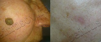
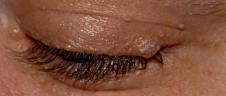
Moles on the back - before surgery. Dermatoscopy:
Doctor: Grishko R.V.
Contraindications
Depending on the method of removal, there are various contraindications to removing formations.
If during the examination the doctor reveals a pathologically altered formation , it is immediately subject to surgical removal, since there is a huge threat to the health and life of the patient. In this case, this is a contraindication for other methods of tumor removal.
If the formation does not pose a threat to the patient’s health, but there are concomitant diseases associated with poor blood clotting, diabetes mellitus and other diseases that may affect the healing of the postoperative wound, it is also worth refraining from the removal procedure.
The medical clinic is equipped with modern, expensive medical equipment, which allows for a high-quality examination of the skin before the removal procedure, such equipment includes:
- Dermatoscope “Heine Delta 20” (Germany) - Leader in the market of hand-held dermatoscopes
- Digital video dermatoscope “FotoFinder ATBM Master” (Germany) - the latest innovative equipment for early diagnosis of malignant skin diseases
- Radio knife “Surgitron” (USA) - used to remove formations using the radio wave method
- Co2 Laser AcuPulse Lumenis (USA-Israel) - used to remove formations using the laser method
Make an appointment
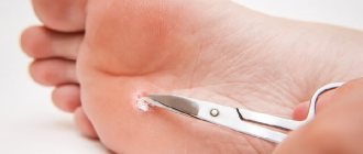
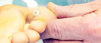
Cryodestruction
Removing moles and warts using liquid nitrogen does not require pain relief. As a result of cold exposure, nerve fibers freeze and sensitivity is switched off. Liquid nitrogen is charged into a mini-cylinder with removable nozzles. The surgeon selects a tip 1 mm larger than the diameter of the tumor and applies it to the mole. The mole is frozen and rejected on its own. The disadvantage of this procedure is that it is difficult to calculate the depth of nitrogen penetration. If the exposure is too strong, healthy tissues are frozen, and if the exposure is weak, the nevus is partially removed. Damage to healthy tissue provokes cosmetic defects and increases the rehabilitation period.
Types of nevi
Dysplastic nevi are pigment spots of an unusual configuration with fuzzy edges, their color varies from reddish-brown to brown on a pink background. They were first noticed because of their unusual appearance and because they are inherited. Dysplastic nevi reach up to 10 mm in diameter.
Borderline pigmented nevus . A special feature is that its melanocytic activity is increased. Outwardly, it looks like a flat black nodule with a diameter of 1 cm with a dry and smooth surface.
An important distinguishing point is the complete absence of hair. Sometimes a border nevus can appear as a spot with smooth, wavy edges. It does not have specific locations. This nevus occurs on the face, neck, and torso. As a rule, borderline nevus is congenital, but can appear even during puberty.
A blue nevus is a blue-blue node of a round shape with a dense consistency. Its smooth surface has no hair.
It is characterized by powerful accumulations of melanin in the dermal layers, hence its bluish color. A nodule measuring 0.5 cm. Blue nevus occurs mainly on the face, limbs, buttocks, and also in the oral cavity.
Blue nevus is especially common among residents of Asian republics. It appears after puberty. Blue nevus is among the melanoma-dangerous ones, because... There have been cases of melanoma developing at the site of this nevus.
Giant pigmented nevus is only congenital. Its height is proportional to the child's height. It can cover the torso, neck and other areas. The surface of the nevus, consisting of bumps, warts and cracks, has hair. The color varies from gray to patchy brown. Development into a malignant tumor occurs in 10% of cases.
Papillomatous nevus is characterized by a surface consisting of tubercles and irregularities. It can often be found on the scalp. It is distinguished by its large size (several centimeters) and irregular outlines. Color ranges from skin-colored to dark brown.
Intradermal nevus . This is the most common birthmark that can be seen on every person. They are found on the skin and mucous membranes. These nevi can be either congenital or can form in the womb. Most people do not consider a birthmark to be a cosmetic nuisance.
Types of pigmented nevus
Histological examination divides pigmented nevi into the following types:
- mixed
- epidermal
- dermal
- spindle cell
- blue
To determine the congenitality of a nevus, it is necessary to conduct an analysis for the presence of nevus cells in the subcutaneous fatty tissue.
From nevus to cutaneous melanoma
Melanoma is a malignant tumor originating from melanocytes. Cutaneous melanoma can develop either from epidermal melanocytes of normal skin or from birthmarks.
Recently, cases of melanoma have become more frequent, especially in women under the age of 40. Melanoma is called the “Queen of Tumors” for its rapid metastasis - the patient dies within a few months.
Melanoma grows above the skin, along its surface and in depth. The deeper the tumor strands penetrate, the worse the prognosis.
Melanoma symptoms
At first, melanoma appears as a dark spot that rises slightly above the surface of the skin. As it grows, it becomes similar to an exophytic tumor, which can subsequently ulcerate.
Melanoma can be recognized by three signs:
- dark color
- shiny surface
- ability to disintegrate
This is explained by the accumulation of pigment, damage to the epidermal layer and the fragility of the formation.
In this case, patients complain of bleeding, burning, itching or pain in the area of the tumor.
Doctor's comment
Every person has pigment spots on their body. The risk of injury or change in the appearance of moles is an indication for their removal. Today there are different ways to remove such formations: destruction using laser, high temperatures, chemicals, as well as surgical excision of the nevus. To choose the method that is most suitable for you, you need to make an appointment with a doctor. Treatment tactics are prescribed taking into account the size of the formation and the presence of signs of malignancy, but for this you should undergo a painless examination. However, if you have moles, especially if they are located in the area of contact with parts of clothing, it is recommended to regularly undergo a dermatological examination; with the help of a simple and painless examination, you can promptly detect signs of malignancy of the nevus. In this case, timely treatment will avoid unwanted consequences. Thanks to the clinic’s equipment with modern equipment, we have the opportunity not only to carry out radical treatment, but also to achieve excellent cosmetic results.
Rehabilitation after removal of dermatological tumors
The duration of rehabilitation after radical and low-traumatic treatment of a dermatological formation depends on its size, morphological characteristics, as well as on the individual characteristics of the patient’s body. To reduce the recovery period, the patient must adhere to the following recommendations:
- During surgical removal, regular wound dressings and examination by a specialist are performed on days 4-7.
- After laser destruction, it is necessary to avoid injury to the resulting crust.
- You cannot peel off the crust yourself; this can lead to infection of the wound and cause the development of a scar. The crust will come off on its own.
- It is necessary to treat the wound with prescribed medications according to the recommended regimen (developed individually).
- After the crust is rejected, the skin at the site of laser exposure must be protected from the sun (use sunscreen and not be in direct sunlight until complete recovery), since these areas are very susceptible to hyperpigmentation.
Reasons for the development of skin tumors
- prolonged exposure to direct ultraviolet rays;
- global disruptions in the immune system (granuloma annulare);
- decreased protective functions of the body (plantar and palmar warts);
- bad habits;
- frequent stress;
- disturbance of metabolic processes in the body (keratomas, fibromas, nevi, xanthomas);
The main danger to human health is due to the fact that many benign tumors are prone to malignancy (malignancy).
Only complete removal of the tumor can prevent its malignancy. Patients of the multidisciplinary medical center have access to a full range of services in the field of detection and treatment of dermatological tumors. Surgical treatment of tumors in our clinic is carried out by highly qualified plastic surgeons and dermatosurgeons; advanced equipment is used during the procedures. During surgical treatment of tumors, our patients are provided with the maximum therapeutic and aesthetic effect.