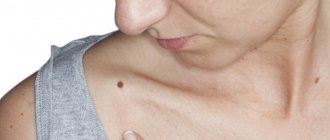
“Decoration on the body” or face in the form of clearly defined pigmented spots on the skin occurs in approximately 90% of the world’s population. Moles or nevi can be congenital or appear during pregnancy, puberty, or frequent sun exposure. In total, more than three hundred types of neoplasms are found on human skin - nevi, warts, papillomas, cysts, keratomas and others. As a rule, they have a benign structure and do not cause concern, but in situations where a mole is constantly injured, begins to change in size, inflammation and itching occur, you should immediately make an appointment with an oncodermatologist. Moles are an area of increased danger - with injury, sun exposure or hormonal imbalances, the process of degeneration of a nevus into a malignant tumor - melanoma can begin. To identify skin cancer in the early stages, the doctor performs a scraping, puncture or dermatoscopy and decides whether to remove the mole.
In recent years, the method of removing nevi with a laser has gained popularity. But in this article I will tell you about a more modern, effective and gentle method - removing moles and other skin growths under the influence of radio waves. The radio wave surgery device was patented by the American radio engineer Irving Ellman in the late 60s of the last century. At first, radio waves were used in the treatment of periodontal disease, but then the scope of application of the device expanded significantly. In modern medicine, the method of radio wave surgery is used to remove a wide range of skin tumors, to treat cervical erosions, to combat ENT diseases and in other areas.
What is removal using the radiosurgical method at the Airport metro station?
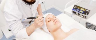
Removal by radiosurgical method is the non-contact removal of unwanted tumors on the human body by exposure to a “beam” of high-frequency radio waves. A device made in the USA, Surgitron, is often used for this purpose. Interesting fact: In our country, radiosurgery began to be used about 15 years ago, but the method appeared in the 70s of the twentieth century. Initially, he was called upon to solve gynecological problems, then otolaryngological ones, and now he has reached cosmetology. With its help, you can remove not only “good” formations, but also malignant ones. The session time range ranges from 3 to 10 minutes.
How is the operation performed?
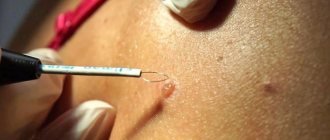
Before the operation to remove the tumor using radio waves, the doctor treats the child’s skin with an antiseptic. Then local anesthesia is administered. The doctor injects an anesthetic into the child's skin using a syringe, or uses a special cream anesthesia. After the skin is anesthetized, the neoplasm is directly removed using the Surgitron radio wave apparatus.
Removal of a papilloma or mole can be done in two ways:
1) removal of the tumor immediately (at the root). In this case, after the formation has been removed, it can be sent for histological examination (this is advisable in cases where specialists have doubts about degeneration into a malignant stage). It is worth noting that this method of radio wave removal of a tumor can lead to the formation of noticeable scars on the child’s skin;
2) removal of skin tumors with smooth fractional movements. This removal technique involves gradually removing the formation tissue layer by layer. In this case, there is no penetration into the deep layers of the skin, which eliminates scarring (or makes it almost invisible).
After the tumor is removed, the doctor removes the remnants of the formation and smoothes the skin using the ball waveguide of the device. This action also allows tissue to coagulate to stop bleeding. After the operation is completed, the wound is treated with an antiseptic solution. The duration of the operation depends on the size of the tumor, but usually takes about half an hour.
The essence of the radiosurgical method of removal
The effect of the method is that under the influence of electric current, skin and soft tissue cells heat up, expand, the cellular fluid boils and evaporates. The result is an incision of the required depth with simultaneous disinfection and blood clotting. For this purpose, a fairly thin electrode is used, also called an electric knife, which does not heat up at all during the procedure. Removal by radio wave is a non-contact method, because the instrument itself does not produce a mechanical effect on the dermis. We can say that the use of radiosurgical method for removing tumors leaves virtually no traces.
HOW TO MAKE AN APPOINTMENT at the PsorMak Institute for Healthy Skin
1. Click the button you see below -
Make an appointment
2. Fill in the fields in the form that appears. Be sure to check the correct phone number so that our specialist can reach you. After filling out, click on the “Submit” button.
3. Wait for our specialist to call. He will answer any of your questions and agree on the date and time of your visit to PsorMak.
The initial appointment includes:
- Visual examination , which will allow the specialist to get a general understanding of the condition of your skin and the pathology itself.
- Collecting anamnesis - finding out information about the development of the disease, living conditions, previous diseases, operations, injuries, chronic pathologies, allergic reactions, heredity, etc. Together with a general examination, this allows you to make a fairly accurate diagnosis and choose a method of treatment and/or prevention.
Make an initial appointment
Areas of use
The scope of application of the method is quite diverse. Its advantages are:
- fairly affordable price;
- minimal probability of relapse (2-3%);
- minimal blood loss (excellent coagulating effect);
- fairly quick recovery after the procedure;
- minimal trauma to the skin surface and mucous membranes;
- minimal likelihood of scarring;
- there is no contact of the device with the skin (the possibility of infection is excluded);
- it is impossible to get burned;
- There is no necrosis of tissue edges.
- no hospitalization required;
- in most cases, anesthesia is not required;
- sterility of the procedure;
- no additional treatment of the treated area with medications is required;
- there are no negative consequences in the form of swelling and pain;
- allowed for use in women who have given birth;
Features of the procedure
Large moles are removed under local anesthesia. When the anesthetic is injected into the skin nearby. Small, red moles can be removed without pain relief. In one session, the doctor will almost always be able to remove all disturbing moles using the radio wave method. The healing time of a wound after radio wave removal is much shorter than with removal by other methods. On the face and neck, small moles only take a few days to heal. Other areas may require more time. It is better to sign up for the procedure in advance so that the doctor does not rush and can prepare the equipment.
Removal by radiosurgical method - indications and contraindications
Recommendations: Indications for radiosurgery:
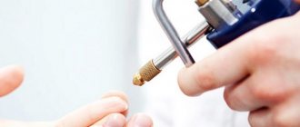
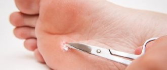
- warts on the sole;
- papillomas;
- Nevi (moles);
- spider veins;
- keratoses;
- vulgar warts;
- Hemangiomas
- Skin basal cell carcinomas
- Ingrown nail
- removal of granulation tissue after surgery;
Attention: Contraindications:
- acute inflammatory processes;
- allergy;
- presence of viral infections;
- feverish condition;
- severe liver and kidney diseases;
- diabetes;
- period of bearing a child;
- herpes
Rehabilitation period
On the first day after surgery, it is necessary to treat the wound with antiseptics that do not contain alcohol. In the following days, a protective crust (scab) forms at the wound site, which protects the wound from bleeding and infections. After about 7 days, the wound is completely healed. During this period, it should be treated with an antiseptic and protected from water and other external influences. You should not try to remove the crust yourself - it will fall off on its own during healing.
After self-(natural) removal of the protective crust, the resulting scar can be lubricated with an anti-scar agent. In the first few weeks after radio wave surgery, the child’s skin should be protected from exposure to direct sunlight and overheating. In some cases, there may be pigment differences in this area of the skin (it will look lighter or darker). But with proper wound care in the postoperative period, this difference will be minimal. The first appointment after surgery is usually scheduled after 3 - 4 weeks, in order to ensure complete removal of the formation. Otherwise, the remainder of the element is corrected.
Recommendations for care after removal of skin tumors:
- After the removal procedure, you should try to avoid being in the sun for a long time;
- If there is a change in color at the removal site or any other significant changes occur, you should immediately consult a doctor;
- When exposed to the sun, it is necessary to protect the place of removal from exposure to rays using special means;
- It is necessary to carefully palpate the removal site for the appearance of a compaction, and also visually inspect for any increase;
- Avoid going to the bathhouse, sauna, swimming pool, or solarium in the first days after the procedure;
- After removing moles, the affected area should not be wet for several days;
- Skin treatment should be carried out according to the doctor’s recommendations (treatment with an antiseptic should be done 3 times a day).
Specialists of the Absolut Med clinic
Chvyrova Tatyana Nikolaevna
Chief physician, director of the Clinic, participant in Russian and international scientific conferences for cosmetologists, dermatologists, allergists. Read more…
Dermatologist, cosmetologist
Deryugina Elena Yurievna
In 1987
Graduated from Vladivostok State Medical Institute. Experience in medicine for more than 29 years. She worked as a dermatologist for 10 years. In aesthetic medicine since 2000. Read more…
Dermatologist, cosmetologist. Doctor of the highest category
Advantages of radio wave surgery:
- The procedure for removing moles is practically painless and does not require hospitalization; it is carried out on the day of treatment;
- No keloid scars form at the site of the removed nevus; there is no need to apply stitches or bandages;
- During radio wave exposure, small vessels are sealed - minimal risk of bleeding and infection;
- Low-traumatic method, convenient for the patient and the doctor.
- Does not require testing;
- Fast tissue healing process;
- Impeccable cosmetic effect.
These advantages make radio wave surgery a leading position in the field of minor surgical interventions.
Summary:
As an oncologist, I recommend removing skin lesions only using radio wave surgery. The method allows you to examine all distant formations and minimizes the risk of developing skin cancer and melanoma.
Sign up for radio wave mole removal
What can be removed by radiosurgery?
- Moles (nevi).
- Neoplasms resulting from human papillomavirus infection. These include: warts (common, plantar), condylomas, papillomas.
- Unaesthetic keloid and hypertrophic scars.
- Hemangiomas (point or star-shaped superficial vascular formations formed by intradermal small veins).
- Tumors and hyperplasia of skin appendages: sebaceous and sweat glands, hair follicles.
- Dermatofibromas are benign skin tumors formed by connective tissue.
- Granulomas are the name given to intradermal nodules that are inflammatory in nature and formed by overgrown connective tissue and an accumulation of immune cells capable of phagocytosis.
- A cutaneous horn is a cone-shaped, horny projection of skin formed by an excessive accumulation of keratinized cells and scales.
- Seborrheic or senile keratosis. This is a local accumulation of epidermal keratinizing cells, with the formation of pigmented protruding plaques-keratomas on the skin.
Rehabilitation after radio wave exposure in gynecology
In the first day after exposure, there may be a slight nagging pain in the lower abdomen. You can take a Nurofen tablet to relieve pain.
Wound healing occurs under the fibrin film within 7-10 days. From 4-5 days, partial rejection of the fibrin film begins. From this time on, the woman may have slight bleeding from the vagina. They should not be abundant and should not have an unpleasant odor. The discharge can last up to 14-20 days, gradually decreasing.
After surgery, it is recommended to insert suppositories with antiseptics into the vagina (for example, Depantol).
After the operation, you cannot have sex, take a hot bath or go to the sauna, play sports, or do heavy physical work for a month.
Contraindications
Considering that the device has a large number of advantages, it may not be suitable for every patient. Therefore, the doctor first conducts a full examination and examination.
Surgitron contraindications:
- the presence of menstruation in the treatment of cervical erosion - radio wave therapy is recommended from 7-10 days of the cycle;
- uterine bleeding of unknown etiology;
- the presence of infectious, bacterial, inflammatory lesions of the genital organs;
- it is strictly prohibited to carry out in the presence of malignant neoplasms of the cervix;
- not prescribed to patients with diabetes mellitus of any type and with blood clotting disorders;
- It is prohibited to remove warts, nevi, moles in case of infectious skin diseases or oncology;
- period of pregnancy and lactation.
If there is even one contraindication, it is strictly forbidden to prescribe this treatment method to the patient. Otherwise, it may cause harm to the body and health.
Pregnant women are strictly not recommended to undergo any surgical intervention. Especially in the first trimester.
Removal of moles and warts using the Surgitron radio wave apparatus
Moles or pigmented nevi are the most common neoplasms of smooth human skin. They are usually benign in nature, so they are eliminated only if there are medical or cosmetic (aesthetic) indications. The Podology Clinic successfully practices the removal of skin tumors (moles and warts) using the Surgitron radio wave apparatus.
How does Surgitron work?
Surgitron, used in the Podology Clinic, is an original portable medical device for radio wave surgery, manufactured by Ellman (USA). It is also called a radio knife, because it is a modern and highly effective alternative to the classic scalpel.
Surgitron uses highly targeted short high-frequency radio waves (3.8 - 4.0 MHz) supplied to tungsten electrodes. They lead to almost instantaneous low-temperature evaporation of intracellular and extracellular fluid and the cells themselves. This is accompanied by local dry coagulation, so that the cut surface is immediately covered with a dense crust and does not bleed. And the antiseptic (disinfecting) effect of radio waves prevents infection of the surgical wound.
The electrodes do not heat up during operation. All processes occur at a temperature of 38 - 40°C, without burns and obvious swelling of surrounding tissues. The incisions for radio wave removal of warts and other formations are very narrow and low-traumatic. Therefore, postoperative wounds heal quickly and painlessly, with the formation of a soft, inconspicuous scar.
Another advantage of the method is the obtaining of high-quality tissue samples for histological examination. This is especially important when removing moles and tumor-like formations with Surgitron.
Contraindications to excision of moles with Surgitron
Excision of moles with a radio knife is contraindicated in the following situations:
- Malignancy (malignancy) of skin formations;
- The presence of chronic inflammatory diseases of the skin;
- Pregnancy, lactation period;
- Active herpetic infection;
- Availability of heart rate sensors.
Before treating a mole with radio waves, the patient undergoes a comprehensive examination, which includes a consultation with an oncologist, a visual examination and laboratory tests. Based on the diagnostic results, the doctor will determine the presence of possible contraindications to the procedure and assess the patient’s health condition.
Types of benign skin neoplasms
Benign skin tumors do not pose a serious threat to humans, but they cause psychological and even physical discomfort, especially when located in areas of the body that are not usually covered by clothing. The main benign neoplasms include:
- Warts and papillomas. Occur on the skin due to infection with the human papillomavirus (HPV). They are painless and are small growths with a height and a diameter of several millimeters. These neoplasms are localized in the folds of the skin, have blood vessels and a loose structure, and can change color and grow.
- Nevi (moles). They are based on melanocytes - cells that contain the main coloring pigment of the body. Most moles are harmless, especially if they are small and do not cause discomfort, that is, they are not located on open areas of the body.