Human papillomavirus infection and pregnancy. Features of diagnosis and management tactics
The prevalence of human papillomavirus infection (PVI) and, accordingly, the pathology associated with it has been steadily increasing over the past decades in many countries of the world, including Russia. According to the literature, up to 3 million new cases of human papillomavirus (HPV) infection are registered annually in the world [1]. Transmission of HPV from person to person can occur in several ways: household contact, vertical, genital, oral, anogenital contact. The high increase in HPV infection in the population, due to its significant contagiousness, the variety of pathologies associated with it, and, most importantly, the ability to transform epithelial cells, triggering the process of carcinogenesis, attract the attention of various specialists to the search for treatment options, timely diagnosis and prevention of diseases associated with HPV [2, 3]. In this case, special attention is paid to PVI of the urogenital tract, which occupies a leading position in prevalence among sexually transmitted infections. The development activity and formation of HPV-associated, as well as other infectious pathologies, are largely determined by the state of the immune system and its ability to adequately respond to the presence of a pathogen. At the same time, HPV, by blocking certain parts of the immune system and exhibiting resistance to TNF-mediated inhibition of proliferation, which is associated with a significant decrease in the expression of TNF receptors, is able to “escape” the immunological surveillance system [4–6]. It has been established that HPV, due to the activation of ubiquitin-mediated proteolysis of the p53 protein, which is a suppressor of carcinogenesis, is able to influence the mechanisms of regulation of the molecular genetic cycle of cell division and block apoptosis [7–9]. It has also been proven that the HPV E7 protein neutralizes the antiviral and antitumor activity of interferon-α2 due to its ability to selectively block most genes induced by interferon [10]. Such multidirectional mechanisms of interaction between HPV and the human immune system contribute to survival, long-term persistence of the virus and a high risk of developing HPV-associated pathology, especially in the presence of trigger factors.
The consistent and holistic picture of the epidemiology and pathogenesis of PVI that has emerged over the past two decades is more clearly presented in women than in men [11]. PVI is registered in 40%, 70% and more than 90% of cases of vulvar, vaginal and cervical cancer, respectively, which are the second leading cause of death in women worldwide [11–14]. Recently, about 20 types of HPV have been associated with cervical canal cancer (95%), among which the most frequently detected are HPV type 16 (50%) and HPV type 18 (10%) [15]. It should be noted that in addition to HPV, a number of associated factors play a determining role in the development of oncogenic transformation. Here, first of all, concomitant infectious diseases of the anogenital area should be highlighted. The combined persistence of HPV with HSV type 2, CMV, EBV, HIV, chlamydia and mycoplasma is unfavorable, especially in terms of the development of cervical dysplasia. Of particular importance, as mentioned above, in the occurrence of infection, the severity of its course, outcome, quality and control of the treatment process for patients with pathologies of the skin and urogenital tract caused by HPV, is the nature of the immune response [16].
One of the features of HPV infection of the urogenital tract is considered to be its wide distribution among young women of reproductive age, mainly under 25 years of age [17], which is due to the low sexual culture of the population, frequent changes of sexual partners, unprotected sex, bad habits (smoking, substance abuse, alcoholism) . There has been an increase in the incidence of anogenital warts in prepubertal children and adolescents, which can be partly explained by an increase in the number of children who become sexually active early. According to sociological surveys, about 15% of girls and 22% of boys noted the presence of sexual contacts in their lives, while 50% of them indicated that their first sexual contact occurred before the age of 15, and for 5% of girls and 20% of boys - up to 12 years [16]. At the same time, it has been noted that spontaneous elimination of HPV occurs more often and faster in adolescents and young women (up to 80% of cases) and regression of existing HPV-associated pathology compared to women of later age. Studies have shown that the average time to clear HPV in adolescents is 8 months (CDC, 1999). According to the observations of S.I. Rogovskaya and V.N. Prilepskaya (2006), in every second patient aged 18–25 years, this period increases to 1.5–2 years.
HPV infection becomes particularly relevant during pregnancy, with the frequency of registration of all types of HPV in pregnant women being 30–65%, and high oncogenic risk types being 20–30% [18].
The main feature of pregnancy is that the fetus in relation to the mother is genetically a half-alien (semi-allogeneic) organism, which is not rejected before the due date. The allogeneity of the fetus lies in the fact that all cells contain, in addition to the haploid set of HLA antigens of the mother, the haploid set of HLA antigens of the father. The maturation of a fertilized egg into a mature fetus in the half-alien body of the mother is carried out due to a suppressor mechanism that develops from the first hours after conception and operates until the development of labor. This mechanism does not allow the mother’s immune system to carry out an immune attack on the fetus with the aim of rejection at all stages of its development [19].
The suppression that develops after conception is multifactorial and is formed both due to products of the endocrine system and due to certain changes in systemic and local immune reactions developed in the process of evolution to protect the semi-allogeneic fetus from the mother’s immune system: the absence of classical antigens of the HLA class I and II; a shift in the functional balance of T helper cells towards type 2 cells and the immunoregulatory role of the placenta, providing a unique immunosuppressive background in the mother’s body [20].
Thus, fertilization itself is of an immune nature. Active processes aimed at local immunosuppression are carried out throughout pregnancy in the fetoplacental complex.
In such a situation, PVI is not only a high risk of developing HPV-associated pathology against the background of physiological immunodeficiency, but also the possibility of its transmission from mother to child during childbirth [21]. In 1989, vertical transmission of the virus was proven, which is confirmed by reports of the detection of HPV in the amniotic fluid of pregnant women and in children born to mothers who are HPV carriers [22]. The possible risk varies according to different authors from 3% to 80% [23]. This scatter is explained by differences in the polymerase chain reaction (PCR) method for detecting HPV DNA. In this case, PVI can be transmitted transplacentally and intranatally (in particular, HPV types 6 and 11). The risk of infection is directly proportional to the severity of the infection (the number of viral particles) and the time of the anhydrous interval during labor, however, studies indicate that delivery by cesarean section does not reduce the risk of infection of the fetus, which indicates predominantly intrauterine infection [22]. Intrapartum infection can lead to juvenile recurrent respiratory papillomatosis (incidence is 1.7–2.6 per 100,000 children and 1 per 1,500 births among women with genital PVI) [24].
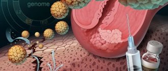
Invasion of the virus occurs through microdamage to the skin and mucous membranes with infection of predominantly immature, dividing cells of the basal layer of the epithelium, which are target cells for HPV. Next, the virus replicates and assembles viral particles in differentiated cells of the surface layer of the epithelium. In this case, HPV can have a productive or transformative effect on the epithelium. With productive exposure, benign neoplasms arise - papillomas, warts and condylomas of the skin and mucous membranes. The result of the transformative effect is dysplasia of varying severity, the progressive development of which leads to cancer [25, 26]. Strong evidence of the acceleration of the development of anogenital cancer and an increase in cancer risk with early exposure to PVI emphasizes the particular importance of not only studying the prevalence of PVI in children, but, most importantly, regular and long-term monitoring of them, taking into account the possibility of developing anogenital neoplasia [27].
There are studies in the literature proving that with PCR-confirmed PVI in the mother, structural damage to the components of the placenta at the end of pregnancy is determined in 76.8% of cases and occurs with morphofunctional signs of chronic placental insufficiency, fetal malnutrition and complications of the neonatal period [28, 29].
Currently, there is an increase and growth of laryngeal papillomatosis in both adults and children. At the same time, transmission of infection prevails in adults through oroanogenital contacts, and in children through passage through the infected birth canal and through household contact.
The manifestation of juvenile respiratory papillomatosis in 75–85% of cases is recorded in the first 5 years of life; in 5–6% of cases in children from 6 months to a year; in 45% of cases in children under 3 years of age [28].
The clinical picture of respiratory papillomatosis consists of voice and breathing disturbances. Most often, when the larynx is damaged in the area of the commissure and the anterior parts of the vocal folds, hoarseness of the voice develops, up to its complete loss. As the lumen of the larynx narrows with papillomas, stenosis develops, and death from asphyxia is possible. The pathological process in childhood is active, it is characterized by prevalence and recurrence rate, and therefore children undergo multiple surgical interventions to remove papillomas. Repeated repeated excision of laryngeal tumors leads to the development of scar complications, the need for tracheostomy, loss of the ability to speak, and worsening chronic respiratory hypoxemia. As the tumor progresses and spreads into the distal respiratory tract, the disease is often fatal [24].
Considering that infection of a child with PVI with the development of juvenile recurrent respiratory papillomatosis is possible not only in the intranatal, but also in the postnatal period, as well as the frequent registration of HPV-associated pathology in newborns and young children, patients should be more actively and thoroughly examined at the planning stage pregnancy and during pregnancy. At the same time, attention should be paid not only to examination for highly oncogenic types of HPV, but also low-oncogenic types, taking into account that, as a rule, in patients suffering from recurrent respiratory papillomatosis, low-oncogenic HPV types 6 and/or 11 are more often found, and they are also often registered and for anogenital warts and genital warts on other areas of the skin and mucous membranes.
The purpose of the study was to assess the frequency of HPV-associated pathology of the skin and mucous membranes of non-genital localization in pregnant women with an established diagnosis of anogenital warts.
Material and research methods
We observed 76 pregnant women (gestational age from 12 to 26 weeks) aged 18 to 42 years with an established clinical diagnosis of anogenital (venereal) warts. The average age of the patients was 26.3 years. In 53 (69.7%) patients, the duration of the disease ranged from 3 weeks to 6 months, in 23 (30.3%) patients - from 6 months to 1 year. 18 (23.7%) patients reported relapses of anogenital warts after previous treatment with destruction methods (laser vaporization, electrosurgical excision, cryodestruction). All patients were referred from antenatal clinics for removal of anogenital warts. Of these, only 2 (2.6%) patients had a combination of genital warts of anogenital and extragenital localization. In 42 (55.3%) patients included in the study, PVI was confirmed by identifying HPV using real-time polymerase chain reaction (RT-PCR): HPV DNA types 16, 31, 33, 35, 52, 58 were identified in 18 (42.9%) patients, HPV types 18, 39, 45, 59 - in 14 (33.3%), HPV types 51, 56 - in 10 (23.8%); Only 6 (14.3%) patients were examined for HPV of the low oncogenic type: types 6 and 11 were found in 5 of them (11.9%). HPV identification by RT-PCR or other laboratory method was not carried out in 34 (44.7%) patients. Results of the study: during a physical examination of the patients at the time they sought medical help, 39 (51.3%) patients were found to have genital warts on the skin and mucous membranes of the extragenital areas: in 14 (35.9%) on the skin of the nipple and peripapillary area; in 11 (28.2%) on the skin of the extremities; in 9 (23.1%) - on the skin of the navel; in 5 (12.8%) - on the oral mucosa. In the anogenital area, papillomatous growths were recorded on the skin of the labia majora - in 5 (6.6%), on the mucous membrane of the vulva - in 30 (39.5%), in the area of the posterior commissure - in 4 (5.3%), on the mucous membrane the membrane of the external opening of the urethra - in 2 (2.6%), on the skin of the perianal and inguinal areas - in 4 (5.3%), on the vaginal mucosa - in 48 (63.2%) patients. Combined damage to several anatomical zones of the anogenital region was observed in 15 (19.7%) patients.
All patients underwent removal of genital warts using the radio wave method. According to our observations, the optimal pregnancy period for this procedure is determined to be after 16 weeks of gestation, when the main stage of placenta formation ends and the immunosuppressive background in the mother’s body is significantly reduced, which sharply reduces the risk of possible relapses.
Thus, the frequent detection of combined HPV-associated pathology of the anogenital and extragenital areas in pregnant women indicates the need for a more thorough examination of such patients in order not only to minimize the risk of complications and recurrence of HPV-associated pathology, but also to exclude infection of the child. A more in-depth examination of pregnant women for HPV, including not only high-oncogenic but also low-oncogenic types, will make it possible to avoid or significantly reduce the risk of developing HPV-associated pathology in children.
Literature
- Nyitray AG, Iannacone MR The epidemiology of human papillomaviruses // Curr Probl Dermatol. 2014; 45(1):75–91.
- Semenov D. M., Danko S. N., Dmitrachenko T. I. Human papillomavirus infection (clinical and pathogenetic features, treatment, prevention). Educational and methodological manual. Vitibsk: State. honey. univ. SPb: Dialect. 2008. 84 p.
- Kiselev V.I. Human papillomaviruses in the development of cervical cancer. M.: , 2004. 180 p.
- Rakhmatullina M. R., Nechaeva I. A., Shalva Mardi. Experience of destructive therapy of anogenital warts // Bulletin of Dermatology and Venereology. 2016. No. 5. pp. 96–101.
- Tavares MC, de Lima Junior SF, Coelho AV et al. // Ann Hum Biol. 2015, Jun; 16; 1–8.
- Prabhavathy D., Subramanian CK, Karunagaran D. Re-expression of HPV16 E2 in SiHa (human cervical cancer) cell potentiates NF-kB activation induced by TNF-α concurrently increasing senescence and survival // Biosci Rep. 2015, Feb; 25; 35(1).
- Aguilar-Martinez E., Morrisroe C., Sharrocks AD The ubiquitin ligase UBE3 A dampens ERK pathway signaling in HPV E6 transformed HeLa cells // PLoS One. 2015. Mar; 27; 10 (3).
- Holloway A., Simmonds M., Azad A., Fox JL, Storey A. Resistance to UV-induced apoptosis by β-HPV5 E6 involves targeting of activated BAK for proteolysis by recruitment of the HERC1 ubiquitin ligase // Int J Cancer. 2015, Jun; 15; 136(12):2831–2843.
- Jing K., Shin S., Jeong S., Kim S., Song KS, Park JH, Heo JY, Seo KS, Park SK, Kweon GR, Wu T., Park JI, Lim K. Docosahexaenoic acid induces the degradation of HPV E6/E7 oncoproteins by activating the ubiquitin-proteasome system // Cell Death Dis. 2014, Nov; 13; 5:1524.
- Sukhikh G. T., Prilepskaya V. N., Rogovskaya S. I. et al. The use of interferon drugs in the treatment of low-grade squamous intraepithelial lesions of the cervix // Effective pharmacotherapy in obstetrics and gynecology. 2009. No. 4. pp. 36–41.
- Batyrshina S.V., Shulaev A.V., Akberova D.R. Human papillomavirus infection: optimization of diagnosis and treatment // Clinical dermatology and venereology. 2015. 14 (5). pp. 67–77.
- Baldwin A., Pirisi L., Creek KE NFI-Ski interactions mediate transforming growth factor beta modulation of human papillomavirus type 16 early gene expression // J Virol. 2004, Apr 1; 78(8):3953–3964.
- Byg L.M., Vidlund J., Vasiljevic N. et al. NF-kB signaling is attenuated by the E7 protein from cutaneous human papillomaviruses // Virus Res. 2012, Oct 1; 169(1):48–53.
- Grulich AE, Jin F, Conway EL et al. Cancers attributable to human papillomavirus infection // Sex Heath. 2010; 7 (3): 244–252.
- Danilova O. V., Yunusova E. I. Tactics for managing papillomavirus lesions of the genitals. Training manual for doctors. Kazan, 2012. 30 p.
- Yunusova E. I., Danilova O. V., Gizatullina R. D. HPV-associated pathology of the urogenital tract. Collection of materials of the V All-Russian scientific and practical conference with international participation “Kazan dermatological readings: synthesis of science and practice. Kazan, 2022. pp. 117–121.
- Van Krogh D., Lacey S.D., Gross D., Baracco R., Schneider A. European guidelines for anogenital warts // STIs. 2002. No. 3. P. 29–37.
- Szepietowska M., Sfodzifski H. et al. Evaluation of frequency of HPV infection during pregnancy // Ginecol Pol. 2002; 73(8):662–665.
- Dolgushina N.V. Immunological aspects of the development of placental insufficiency and miscarriage in patients with chronic viral infections // Obstetrics and Gynecology. 2008. No. 4. pp. 16–19.
- Wicherek L., Basta P., Sikora J. at al. RCAS1 decidual immunoreactivity in servere pre-eclampsia: Immune cell pre-cence and activity // Amer. J.Period. Immunol. 2007. V. 68, No. 4. P. 358–366.
- Woodhall UK, Jeet M, Soldan K et al. Effect of genital warts: the loss of quality of life and cost of treatment in eight sex clinics in the UK // Sex and the transmissions. 2011; 87:458–463.
- Sedlacek TV, Lindheim S. et al. Mechanism for HPV transmission at birth // Am J Obstet Gynecol. 1989; 161:55–59.
- Watts DH, Koutsky LA et al. Low - risk perinatal transmission of HPV: results from a prospective cohort study // Am J Obstet Gynecol. 1989; 178:365–373.
- Green GE, Bauman NM, Smith RJ Pathogenesis and treatment of juvenile onset recurrent respiratory papillomatosis // Otolaryngol Clin North Am. 2000; 33(1):187–207.
- Rogovskaya S.I. Human papillomavirus infection in women and cervical pathology. M.: GEOTAR-Media, 2005. pp. 15–17.
- Yunusova E.I., Yusupova L.A., Mavlyutova G.I., Garayeva Z.Sh. Flat warts: features and treatment options // Treating Doctor. 2016. No. 5. pp. 52–55.
- Kokolina V.F., Malinovskaya V.V. Human papillomavirus infection. A manual for doctors. M., 2008. 44 p.
- Vorobtsova I.N., Tapilskaya N.I., Gaidukov S.N. Results of examination of newborns born to mothers with various forms of human papillomavirus infection // Pediatrician. 2011. T. II. No. 4. pp. 72–75.
- Chistyakov M.A. Pathomorphology of human papillomavirus infection in the “mother-placenta-fetus” system. Author's abstract. dis. ... Ph.D. M., 2008. 25 p.
E. I. Yunusova1, Candidate of Medical Sciences O. V. Danilova, Candidate of Medical Sciences L. A. Yusupova, Doctor of Medical Sciences, Professor G. I. Mavlyutova, Candidate of Medical Sciences Z. Sh. Garayeva, Candidate of Medical Sciences
GBOU DPO KSMA Ministry of Health of the Russian Federation, Kazan
1 Contact information
Human papillomavirus infection and pregnancy. Features of diagnosis and management tactics / E. I. Yunusova, O. V. Danilova, L. A. Yusupova, G. I. Mavlyutova, Z. Sh. Garayeva For citation: Attending physician No. 5/2018; Page numbers in the issue: 56-59 Tags: human papillomavirus infection, skin lesions, sexual transmission
Why do papillomas grow during pregnancy?
The occurrence of papillomas during pregnancy is a common occurrence. With what it can be connected? As is known, the main cause of the appearance of such formations is human papillomavirus infection, but this does not mean that pregnant women are more likely to become infected with HPV. The fact is that during pregnancy, latent infection is activated, that is, viruses that seem to be in a dormant, inactive state. Many physiological changes occur in the body of a pregnant woman, including a weakening of the immune system and changes in hormonal levels. All this can lead to the appearance of papillomas, warts, condylomas on the face, body, and genitals.
Routes of infection
The human papillomavirus is weak in external conditions. Infection occurs through direct interaction. The main route of infection is sexual contact. The source of the virus is the skin cells and mucous membrane of a sick person. The changes in the infected cell may not yet be obvious, but the virus has already entered.
The penetration of the virus through household means (common areas, household items) has been little studied and is extremely unlikely. HPV infection can occur when the skin is damaged - microtraumas, cracks.
Prevention methods
There are methods to prevent the appearance of papillomas. All of them are divided into specific and nonspecific.
Specific prevention
Specific prevention involves vaccination. To do this, you need to plan your pregnancy and get vaccinated in advance. To date, two vaccines have been registered to prevent human papillomavirus infection:
- Cervarix – against HPV types 16 and 18;
- Gardasil – against HPV types 6, 11, 16 and 18.
Girls under 26 years of age are subject to vaccination; the course consists of 3 vaccinations.
Nonspecific prevention
Methods of nonspecific prevention are aimed at preventing infection with papillomavirus before pregnancy and strengthening the immune system during pregnancy. To prevent infection, you need to use barrier methods of contraception, observe personal hygiene rules, wash your hands frequently, and do not share towels and slippers. To strengthen the immune system, you need to eat right, move more, and treat concomitant diseases.
Reasons for the appearance of papillomas in intimate places in a pregnant woman
The main cause of papillomas in pregnant women is the presence of HPV in the blood
. In addition, there are a number of factors that can trigger the development of the virus with its subsequent manifestation in the form of epidermal formations.
Let's consider the factors that cause the occurrence of papillomas during pregnancy in intimate places:
- Weakening the body's overall immune response;
- Exacerbation of chronic diseases;
- A surge of hormones, which provokes a quantitative increase in certain cells in the epidermal layers;
- Friction and trauma to the skin, which inevitably accompanies an increase in body weight and volume due to pregnancy;
- Presence of diabetes mellitus.
Vitamin deficiency, hypothermia, psychological and physical stress, and severe fatigue can also indirectly affect the intensity of the development of the virus.
In general, any “shake-up” for the body is a reason for the activation of the papillomavirus. And pregnancy is a powerful stress factor for the female body, which gives a chance for papillomas to appear.
ethnoscience
Since pregnant women are not recommended to use medications, you can use traditional medicine recipes, which not only have a positive effect, but are also safe for the health of the mother and the unborn baby.
- Coat the growth with liquid vitamin A, which is sold in pharmacies in capsule form.
- Apply fresh potato pulp to the papilloma 2-3 times a day.
- Small growths may go away within 5-7 weeks if a banana peel is applied to them.
- Make a paste of several cloves of garlic and lubricate the papillomas with it. Next, cover the formation with a band-aid for 3 days. With this method, papilloma comes out from the root and does not form in the future.
- Apply a cotton wool soaked in apple cider vinegar to the growths several times a day. Keep the cotton wool for about 10-20 minutes. For convenience, it can be secured with adhesive tape.
Do not be upset if papillomas appear during pregnancy. They can be removed quite easily without causing harm to the health of yourself and the unborn child. It is enough to contact a gynecologist or obstetrician to receive all the necessary recommendations for effective removal of formations.