Why does mastocytosis occur?
To begin with, mastocytosis is caused by an excessive accumulation of mast cells. What are mast cells? Mast cells are a type of white blood cell that play a critical role in the development of inflammation and allergic reactions. When there is some kind of threat to the body, for example, an infection, an allergen or a toxin, etc., mast cells release special mediators - biologically active substances that trigger a protective reaction in the body. For example, one of these “defenders” is histamine, which triggers an immediate allergic reaction: itching, swelling and redness.
Mastocytosis causes an excess of mast cells, which abnormally multiply and accumulate in tissues and organs. Why this happens is still completely unknown.
Trigger factors for the development of mastocytosis can be any reason that can cause an allergic reaction, including stress, cold, heat, and excess ultraviolet radiation. An excessive number of mast cells leads to the release of a large number of mediators, including histamine.
Mastocytosis in adults - symptoms and treatment
Many systemic and cutaneous forms are generally treated the same way. Therapy consists of two large parts: symptomatic and cytostatic (chemotherapeutic). The choice of treatment for mastocytosis depends on the aggressiveness of the process:
- Patients with indolent forms of mastocytosis, as well as mast cell activation syndrome, require symptomatic therapy [18]. Basic measures are taken to improve the patient's quality of life.
- In case of aggressive mastocytosis, along with symptomatic mastocytosis, antitumor treatment is also carried out.
Symptomatic therapy
Symptomatic treatment is aimed at preventing attacks and reducing symptoms of mastocytosis. As a rule, patients themselves know what factors cause attacks of the disease and avoid them.
To reduce the negative impact of mediators on the body and reduce the frequency and severity of attacks, constant use of antihistamines and mast cell membrane stabilizers is necessary:
- Antihistamines block histamine receptors. It is optimal to use “daytime” antihistamines with a long action. These are ebastine, cetirizine, fexofenadine, loratadine, etc. [19]
- Mast cell membrane stabilizers are drugs that block not only histamine in the blood, but also the release of all mediators in general. Such drugs include cromolyn sodium and ketotifen [19]. The disadvantage of cromolyn sodium is the low availability of tablet forms in the Russian Federation. In addition, taking the drug 3-4 times a day can be psychologically difficult for some patients. Unpleasant side effects of ketotifen include drowsiness.
Patients usually tolerate treatment with antihistamines and mast cell membrane stabilizers well, so they are suitable for long-term, even multi-year, use.
The use of antihistamines in pregnant women. Most antihistamines in Russia are not approved for use in pregnant women. However, studies show that drugs such as cetirizine and loratadine are safe for the fetus, so they can be used during pregnancy [23][24].
To relieve labor pain, epidural or general anesthesia can be used according to surgical protocols for mastocytosis. Before delivery, it is extremely important to administer an antihistamine and a glucocorticosteroid, since labor itself and stress can lead to the release of mast cell mediators [8].
To reduce itching , in certain groups of patients, ointments with glucocorticosteroids and PUVA therapy are used - irradiation of the skin with safe ultraviolet rays with the intake of a photosensitizer, which increases the sensitivity of the skin to ultraviolet rays [15]. Many patients note an improvement in their well-being after a light tan [22].
To slow down the process of bone demineralization and reduce the risk of fractures, constant intake of antihistamines, mast cell membrane stabilizers and calcium supplements is necessary. If osteoporosis develops, then the full range of drugs is used, including bisphosphonates, RANKL receptor blockers and low doses of interferon [10][20][21].
To cope with heartburn, antihistamines that act in the gastrointestinal tract are used: famotidine, ranitidine.
In case of anaphylactic shock, a subcutaneous injection of adrenaline is helpful as an emergency measure, so all patients with mastocytosis should have this drug with them. Adrenaline raises blood pressure and gives the patient time to ask for help. It is optimal to use special syringe pens with adrenaline (Anakit, Anahelp, etc.). If such a syringe pen is not available, simple adrenaline from an ampoule in a dose of 0.3 mg will do [19].
When people with mastocytosis seek medical care for other reasons, doctors should pay special attention to them because of the increased risk of anaphylaxis. For example, one study analyzed the incidence of complications after general anesthesia. It was found that in patients with mastocytosis, the incidence of severe complications of general anesthesia is 40 times higher than in patients without mastocytosis [8]. Standard drug preparation, consisting of a combination of an antihistamine and a corticosteroid, montelukast and a benzodiazepine, led to a decrease in the number of complications after anesthesia [8].
As with other rare diseases, it is important to educate patients and physicians about the risks of mastocytosis. For this purpose, regular meetings between patients and specialists are held and brochures are published. A Russian-language manual for patients with mastocytosis and their loved ones, which describes actions during vaccination and general anesthesia, is given in the list of references [38][39].
Correctly selected symptomatic treatment allows the patient to feel better and live a full life.
Cytostatic treatment
The developed treatment options are aimed at reducing the tumor mass - cytoreduction, which in this case is carried out using chemotherapy. Chemotherapy is indicated only for patients with aggressive mastocytosis. In extremely rare situations, cytostatics are also used in patients with severe mediator reactions, but this approach is not supported by all specialists.
Treatment of patients with the KIT D816V mutation. When choosing therapy, the variant of the KIT gene mutation is important. If the D816V mutation is detected in the KIT gene, then cladribine and interferon-alpha are used as the main drugs. Their effectiveness is approximately the same and is about 50% [18].
- Interferon-alpha is not a chemotherapy drug. This is an immunodrug that is devoid of many side effects of cytostatics (hair loss, immune suppression, etc.). Most often, on the days of administration, interferon causes flu-like symptoms: aching joints and bone pain, but these quickly pass. Interferon was invented a long time ago and is still successfully used to treat aggressive forms of mastocytosis. It must be administered subcutaneously. In general, the drug is well tolerated, so treatment is often started with it. However, sometimes you have to wait several months for results.
- Cladribine is used when it is necessary to quickly remove the maximum number of tumor cells. The risk of severe complications with cladribine is slightly higher than with other drugs, although the drug is generally well tolerated. Its effectiveness is about 55%, but it acts much faster than other drugs [26].
In the treatment of aggressive mastocytosis, tyrosine kinase inhibitors work well - a group of drugs with a “targeted” effect on the enzyme tyrosine kinase, the activity of which has a significant contribution to the development of the disease. Point-of-care drugs are also called “targeted” (from the English target - “target”). Dasatinib, nilotinib and mazitinib were tried for mastocytosis, but they were ineffective and were abandoned in daily practice [33]. The most effective drugs in this group are imatinib mesylate, midostaurin and avapritinib.
- Midostaurin gives a positive antitumor effect in 60% of patients, which is accompanied not only by a decrease in tumor mass and improved analysis, but also by a reduction in attacks and an improvement in general well-being [27]. The drug was recently registered in Russia and is beginning to enter clinical practice for the treatment of patients with mastocytosis.
- Avapritinib will be available in the coming years . According to the results of clinical studies, it turned out to be more effective than midostaurin, both in terms of antitumor effect and symptom reduction [32].
Treatment of patients without the KIT D816V mutation. Up to 15% of patients with systemic mastocytosis do not have the KIT D816V mutation. Imatinib is effective in such patients [29][30]. This drug is able to suppress some enzymes of tumor cells and completely cure a number of diseases. The drug is quite safe, but only patients who do not have the KIT D816V mutation are sensitive to it [29].
Allogeneic hematopoietic stem cell transplantation is the only method that can cure mastocytosis. However, the procedure is associated with risks, so it can only be used in a situation where all other methods of therapy have already been tried.
Forms and symptoms of the disease
The disease most often appears before the age of 3 years.
Mastocytosis appears differently depending on the form of the disease.
The most common form of cutaneous mastocytosis in children and adults is urticaria pigmentosa , also known as maculopapular mastocytosis.
Maculopapular cutaneous mastocytosis accounts for 70-90% of all cases of cutaneous mastocytosis in children. At the same time, 80% of children with urticaria pigmentosa develop initial skin lesions in the first year of life, the disease is diagnosed by 2 years.
The following symptoms are typical for urticaria pigmentosa:
- the presence of yellow-brown or reddish-brown spots, or slightly raised papules, which may at first be mistaken for freckles;
- as a rule, the upper and lower extremities, chest and abdomen are affected;
- childhood mastocytosis is characterized by damage to the face and scalp;
- in adults, areas of the skin such as palms, soles, scalp and others exposed to the sun do not develop rashes;
- itching
The following factors can aggravate the course of urticaria pigmentosa:
- temperature changes can provoke and aggravate skin itching,
- physical exercise,
- hot shower,
- local friction,
- drinking hot drinks,
- spicy food,
- ethanol,
- emotional stress,
- some types of medicines.
Systemic mastocytosis , another form of mastocytosis that affects other organs, is relatively less common: approximately 10% of all cases of mastocytosis in children.
Symptoms of systemic mastocytosis:
- skin rash,
- changes in clinical blood test,
- enlargement of the liver and spleen,
- enlarged lymph nodes,
- A blood test will show elevated serum total tryptase, which is a major marker of mast cell activation.
If the diagnosis does not show an enlargement of the liver and spleen, as well as lymph nodes, laboratory tests are normal, then observation of this condition is required.
In some cases, in young children, enlarged lymph nodes may be the norm, just like thrombocytosis - these indicators can normalize over time.
Mast cell accumulation factors:
- Medicines: non-steroidal anti-inflammatory drugs; iodine-containing contrast agents; vancomycin and muscle relaxants, which are used in anesthesia; as well as opioids and narcotics.
- Physical: massage, friction; physical exercise; sudden changes in temperature, including the application of extreme temperatures to the skin; spicy food.
- Surgical interventions, including biopsy and endoscopy.
- Infections: viral, bacterial, parasitic.
- Emotional stress.
- Hymenoptera bites.
- Toxic effects: jellyfish, snake stings.
Also, patients with mastocytosis may experience concomitant allergic diseases:
- allergic rhinitis,
- bronchial asthma,
- food or drug allergies,
- allergic reactions to Hymenoptera bites.
Mastocytosis is characterized by the presence of daily symptoms that significantly reduce the child’s quality of life.
These symptoms are:
- itching, turning into hyperemia, erythema, swelling and dermatographism;
- with diffuse skin lesions and mastocytoma in children, blisters and blisters may occur;
- With systemic mastocytosis, daily symptoms include diarrhea, abdominal pain, and low blood pressure.
Important! A mastocytosis rash should not be rubbed or scratched, as this can cause even more mast cells to be released, leading to generalized redness and hives!
The diagnosis of mastocytosis is often made based on the history and visible skin lesions. Rubbing or trauma to the affected skin causes a blister and reddening of the skin (Darier's sign) in more than 90% of patients. In all cases, a biopsy is indicated to confirm the diagnosis.
Measurements of the levels of released mediators (histamine, prostaglandin D2, tryptase) and their metabolites (eg, N-methyl histamine) can be used to confirm the diagnosis, although none of these tests are 100% specific. Most laboratories measure urinary N-methylhistamine (NMH) and serum tryptase. It was confirmed that NMH indicators depending on age were significantly higher in the group of children with active mastocytosis than in the control group. There was a significant difference, but also overlap, in NMH rates in the groups of children with diffuse cutaneous mastocytosis, active PC and active mastocytomas. There is less overlap in adults. It is recommended that urinary NMH levels be measured initially at the time of diagnosis and then repeated at follow-up only in cases where levels were initially elevated or systemic signs developed.
Further diagnostic procedures to rule out systemic involvement are performed in children with very large skin lesions and high urinary NMH or high serum tryptase levels, and in children with signs of other organ involvement (including hematemesis, tarry stools, severe bone pain, and hematologic abnormalities such as anemia, leukopenia or the presence of mast cells in the peripheral blood). Diagnostic testing of internal organs is performed in adults when there are abnormalities in functional tests or systemic signs.
A complete peripheral blood smear and blood chemistry panel are performed routinely and repeated to rule out associated hematologic diseases and systemic involvement in mastocytosis. Anemia, leukopenia, leukocytosis or thrombocytopenia may indicate bone marrow damage. New research suggests measuring α-protryptase, which may be an even more sensitive screening test than bone marrow biopsy for suspected systemic mastocytosis.
Other invasive diagnostic procedures are limited to patients with specific symptoms suggestive of systemic mastocytosis. Abdominal pain may necessitate abdominal ultrasound, contrast studies and/or endoscopy. A bone scan may be required if a bone lesion is suspected. The usefulness of skeletal examination should be carefully weighed because skeletal lesions may be transient and no correlation has been found between skeletal abnormalities and systemic involvement.
Diagnostic criteria for cutaneous mastocytosis
- Large criteria:
- characteristic clinical picture of the rash
- positive Daria-Unna symptom
- histological examination of the skin
- DNA diagnostics of c-KIT gene mutations in the skin
Diagnostic criteria for systemic mastocytosis
- Main - typical clinical manifestations
- Additional
- The main one is multifocal dense infiltrates of mast cells in a biopsy of bone marrow and/or other organ(s) with immunohistochemical study.
- Minor:
- more than 25% mast cell infiltrates in bone marrow areas or other organs or the presence of more than 25% atypical mast cell infiltrates in bone marrow cells;
- detection of a c-KIT point mutation at codon 816 in bone marrow or blood or biopsy of organ(s)
- c-KIT gene + mast cells in bone marrow or blood or organ together with expression of CD117, CD2, CD25
- serum tryptase concentration more than 20 ng/ml
To make a diagnosis, it is necessary to have a main criterion and two additional ones.
Diagnosis of mastocytosis and its treatment
Diagnosis of cutaneous mastocytosis includes a history and physical examination, and laboratory tests to see if there is damage to other organs.
Diagnosis of mastocytosis may include the following:
- physical examination for rashes, enlarged lymph nodes, enlarged liver and spleen;
- detailed clinical blood test;
- skin biopsy if necessary;
- It is possible to prescribe an abdominal ultrasound or CT scan.
As a rule, children with cutaneous mastocytosis have a favorable prognosis if it appears in the first 2 years of life, because spontaneous resolution of the disease occurs most often in this age group after several years, before puberty.
Manifestations on the skin begin to disappear after the child is 5-6 years old, the average duration of the disease is 10 years.
Mastocytomas can go away on their own after a few years.
If cutaneous mastocytosis develops after 2 years of age or older, as well as in adulthood, it usually persists. Despite this, in more than 80% of children with cutaneous mastocytosis, the disease resolves on its own.
The treatment of cutaneous mastocytosis is carried out by a pediatrician, if necessary, involving more specialized specialists: a dermatologist, hematologist, allergist and others.
Mastocytosis: clinical manifestations, diagnostic methods and patient management tactics
Mastocytosis is a group of diseases caused by the accumulation and proliferation of mast cells in tissues [1]. It was first described by E. Nettleship and W. Tay in 1869 as a chronic urticaria that left brown spots. In 1878, A. Sangster proposed the term “urticaria pigmentosa” to refer to such rashes. The nature of these rashes was revealed in 1887 by the German dermatologist P. Unna as a result of histological studies. In 1953, R. Degos introduced the term “mastocytosis.”
Mastocytosis is a fairly rare disease. In the Russian Federation, there are 0.12–1 cases of mastocytosis per 1000 patients [2]. Pediatric dermatologists see these patients more often. Thus, in the international guide to dermatology “Andrews' Diseases of the Skin. Clinical Dermatology” indicate a ratio of 1 case per 500 pediatric patients [3]. Perhaps in the Russian Federation there is an underdiagnosis of mastocytosis. Both sexes are affected equally often. Mastocytosis in children accounts for a significant portion, and in children, mastocytosis is usually limited to skin lesions, while in adults, systemic mastocytosis (SM) more often develops. Knowledge of the manifestations of SM and the management tactics of such patients helps to prevent possible serious complications that can accompany not only aggressive SM, but also cutaneous, non-systemic mastocytosis, which occurs benignly. Such complications include anaphylaxis, urticaria and angioedema, gastrointestinal disorders, etc.
Biology of mast cells and etiopathogenesis of mastocytosis
The etiology of the disease is unknown. Mast cells were first described by P. Ehrlich in 1878 and named so because of the special coloring of large granules. The appearance of these granules led scientists to mistakenly believe that they existed to feed surrounding tissues (hence the cells' name "Mastzellen", from the German Mast, or "fattening" of animals). Currently, mast cells are considered to be very powerful cells of the immune system, involved in all inflammatory processes and especially IgE-mediated mechanisms.
Mast cells are widespread in almost all organs. They are located close to blood and lymphatic vessels, peripheral nerves and epithelial surfaces, which allows them to perform various regulatory, protective functions and participate in inflammatory reactions. Mast cells develop from pluripotent bone marrow progenitor cells that express the CD34 antigen on their surface. From here they disperse as precursors and undergo proliferation and maturation in specific tissues. Normal mast cell development requires interactions between mast cell growth factor, cytokines, and c-KIT receptors, which are expressed on mast cells at various stages of their development. Mast cell growth factor binds the protein product of the proto-oncogene c-KIT. In addition to stimulating mast cell proliferation, mast cell growth factor stimulates melanocyte proliferation and melanin synthesis. This is associated with hyperpigmentation of skin rashes with mastocytosis. Mast cells can be activated by IgE-mediated and non-IgE-dependent mechanisms, resulting in the release of various chemical mediators that accumulate in secretory granules; at the same time, the synthesis of membrane lipid metabolites and inflammatory cytokines occurs (tryptase, histamine, serotonin; heparin; thromboxane, prostaglandin D2, leukotriene C4; platelet activating factor, eosinophil chemotaxis factor; interleukins-1, 2, 3, 4, 5, 6; and etc.) [4]. The episodic release of mediators from mast cells that have undergone excessive proliferation leads to a wide range of symptoms. Such hyperproliferation may represent reactive hyperplasia or a neoplastic process. Disturbances of c-KIT receptors or overproduction of their ligands possibly lead to disordered cell proliferation. A mutation in the c-KIT gene locus causes constitutional activation and increased expression on mast cells. It is this clonal proliferation that is believed to play a major role in the pathogenesis of mastocytosis [5].
Two types of mutations are known that lead to the development of mastocytosis in adults: mutation of the c-KIT proto-oncogene (most often) and other mutations (Table 1). The protein of this gene is a transmembrane receptor tyrosine kinase (CD117), the ligand of which is a stem cell factor (mast cell growth factor). A mutation in codon 816 of the named proto-oncogene leads to tumor transformation of mast cells. Rarely, other c-KIT mutations can be found [1] (Table 1).
Another mutation may occur on chromosome 4q12 in the form of a deletion of this region of the chromosome. This leads to pathological juxtaposition of the platelet-derived growth factor receptor alpha gene and the FIP1L1 gene. As a result of the fusion of these genes, activation of hematopoietic cells and hyperproliferation of mast cells and eosinophils occurs. The same mutation causes the development of hypereosinophilic syndrome.
The above gene mutations are rarely observed in children. The disease, as a rule, is not familial, with the exception of rare cases of autosomal dominant inheritance with reduced expressivity (Table 1). Mastocytosis in children is associated with spontaneous cases of cytokine-mediated mast cell hyperplasia, c-KIT gene mutations other than codon 816, or other as yet unknown mutations.
According to the 2005 Consensus on Mastocytosis Standards and Standardization [1], the following markers should be determined in the biopsy specimen:
1) CD2 - T-cell surface antigen (normally found on T-lymphocytes, natural killer cells, mast cells). The absence of this antigen on mast cells suggests that mast cell infiltration is not associated with mastocytosis;
2) CD34 is an adhesion molecule marker expressed on mast cells, eosinophils, and stem cells;
3) CD25 is the alpha chain of interleukin-2, expressed on activated B and T lymphocytes, on some tumor cells, including mast cells. CD25 is a marker of SM;
4) CD45 is a common leukocyte antigen present on the surface of all representatives of the hematopoietic series, except for mature erythrocytes. Normally located on the surface of mast cells;
5) CD117 - transmembrane receptor c-KIT, located on the surface of all mast cells;
6) antibodies to tryptase.
Classification of mastocytosis
The modern classification of mastocytosis was proposed by C. Akin and D. Metcalfe, which is considered the WHO classification (2001) (Table 2) [3].
Clinic and diagnosis of mastocytosis
There are cutaneous and systemic mastocytosis. The cutaneous form affects mainly children and rarely adults. Infantile mastocytosis is divided according to its prevalence into the following three categories: the most common form (60–80% of cases) is urticaria pigmentosa; cases of solitary mastocytoma are observed less frequently (10–35%); even rarer forms are diffuse cutaneous mastocytosis or telangiectatic type. The disease usually occurs during the first two years of a child's life (75% of cases). Fortunately, cutaneous mastocytosis in children is prone to spontaneous regression [2]. A significant proportion of adult patients have SM, as they typically have clonal proliferation of mast cells from the bone marrow. Among adults with SM not associated with hematologic disease, 60% have indolent disease and 40% have aggressive mastocytosis (such patients usually do not have cutaneous manifestations). Symptoms of SM are determined depending on the location of infiltrates and mediators released by mast cells, and include: itching, flushing (sudden redness of the skin, especially the face and upper body), urticaria and angioedema, headaches, nausea and vomiting, paroxysmal abdominal pain , diarrhea, duodenal and/or gastric ulcers, malabsorption, asthma-like symptoms, presyncope and fainting conditions, anaphylaxis. These symptoms may occur spontaneously or be the result of factors that promote mast cell degranulation (eg, drinking alcohol, morphine, codeine, or rubbing large areas of skin). Insect bites can often cause anaphylaxis in such patients. Hyperreactivity to some nonspecific factors (for example, taking Aspirin and other non-steroidal anti-inflammatory drugs, cold, contact with water), causing pronounced manifestations of acute recurrent or chronic urticaria, may also be a manifestation of SM. In the blood of such patients there is no increase in the level of total IgE and specific IgE antibodies are rarely detected, since such patients may not have allergies more often than the general population. At the same time, a consistently elevated level of tryptase in the blood is a sign of SM. Since mast cells produce heparin, this can lead to nosebleeds, hematemesis, melena, and ecchymosis. Spontaneous fractures due to osteoporosis are more common in patients with SM. Osteoporosis is probably caused by an imbalance between osteoblasts and osteoclasts towards activation of the latter under the influence of heparin [6].
Some authors propose to include another disease in the classification of mastocytosis - “bone marrow mastocytosis”. With this isolated variant of mastocytosis, there is a low content of mast cells in other tissues, a low level of tryptase in the blood and a good prognosis. This disease can be suspected in cases of unexplained signs of anaphylaxis, osteoporosis of unknown etiology, unexplained neurological and constitutional symptoms, unexplained intestinal ulcers or chronic diarrhea [1].
The differential diagnosis of mastocytosis is very wide and depends on the manifestations of the disease (Table 3) [7].
Let us dwell in more detail on the cutaneous form of mastocytosis.
Cutaneous mastocytosis
Typically, the diagnosis of cutaneous mastocytosis is not difficult for an experienced dermatologist. However, the author of the article has repeatedly encountered misdiagnosis of this disease in both children and adults. Cutaneous mastocytosis in children manifests itself in three forms: solitary mastocytoma; urticaria pigmentosa and diffuse cutaneous mastocytosis (the latter is extremely rare). A combination of these forms in the same child is possible. In children, as a rule, the diagnosis is made on the basis of the clinical picture, without histological examination. This is justified by the fact that in children, cutaneous mastocytosis usually resolves spontaneously within several years. However, since we are talking about a proliferative disease of a hematological nature, it is always better to conduct a histological and immunohistochemical examination of the biopsy specimen. It is especially important to carry out such an analysis in cases where the rash appeared after the age of 15 years (manifestation of SM). P. Valent et al. [1] indicate the following criteria for the diagnosis of cutaneous mastocytosis: typical clinical manifestations (major criterion) and one or two of the following minor criteria: 1) monomorphic mast cell infiltrate, which consists of either aggregates of tryptase-positive mast cells (more than 15 cells per cluster), or - scattered mast cells (more than 20 in the field of view at high (×40) magnification); 2) detection of c-KIT mutation at codon 816 in biopsy tissue from the lesion.
P. Vaent et al. [1] proposed to determine the severity of cutaneous manifestations of mastocytosis. In addition to assessing the area of skin lesions, the authors propose to distribute the severity of rashes into five degrees, depending on the presence of concomitant symptoms that may accompany skin manifestations - itching, flushing and blistering (Table 4).
| Rice. 1. Urticaria pigmentosa in a child (a surface resembling an orange peel is noticeable) |
| Rice. 2. Urticaria pigmentosa in the same child (in the area of the right shoulder blade - erosion after a burst bladder) |
Children's type of generalized rash (urticaria pigmentosa)
This form of cutaneous mastocytosis occurs in 60–90% of cases of mastocytosis in children. In this case, the rash appears during the first weeks of the child's life and appears as pink, itchy, urticarial, slightly pigmented spots, papules or nodules. The rashes are oval or round in shape, ranging in size from 5 to 15 mm, and sometimes merge with each other. The color varies from yellow-brown to yellow-red (Fig. 1, 2). Occasionally, the rashes may be pale yellow in color (they are also called “xanthelasma-like”). The formation of vesicles and blisters is an early and fairly common manifestation of the disease. They may be the first manifestation of urticaria pigmentosa, but never last more than three years. At older ages, vesiculation is very rare.
Usually, at the beginning of the disease, the rash looks like urticaria, with the difference that urticaria are more persistent. Over time, the rash gradually turns brown. When the skin is irritated in the area of the rash, urticaria appear on an erythematous background or blisters (positive Darier's sign); A third of patients have urticarial dermographism. Hyperpigmentation remains for several years until it begins to fade. All manifestations of the disease usually disappear by puberty. Rarely do rashes persist into adulthood. Although systemic involvement is possible, malignant systemic disease is extremely rare in this form of mastocytosis [3].
Solitary mastocytoma
Between 10% and 40% of children with mastocytosis have this form of the disease. Solitary (single) rashes may be present at birth or develop during the first weeks of a child's life. As a rule, these are brownish or pink-red edematous papules that form a blister when the skin is irritated (positive Darier's sign). It is not uncommon to see several mastocytes on a child’s skin (Fig. 3). Mastocytomas may also appear as papules, raised round or oval plaques, or as a tumor. The sizes are usually less than 1 cm, but can sometimes reach 2–3 cm in diameter. The surface is usually smooth, but may have an orange peel appearance. Mastocytoma can be located in any location, but the dorsal surface of the forearm, near the wrist joint, is more common. Edema, urticaria, vesiculations, and even blisters may be found along with mastocytoma. A single mastocytoma may produce systemic symptoms. Within three months from the date of the first mastocytoma, such rashes may spread. Mastocytomas can be combined with urticaria pigmentosa in the same child (Fig. 4). Most mast cellomas regress spontaneously within ten years. Individual formations can be excised. It is also recommended to protect rashes from mechanical stress using hydrocolloid dressings. Progression to malignancy does not occur.
| Rice. 3. Multiple mastocytomas | Rice. 4. Urticaria pigmentosa in the same child (with mastocytomas) |
Diffuse cutaneous mastocytosis
The disease is rare, manifests itself in the form of a continuous infiltrated surface of the skin of a special orange color, called “home orange” (French “orange man”). Upon palpation, a doughy consistency is determined, sometimes lichenification. This is due to diffuse infiltration of the dermis by mast cells. In infancy, widespread blistering rashes are possible, which are mistakenly diagnosed as congenital epidermolysis bullosa or other primary blistering dermatoses. This phenomenon is called “bullous mastocytosis”.
Clinical forms of cutaneous mastocytosis in adults
Typically, cutaneous manifestations of mastocytosis in adults are part of a sluggish (indolent) SM [1]. The exception, perhaps, is macular eruptive persistent telangiectasia, especially if it appeared in childhood, which most often is a dermatosis without systemic manifestations. However, even in this case, the patient should be examined for SM. In addition to routine examination, histological and immunohistochemical examination of the skin from the lesion, bone marrow examination, determination of serum tryptase levels, abdominal ultrasound and chest x-ray should be performed. If lymph node involvement is suspected, it is advisable to use positron emission computed tomography.
Generalized cutaneous mastocytosis, adult type
The most common cutaneous form of mastocytosis in adults. The rashes are generalized, symmetrical, monomorphic, represented by spots, papules or nodules of dark red, purple or brown color (Fig. 5, 6). Rarely, they may resemble common acquired melanocytic nevi. There are no subjective symptoms. A positive Daria sign is possible.
| Rice. 5. Generalized cutaneous macular mastocytosis of adults | Rice. 6. Generalized cutaneous macular mastocytosis of adults |
Erythrodermic form of mastocytosis
Erythroderma, which has the appearance of “goose bumps”. Unlike diffuse cutaneous mastocytosis in children, the skin color does not have the characteristic orange color and infiltration is less pronounced. Blisters occur in various parts of the body.
| Rice. 7. Macular eruptive persistent telangiectasia. Multiple small hyperpigmented spots against a background of erythema without subjective sensations |
Telangiectasia macular eruptive persistent
Rash in the form of generalized or widespread erythematous spots that are less than 0.5 cm in diameter, with a slight red-brown tint. Despite the name, there is little or no telangiectasia in this form of mastocytosis (Fig. 7). The rash is not accompanied by subjective sensations; Daria's sign is negative. Unlike other cutaneous manifestations of mastocytosis in adults, this disease is rarely associated with SM. However, as with other forms of cutaneous mastocytosis in adults, screening to rule out systemic involvement, including a bone marrow biopsy, is necessary.
Systemic mastocytosis
For laboratory diagnosis of SM, it is necessary to have at least one major and one minor criterion or 3 minor criteria from the following.
The main criterion is a dense infiltration of mast cells (15 or more cells) in the bone marrow or tissue other than the skin.
Minor criteria:
1) atypical mast cells;
2) atypical mast cell phenotype (CD25+ or CD2+);
3) tryptase level in the blood is higher than 20 ng/ml;
4) the presence of a mutation in codon 816 of c-KIT in peripheral blood cells, bone marrow or affected tissues [1].
Despite the fact that mastocytosis is usually limited to skin lesions in children, the level of tryptase in the blood should be examined at least once in each child in connection with the possibility of developing SM. In this case, you should make sure that the child has not had an immediate allergic reaction within 4-6 weeks. If the serum tryptase level is from 20 to 100 ng/ml, without other signs of SM, indolent SM should be assumed and the child should be followed until puberty for this diagnosis. In this case, the child does not need a bone marrow biopsy. If the tryptase level is above 100 ng/ml, it is necessary to conduct a bone marrow test. In cases where it is not possible to examine the level of tryptase in the blood, the decisive criterion may be ultrasound data of the liver and spleen: the presence of enlargement of the liver and/or spleen should serve as a basis for a bone marrow examination. Of course, testing the level of tryptase is a more objective indicator that should be preferred for diagnosis.
Sluggish (indolent) systemic mastocytosis
The most typical form of SM in adults is indolent systemic mastocytosis. These patients do not have manifestations of hematological diseases associated with SM, as well as organ dysfunction (ascites, malabsorption, cytopenia, pathological fractures) or mast cell leukemia. The skin rashes described above are common, and systemic symptoms may sometimes occur, especially when exposed to mast cell triggers. This disease is diagnosed based on clinical, histological and immunohistochemical findings of the affected skin, as well as monitoring of serum tryptase levels. Organ damage is indicated by bone marrow infiltration, where at least 30% are mast cells, blood tryptase levels greater than 200 ng/ml, and hepatosplenomegaly [3].
Systemic mastocytosis associated with hematologic disease (non-mast cell)
As a rule, patients with SM associated with hematological pathology are elderly people with various systemic symptoms (~30% of cases of SM diseases). Hematological pathology may include: polycythemia vera, hypereosinophilic syndrome, chronic myelo- or monocytic leukemia, lymphocytic leukemia, primary myelofibrosis, lymphogranulomatosis.
The most common concomitant pathology is chronic monomyeloid leukemia. Less commonly, lymphoid neoplasia (usually B-cell, for example, plasma cell myeloma). Typically, such patients do not have skin rashes. The prognosis depends on the concomitant disease, but the presence of SM worsens the prognosis.
In the case of SM with eosinophilia, in which a constant increase in the number of eosinophils in the peripheral blood is determined (more than 1500 in one μl of blood), the final diagnosis can only be made on the basis of DNA analysis. Other signs are of auxiliary importance. For example, the presence of the FIP1L1/PDGFRA gene (two genes that have undergone fusion with each other) and/or deletion of the CHIC2 gene makes it possible to diagnose “SM with chronic eosinophilic leukemia.” In patients with clinical signs of chronic eosinophilic leukemia, in whom the above clonal disorders have not been confirmed, the diagnosis is changed to “SM with hypereosinophilic syndrome.” When assessing the clinical picture of the disease, it should be borne in mind that SM with eosinophilia can lead to fibrosis of the lungs and myocardium only at a very late stage, in contrast to hypereosinophilic syndrome [1]. This confirms how difficult the diagnosis of SM is, especially in the context of limited laboratory diagnostic capabilities in the periphery.
Aggressive systemic mastocytosis
Aggressive adult SM has a more fulminant course, with target organ dysfunction due to mast cell infiltration (bone marrow failure, liver dysfunction, hypersplenism, pathological fractures, gastrointestinal involvement with malabsorption syndrome and weight loss). These patients have a poor prognosis.
Mast cell leukemia
Mast cell leukemia is detected when there are 10% or more atypical mast cells (cells with multilobed or multiple nuclei) in the peripheral blood and 20% or more in the bone marrow. The prognosis is bad. The life expectancy of such patients is usually less than one year.
Mast cell sarcoma
Mast cell sarcoma is an extremely rare form of mastocytosis. To date, only isolated cases of this disease have been described in the world. This is a destructive sarcoma consisting of very atypical mast cells. In these cases, no systemic lesion was found at diagnosis. However, secondary generalization involving internal organs and hematopoietic tissue has been described. In the terminal stage, mast cell sarcoma may be indistinguishable from aggressive SM or mast cell leukemia. The prognosis for patients with mast cell sarcoma is poor.
Mast cell sarcoma should not be confused with extracutaneous mastocytomas, rare benign mast cell tumors without destructive growth.
Prevention of systemic complications of mastocytosis
Since systemic complications associated with the release of biologically active substances from mast cells are possible not only in patients with SM, but also in any skin form, with the exception of persistent macular eruptive telangiectasia (if it is not combined with SM), such patients should adhere to the following rules [8]:
- In the event of anaphylaxis, it is advisable to have two or more automatic syringes with epinephrine, especially if you plan to go outdoors (unfortunately, there are no such syringes in Russia).
- Patients with IgE-mediated allergies, if indicated, receive allergen-specific immunotherapy to reduce the risk of developing allergic reactions.
- You should avoid eating foods that can cause mast cell degranulation: seafood (squid, shrimp, lobster); cheese, alcohol, hot drinks, spicy foods.
- The following medications should be avoided if possible: non-steroidal anti-inflammatory drugs, especially Aspirin, as they promote direct degranulation of mast cells; antibiotics - vancomycin, polymyxin (including drops for external treatment), amphotericin B; dextran (Reopoliglucin solution for intravenous administration, as well as a component of eye drops for moisturizing the cornea); quinine (antiarrhythmic drug); narcotic analgesics (including codeine in cough tablets, morphine, etc.); vitamin B1 (thiamine); scopolamine (in eye drops to treat glaucoma). The use of certain drugs during anesthesia is undesirable. In particular, succinylcholine and cisatracurium have the least ability to degranulate mast cells; aminosteroids (vecuronium, rocuronium, rapacuronium) - moderate activity; Atracurium and mivacurium are the most active in this regard and therefore their use is not advisable in such patients. Inhalational anesthetics are safe for patients with mastocytosis. Among intravenous anesthetics, ketamine has little effect on mast cell membrane stability, and the use of propofol and thiopental should be avoided [9]. When performing local anesthesia, benzocaine and tetracaine should not be prescribed (lidocaine or bupivacaine can be used). It is undesirable to use intravenous radiopaque iodine-containing preparations and gallium preparations; reserpine; beta-adrenergic blockers (propranolol, metaprolol, etc.). If the use of the above drugs is unavoidable, the patient should at least be given antihistamines first to prevent unwanted side effects. If it is necessary to undergo surgery or an X-ray examination using iodine-containing radiopaque agents, corticosteroids are additionally administered (for example, prednisolone 1 mg/kg body weight - 12 hours before the procedure, followed by a gradual reduction in the dose over three to five days).
- You should pay attention to the composition of cosmetics and detergents, where methylparaben may be used as a preservative. This substance can also cause mast cell degranulation.
Treatment of mastocytosis
Cutaneous mastocytosis in children, if it is not accompanied by systemic symptoms, usually does not require treatment, since it tends to heal itself. Measures to prevent mast cell activation are important here. In case of systemic symptoms, antihistamines are the mainstay of therapy. Since skin symptoms (redness, itching, urticaria) are mediated primarily through H1 receptors, they can be controlled with antihistamines. H1 antagonists may also relieve gastrointestinal spasms. H2 receptor antagonists suppress excessive acid secretion in the stomach, which is an important factor in the development of gastritis and peptic ulcers. Although there is no specific evidence regarding which antihistamines provide significant benefits, combining H1 and H2 blockers appears to be more effective in inhibiting the effects of histamine. H2 antihistamines are often ineffective in controlling diarrhea. In this case, anticholinergic drugs or cromones may provide relief (Tables 4 and 5). Cromones prescribed orally, in addition, relieve skin symptoms, disorders of the central nervous system, and reduce abdominal pain.
Like cutaneous mastocytosis, indolent SM is treated with antihistamines.
Systemic corticosteroids may help with severe skin rashes, intestinal malabsorption, or ascites. Topical corticosteroids, especially as occlusive dressings for a limited period of time, or intralesional corticosteroid injections may temporarily reduce the number of mast cells and relieve symptoms. Such methods are used for mastocytomas in some cases. Photochemotherapy (ultraviolet irradiation of the spectrum A in combination with a photosensitizer, PUVA therapy) leads to a decrease in itching and the disappearance of rashes, but after cessation of therapy, symptoms resume. Leukotriene receptor inhibitors are used to relieve itching, but there is little data on the effectiveness of such therapy. Interferon alfa may control symptoms of aggressive SM, especially when combined with systemic corticosteroids. Interferon alpha is also used to treat osteoporosis caused by SM [7]. Relatively recently, a tyrsine kinase receptor inhibitor (Imatinib mesylate) has appeared, which can be used in aggressive SM, but in Russia this medicine is only allowed to be used for the treatment of chronic myeloid leukemia. If hematological diseases are present, patients should receive appropriate treatment.
Treatment for mast cell leukemia is similar to that for acute myeloid leukemia, but no effective treatment has yet been found.
Literature
- Valent P., Akin C., Escribano L. et al. Standards and standardization in mastocytosis: Consensus Statements on Diagnostics, Treatment Recommendations and Response Criteria // EJCI, 2007; 37:435–453.
- Skin and venereal diseases: A guide for doctors. In 4 volumes. T. 3. Ed. Yu. K. Skripkina. M.: Medicine, 1996. 117–127.
- James WD, Berger TG, Elston DM Andrews' Diseases of the Skin. Clinical Dermatology, 10th Edition // Saunders/Elsevier, 2006: 615–619.
- Bradding P. Human Mast Cell Cytokines // CEA, 1996; 26 (1): 13–19.
- Golkar L., Bernhard JD Biomedical Reference Collection: Comprehensive Seminar for Mastocytosis // Lancet, 1997; 349(9062):1379–1385.
- Handschin AE, Trentz OA, Hoerstrup SP et al. Effect of Low Molecular Weight Heparin (Dalteparin) and Fondaparinux (Arixtra®) on Human Osteoblasts in Vitro // BJS, 2005; 92: 177–183.
- Valent P., Akin C., Sperr WR et al. Mastocytosis: Pathology, Genetics, and Current Options for Therapy // Leukemia and Lymphoma. 2005, Jan; 46 (1): 35–48.
- Khaliulin Yu. G., Urbansky A. S. Modern approaches to the diagnosis and drug therapy of dermatoses (a textbook for the system of postgraduate and additional professional education of doctors). Kemerovo: KemGMA, 2011: 130–131.
- Ahmad N., Evans P., Lloyd-Thomas AR Anesthesia in Children with Mastocytosis - a Case Based Review // Pediatric Anesthesia. 2009; 19:97–107.
Yu. G. Khaliulin, Candidate of Medical Sciences, Associate Professor
GBOU VPO KemSMA Ministry of Health and Social Development of Russia, Kemerovo
Contact information about the author for correspondence
Fat adversity
In addition to the common diseases that everyone knows, there are many diseases that are rare and manifest themselves atypically. And they bring a lot of suffering to patients and headaches to doctors. Such diseases include mastocytosis, an oncological lesion of the bone marrow, which is often detected and treated when time has already passed. Together we will understand what it is, how it appears, what it is like and how to treat it correctly.
With mastocytosis, the production of mast cells (mast cells, one of the types of white blood cells) increases. They enter the blood, accumulate in the connective tissue of various organs (diffusely or in the form of aggregates) and are activated. These cells serve as an important source of inflammatory mediators and vasoactive substances, and also take part in wound healing, the formation of new blood vessels and allergic reactions.
Mastocytosis is rare - according to various sources, from one case per 8,000–20,000 inhabitants of the Earth, regardless of gender and ethnicity.
The actual prevalence may be higher due to underdiagnosis or misdiagnosis.
About 65 percent of cases develop in children, although it can occur at any age. Mastocytosis can manifest itself in cutaneous and systemic forms. Cutaneous is much more common, especially in children (about 90 percent of all cases). You can download blood tests and find out if there are abnormalities in them that may indicate systemic mastocytosis on the Hemogid
. There you will receive a free information consultation from a specialist.
Share
Cutaneous manifestations of mastocytosis were first described by British researchers Edward Nettleship and Warren Tay in 1869. Nine years later, A. Sangster called these manifestations urticaria pigmentosa.
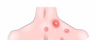
An example of skin lesions in systemic mastocytosis
Share
A year later, the famous German immunologist, bacteriologist and chemist Paul Ehrlich discovered mast cells. And in 1887, his compatriot and namesake Unna (Paul Unna), based on clinical and histological data, presented a detailed description of the disease.
In 1936, the Frenchman Albert Sézary and co-authors coined the term "mastocytosis", popularized in 1953 by Robert Degos. 13 years after Cesari's publication, J. M. Ellis first described the systemic form of the disease. The World Health Organization (WHO) approved the terminology, classification and diagnostic criteria for mastocytosis only in 2001.
WHO classification of mastocytosis, 2016
Cutaneous mastocytosis.
- Maculopapular cutaneous mastocytosis, or urticaria pigmentosa.
- Diffuse cutaneous mastocytosis.
- Skin mastocytoma.
Systemic mastocytosis.
- Sluggish (indolent, indolent) systemic mastocytosis.
- Sluggish (smoldering) systemic mastocytosis.
- Systemic mastocytosis associated with a hematologic disease (non-mast cell).
- Aggressive systemic mastocytosis.
- Mast cell leukemia.
Mast cell sarcoma.
Share
The next paragraph contains a detailed description of what is happening in the patient’s body from a biological point of view. If you are interested in finding out how to notice this disease and what, besides alcohol and spicy foods, can cause symptoms, scroll directly to the subheading “System diagnosis”
.
What happens in the patient's body
Despite the relatively long history of research on mastocytosis, the mechanisms of its occurrence and development have not yet been fully studied. However, with the improvement of diagnostic techniques, new data on this issue are emerging.
Mastocytosis is almost always caused by a point mutation in the gene encoding proto-oncogenic receptor tyrosine kinase c-kit (CD117, SCFR), which binds to stem cell factor (SCF). In simple terms, a more active receptor on the surface of mast cells and their precursors in the bone marrow causes uncontrolled increased growth and maturation of this type of cell.
The most common mutation (more than 80 to 95 percent of patients with systemic mastocytosis) is D816V, but other c-kit polymorphisms can cause the disease; at the moment, about 20 of them are known. Concomitant mutations of other genes, such as TET2, SRSF2, ASXL1, RUNX1, JAK2, CBL, NRAS, KRAS and others, can also contribute to the development of various forms of mastocytosis and their severity. In the vast majority of cases, such genetic defects arise in somatic (non-reproductive) cells after conception, therefore they are absent from relatives and are not passed on to offspring
. In isolated cases, the disease is familial in nature and is inherited in an autosomal dominant manner, that is, a person with one defective copy of the gene will pass it on to a child with a 50 percent probability.
Mast cells with similar mutations (degenerated, neoplastic), in addition to the “native” membrane proteins—mast cell tryptase (MCT) and CD117—often express unusual CD25, CD2 and, less commonly, CD30, CD33, CD52 and/or CD123. Detection of these proteins contributes to the diagnosis of the disease. In addition, the altered cells synthesize drug resistance surface antigens such as CD47, CD243 and CD274, which could become targets for new drugs
Immunohistochemical analysis of bone marrow biopsy (400x magnification): aggregates of mast cells expressing the membrane protein CD25
Malisha R Johnson et al. / Modern Pathology, 2009
Share
Bone marrow aspirate smear (1000x magnification, Wright-Giemsa stain): atypical mast cell morphology, including spindle-shaped, degranulated, and hypogranular forms
Malisha R Johnson et al. / Modern Pathology, 2009
Share
Immunohistochemical analysis of bone marrow biopsy (40x magnification): multifocal aggregates of mast cells containing tryptase
Malisha R Johnson et al. / Modern Pathology, 2009
Share
Bone marrow biopsy (200x magnification, hematoxylin and eosin staining): large aggregate of mast cells (more than 15) with an admixture of lymphocytes and eosinophils
Malisha R Johnson et al. / Modern Pathology, 2009
Share
The further scenario, as follows from the definition of mastocytosis, is obvious: uncontrollably multiplying cells enter the blood and accumulate in various organs. There they release vasoactive substances and inflammatory mediators: histamine, heparin, prostaglandins, leukotrienes, tumor necrosis factor α and many others, causing tissue damage. Compression by accumulated mastocytes also contributes to organ dysfunction.
In the more common cutaneous form, their “invasion” is limited to the skin, which is already quite unpleasant. In the much rarer, more severe and difficult-to-diagnose systemic form, neoplastic mast cells accumulate outside the skin, although they usually involve it too. This form is often “masked” as skin diseases or diseases of individual organs. Therefore, we will consider systemic mastocytosis in more detail.
System diagnostics
Most often, systemic mastocytosis affects the skin (manifestations are similar to the cutaneous form) and the gastrointestinal tract (inflammation; digestive disorders, liver and pancreas function). Less commonly - the heart (tachycardia, palpitations, hypotension), lungs (shortness of breath, asthma-like syndrome), nervous system (cognitive disorders, impaired concentration and short-term memory, headache, depression) and bones (osteoporosis and fractures). The number of leukocytes in the peripheral blood may also decrease, which leads to weakened immunity and, as a result, frequent infections.
Typical symptoms of systemic mastocytosis are nonspecific and include:
- redness and itching of the skin, age spots, urticaria;
- Darier's symptom (urticaria in response to mechanical irritation of the skin);
- discomfort, bloating and pain in the abdomen (including due to the formation of ulcers in the stomach due to excess acid production);
- digestive disorders;
- nausea and vomiting;
- diarrhea;
- enlargement of the liver, spleen and lymph nodes;
- disturbances of smell;
- inflammation of the ENT organs;
- pain and discomfort in the eyes, lacrimation;
- dizziness and fainting;
- headache;
- depression;
- cognitive impairment;
- pain in the musculoskeletal system;
- bleeding;
- anaphylaxis.
Share
These manifestations can occur in any combination
depending on the damage to specific organs and systems. Triggers for the onset of symptoms can be various things, the most common being skin irritation, exercise, alcohol or spicy foods, insect bites and certain medications.
Diagnosis of systemic mastocytosis is based on a detailed interview.
It is needed to identify symptoms. Here, the patient’s attention to his body and well-being is extremely important - it will help to suspect in time that something is going wrong and meaningfully tell a specialist about it.
Symptom diagram
Share
Instrumental methods most often include bone marrow and target organ biopsies
(if their damage is suspected) to search for mast cell agglomerates and determine the morphology of these cells. Biopsy analysis may include immunohistochemical studies to examine the expression of specific surface proteins (CD25, CD2, and others).
Molecular diagnostic methods are also
: real-time sequencing and various modifications of the polymerase chain reaction. With their help, mutations that lead to the development of the disease and determine the severity of its course are determined.
A biochemical blood test can help identify elevated tryptase levels, characteristic of systemic mastocytosis, as well as markers of organ damage
(for example, increased concentrations of liver enzymes). A general blood test can show a large number of mast cells, including altered shapes, and a decrease in the concentration of other formed elements.
Radiation methods help assess target organ damage
(ultrasound, radiography, computed tomography and magnetic resonance imaging)
and endoscopy
(with or without biopsy)
Diagnostic criteria for systemic mastocytosis
The main criterion is multifocal accumulation and clustering of mast cells (15 or more per cluster) in the bone marrow or tissues other than skin.
Minor criteria:
- abnormal morphology (eg, spindle-shaped) of more than 25 percent of the mast cells in the biopsy specimen;
- expression of CD2 and/or CD25 in mast cells of bone marrow, blood or internal organs;
- expression of an activating mutation of codon 816 of the KIT
in the bone marrow or internal organs; - basal serum tryptase level more than 20 nanograms per milliliter (not significant with concomitant hematological diseases).
The diagnosis is made based on the presence of either a major and one minor criteria or three minor criteria.
Share
Compounding the diagnostic challenge is how different the different forms of systemic mastocytosis are. Read more about them in the next paragraph.
Form, form, discord
The listed criteria make it possible to make a diagnosis, however, individual forms of systemic mastocytosis have their own specific characteristics.
Sluggish forms of systemic mastocytosis
They occur most often, are relatively mild and progress slowly. Indolent differs only in relatively less damage to organs. These forms are characterized by skin symptoms; there are no concomitant hematological diseases.
Systemic mastocytosis associated with hematological disease
(approximately 20 percent of all cases), in addition to the general diagnostic criteria, has signs of proliferative or dysplastic bone marrow lesions. It is more severe and develops faster than sluggish forms. Skin lesions are rare.
Aggressive systemic mastocytosis
It is rarely observed, is severe, affects many organs and systems, and progresses quickly. Skin manifestations are usually absent.
Mast cell leukemia
- the most rare and malignant form of systemic mastocytosis. Characterized by a high content of immature mast cells in the bone marrow (more than 20 percent) and peripheral blood (more than 10 percent in the leukemic variant and less in the aleukemic variant).
Treat effectively
Unfortunately, there is no radical treatment for systemic mastocytosis yet.
, however, available methods can significantly improve well-being and slow down the course of the disease. Therapy is divided into symptomatic (eliminates individual manifestations of the disease) and cytoreductive (reduces the number of neoplastic mast cells). The choice of individual drugs depends on the specific form of the disease and its severity.
Means for symptomatic therapy include:
- antihistamines (block histamine receptors, reducing its effects and, as a consequence, allergic manifestations);
- mast cell membrane stabilizers (reduce the release of cytokines and vasoactive substances by mast cells, used when antihistamines are insufficiently effective);
- leukotriene inhibitors (block the action of the corresponding mediators of allergy and inflammation);
- glucocorticosteroids (anti-inflammatory hormonal drugs are stronger than antihistamines and antileukotriene drugs, although with more pronounced side effects, are used for aggressive forms of the disease either locally or systemically - in the form of tablets or injections);
- adrenaline, also known as epinephrine (administered together with glucocorticoids, antihistamines and infusion therapy for emergency treatment of anaphylactic shock); for uncontrolled anaphylaxis, the immunoglobulin E antagonist omalizumab is used;
- other symptomatic drugs are prescribed for lesions of specific organs, for example, to reduce the acidity of gastric juice and dilation of the bronchi.
Cytoreductive therapy uses:
- interferon-α (an immunotherapeutic drug that reduces the degree of infiltration of the bone marrow by mast cells and, accordingly, reduces organ damage);
- cladribine (a chemotherapy agent that is more effective than interferon, quickly inhibits the growth of mast cells and, undesirably, some other types of cells);
- tyrosine kinase inhibitors (selectively block the corresponding group of enzymes necessary for the production of mast cells, used for aggressive forms of systemic mastocytosis, including those not associated with mutations of the C-KIT
).
Share
Rarely, in case of ineffectiveness of the listed methods of therapy, donor bone marrow transplantation is resorted to. To do this, a suitable donor is selected, the patient's own bone marrow is destroyed with chemotherapy drugs (sometimes with the addition of radiation therapy) and replaced with normally functioning donor cells that do not contain mutations.
All forms of mastocytosis are treated by hematologists, and ideally by oncohematologists. They should be contacted in case of suspicious symptoms described above. At the same time, most people with systemic mastocytosis go to therapists and struggle for a long time and unsuccessfully with individual manifestations
without receiving a correct diagnosis.
The easiest way to find a good specialist is in large hematology centers and clinics; People who have already undergone treatment and patient organizations can help with this. It would also be useful to get a “second opinion” - visit several specialists and listen to their recommendations.
You can find a suitable clinic linked to a map, as well as get information about patient organizations on the Gemogid
.
Share
With proper treatment, patients with indolent forms of systemic mastocytosis usually live at least as long as those who do not suffer from this disease. In more aggressive forms of the disease, life expectancy and its quality depend on the degree of organ damage; how quickly a person went to the doctor and received the correct diagnosis; the effectiveness of treatment and the competence of the attending physician.
If everything goes well, “fat adversities” will not prevent you from living a long and fulfilling life. But here it is important to mention that the main factor that allows you to save a life and improve its quality is the patient’s attention to his body and the fear of finding out what is happening to him if the prescribed treatment does not help.
Oleg Lishchuk
198535/HEMA/media/04.21/1 N+1
Mastocytosis (mast cell leukemia)
There are 4 forms of mastocytosis
:
- cutaneous mastocytosis
of infancy or early childhood, in which skin lesions resolve during puberty, systemic changes are usually absent, and progression with the development of a systemic form occurs extremely rarely. - cutaneous mastocytosis
of adolescents and adults with frequent systemic lesions, which is often accompanied by systemic disorders, but they usually do not progress. Spontaneous regression in adults, unlike in children, has not been described. Sometimes this form progresses, turning into systemic mastocytosis with progressive damage to internal organs; - systemic mastocytosis
; - malignant mastocytosis
(mast cell leukemia) is a very rare form, characterized by the presence of cytologically malignant mast cells in many organs and tissues, especially in the bones and peripheral blood, with a rapid fatal outcome; usually not accompanied by skin manifestations.
More than half of patients with mastocytosis
complaints are limited to skin lesions. About 1/3 of patients complain of itching, attacks of skin redness, paroxysmal tachycardia, drops in blood pressure, periodic increases in body temperature, etc. These symptoms are caused by degranulation of mast cells and are observed in common forms of mastocytosis of the skin or internal organs.
For mastopitosis
There are five types of skin lesions (the first two of which are observed in both infants and adults).
Maculopapular type of mastocytosis
, represented by tens or even hundreds of small red-brown hyperpigmented spots and papules, which after friction (Darier-Unna test) acquire an urticarial character, is the most common.
Multiple nodular type of mastocytosis
presented by multiple dense hemispherical nodes of pink, red or yellow color, 0.5-1 cm in diameter with a smooth surface, sometimes the nodes merge into plaques, the Daria-Unna phenomenon is weakly expressed, especially in adults.
Type of large solitary node
, or mastocytoma, manifests itself as a node with a diameter of 2-5 cm, having a smooth or wrinkled surface, like an orange peel, and a rubbery consistency. Occasionally there are up to 3-4 nodes. Nodes occur exclusively in infants, most often in the neck, torso, and forearms. The Daria-Unna test is positive. Trauma leads to the formation of vesicles, pustules or blisters on the surface of the node, as well as a tingling sensation. Mastocytomas usually regress spontaneously, with them receding and flattening. In some cases, the nodes are associated with rashes characteristic of the maculopapular type of mastocytosis.
Diffuse (erythrodermic) type of mastocytosis
always begins in early childhood. It is characterized by large itchy foci of yellow-brown infiltration, usually in the armpits and intergluteal folds. The lesions have an irregular shape, clear boundaries, a dense (to the point of woody) consistency, and ulcerations, cracks, and excoriations easily occur on their surface. Progression of the pathological process can lead to erythroderma. In this case, the skin acquires a doughy or dense consistency, its color varies from pink-red with a yellow-brown tint to dark brown. The Daria-Unna test is positive, and even mild trauma to the lesion leads to the appearance of blisters, often accompanied by intense itching. When blisters predominate in the clinical picture, they speak of bullous mastocytosis. The course of the disease gradually improves, progression to systemic mastocytosis occurs rarely, and death occurs only in infants as a result of histamine shock.
Telangiectatic type of mastocytosis
(a type of persistent macular telangiectasia) is a rare form of mastocytosis that occurs predominantly in adults, mainly in women. It appears as widespread brownish-red spots of various shapes, consisting of bright telangiectasia. located on a pigmented background. There is a slight tendency for blisters to appear in areas of friction or spontaneously. The skin of the chest and limbs is most often affected. Itching is characteristic. In some cases, bone damage and peptic ulcers occur.
Systemic mastocytosis
characterized by damage to internal organs in combination with or without cutaneous mastocytosis, occurs equally often in men and women. It is observed in 10% of patients with skin manifestations of mastocytosis. In most cases, skin lesions precede signs of a systemic process. Skin changes in systemic mastocytosis are represented by maculopapular, multiple nodular or diffuse (erythrodermic) types.
During the process of mastocytosis
Any organs and systems may be involved. Most often - bones, liver, spleen, lymph nodes, gastrointestinal tract, central nervous system. 20-30% of patients with mastocytosis have bone lesions, which on X-ray examination manifest as osteoporosis and osteosclerosis. Osteolytic changes are accompanied by pain. About 1/4 of bone lesions are mastocytomas, but they are uncommon in children. Liver lesions present with asymptomatic mast cell infiltrates and fibrosis with hepatomegaly and diffuse mastocytosis.
Systemic mastocytosis
along the course it can be benign and malignant. Malignant mastocytosis includes mast cell leukemia, mast cell sarcoma, and variants of systemic mastocytosis associated with lymphomas, solid neoplasms, or malignant blood diseases.