Atheroma is harmless, it only affects the appearance, so it can be treated at home. It is a benign tumor that occurs when the sebaceous gland is blocked. In appearance it resembles a dense formation.
The site provides reference information. Adequate diagnosis and treatment of the disease is possible under the supervision of a conscientious doctor. Any medications have contraindications. Consultation with a specialist is required, as well as detailed study of the instructions! Here you can make an appointment with a doctor.
Benign atheroma - treatment at home
If you don't treat it right away, it can fester. This disease must be treated from the first moment of detection. Folk remedies that can effectively get rid of this problem can come to the rescue.
The most effective recipes and methods for treating atheroma at home:
- Lamb fat. It needs to be melted and poured into a container convenient for use. Every day, rub the fat into the area of formation several times. It's better to do this as often as possible. To increase efficiency, you can add vegetable oil or garlic juice to lamb fat.
- Onion mask. If a tumor appears on the face, a mask made from ground laundry soap and baked onions will help get rid of it. The mixture must be mixed, applied over the wen, and wrapped tightly. Apply a fresh mask several times a day, removing the old one.
- Sour cream with honey. To prepare the ointment, mix honey, sour cream and salt in equal proportions in a convenient container and close with a tight lid. Rub into the area of atheroma on slightly warmed skin. After half an hour, rinse with warm water.
- Aloe. Rub aloe juice 3-4 times every day. Effectively fights disease in the earlobes or behind the ears.
- Coltsfoot. Apply fresh leaves to the tumor and wrap it. Do not remove for a day, then change to a new sheet. After 7-9 days the disease will disappear.
- Peony and wormwood. To prepare, mix ammonia diluted with water with 3-4 tablespoons of crushed peony roots and 3 tablespoons of wormwood. Before adding, boil the peony root and wormwood for a few minutes and let it brew for an hour. Apply lotions several times a day for 40-60 minutes.
- Garlic. Mix crushed garlic and vegetable oil in equal quantities and rub the skin on the affected area. If itching occurs, wash off and apply the mixture again.
Causes
Basically, the occurrence of such a wen is influenced by poor ecology, living conditions, hormonal imbalance, and metabolism.
Find the answer
Are you having any problem? Enter “Symptom” or “Name of the disease” into the form, press Enter and you will find out all the treatment for this problem or disease.
The causes of atheroma will be any condition that leads to blockage of the sebaceous gland ducts.
Causes:
- Swelling of the hair follicle;
- Gland duct injuries;
- Blockage of the gland duct;
- Hyperhidrosis.
The main factors that provoke inflammation:
- Increased testosterone levels;
- High oily skin;
- Lack of personal hygiene;
- Metabolic disorders in the body;
- Frequent use of deodorants;
- Unfavorable, harmful work.
Symptoms and clinical signs
Atheroma occurs on the skin where there are many sebaceous glands. Common locations include the scalp, neck, back, chin, armpits, and groin area.
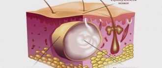
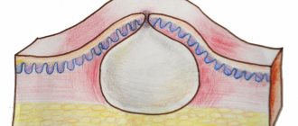
Inside the wen there is a thick epidermis, the secretion of the sebaceous glands. In appearance, this substance resembles a viscous white mass.
Clinical signs of atheroma are:
- A rounded formation under the skin;
- Smooth surface of formation;
- Mobility;
- Density and clear boundaries;
- No pain during touching;
- In the center of the formation there is an edematous excretory duct, slightly increased in size.
When such a tumor occurs, pain rarely occurs. Only a specialist can determine the presence of atheroma. Often an infection gets inside, which provokes the onset of an inflammatory process and the formation of pus inside. It is painful and requires immediate treatment.
Complications
Delaying the treatment of atheroma is fraught with suppuration of the cyst, when its capsule grows in size, swells, turns red, the skin over it becomes tense and causes pain in the patient. Independent attempts to squeeze out the contents of the formation often end with the transition of the disease to a new stage - abscessing atheroma, accompanied by swelling of nearby tissues and lymph nodes, severe pain, and general intoxication of the body. The most unfavorable outcome of the developing situation may be the entry of infection from the purulent sac into the patient’s circulatory system, which can cause sepsis.
If you are looking for where to remove a wen, contact the surgical department of the Miracle Doctor clinic. Experienced surgeons will diagnose and treat using the optimal method.
Make an appointment on the clinic’s website in the “Schedule” section or by calling the hotline.
Removal by laser and radio wave method
Radio wave surgery is considered one of the best methods for removing atheroma.
The method is considered effective and safe.
There are several main advantages of radio wave surgery:
- The risk of bleeding is completely eliminated;
- There is no chance of relapse, because the tumor is removed once and for all;
- Painless removal procedure;
- There is no need for stitches after the operation;
- There are no traces left on the body;
- Does not affect the patient's ability to work.
The duration of the procedure is 20-30 minutes. During such an operation, the surrounding tissues are not damaged, so no traces of the intervention remain on the patient’s body.
The atheroma is burned out by radiation of radio waves, which leaves only a small depression after the cyst, which is covered with a bandage until it disappears completely.
Laser removal is also a popular method of combating it. The procedure itself takes little time, and the results appear immediately. This procedure is carried out if the size of the wen is small.
After such an operation, no traces are left on the body, so it is often performed on the face or open areas of the body. If a cyst appears on the head, then during laser surgery there is no need to shave off the hair around the tumor.
The duration of the entire procedure is less than 20 minutes; after it is completed, the return of such a disease is excluded. If the size is large, then before laser removal it is cut with a scalpel, only then the laser is used. The procedure requires a rehabilitation period of 1-2 weeks.
Correcting the problem surgically
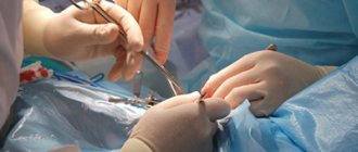
Large cysts are removed surgically. The contents of the capsule and its shell are removed under sterile conditions to prevent infection.
The removal process takes place in several stages:
- From the very beginning, the patient undergoes the necessary diagnostic tests, laboratory or other examinations. You cannot eat for 4 hours before surgery. The length of surgical removal depends on the size and complexity of the cyst, but the procedure usually does not take more than an hour.
- The patient goes to the operating room, where he is given painkillers. Often, using local anesthesia, the medicine is injected near the tumor.
- The skin over the cyst is cut with a scalpel, and the cyst along with the capsule is removed. Sometimes, the contents are removed first, and only after that the capsule shell is removed. The latter option is used more often, because after it the seam will be smaller and therefore less noticeable.
- The incision site is treated with an antiseptic and sutured. Then a tight bandage is applied. The type of suture that is placed on the wound depends on the location of the cyst. If it has formed on the face, then it is stitched up with a cosmetic one, and if in places where the muscles move much more actively, a reinforced suture is used.
- After the operation, the patient is transferred to the ward where rehabilitation will take place. The dressing on the wound is changed every day, checking its condition. If everything went well and no problems arose, the stitches are removed after a week.
- The duration of wound healing depends on the location of its formation. Healing can take a month, and sometimes it only takes a week.
Often, a scar remains after surgery. Its size and visibility depend on the size of the extracted cyst, the method of treatment, and the professionalism of the attending physician. Sometimes the scar completely resolves, but this depends on the individual patient’s body.
Features of treatment of atheroma on the face and ear without surgery
It often occurs on the face, earlobes, or behind the ears.
A cyst on the face often appears on the following parts:
- Forehead;
- Chin;
- Nose;
- Below the cheeks.
She almost never grows big. But it often becomes inflamed, because the face constantly becomes oily, there are a lot of microbes on it, so the infection often gets inside.
If the atheroma on the face is inflamed, it should be removed as quickly as possible. It is removed with a laser or radio waves so that there are no marks or scars left. If the tumor is noticed early, then traditional medicine will help: ointments, lotions, compresses, rubbing.
The cyst is often localized on the earlobe; in rare cases, it occurs on the auricle. Often only one neoplasm appears, which takes a long time to develop and can reach large sizes - 4 cm in diameter.
On the earlobe, atheroma often becomes inflamed and festers, which leads to swelling and redness. Touching her is painful. If an inflammatory process occurs, then such a cyst is removed using conservative or surgical treatment.
The formation behind the ear reaches 0.4-0.5 mm in diameter and may not manifest itself for a long time. When inflammation begins, the cyst increases significantly in size, there is a feeling of itching, burning, swelling, and the formation of free fluid to the touch.
If the patient’s body is healthy and can withstand immunity, then after inflammation the abscess opens on its own, all its contents come out. It's not scary, but small, almost invisible scars may remain on the skin. If the cyst has not opened, then you need to seek help and remove it.
Types of purulent atheromas
The cyst begins to become inflamed in the case of a long course of the disease and occurs in a septic or aseptic way. Inflammation becomes aseptic when the cyst capsule is irritated by the underlying tissues. This is possible when clothing rubs against the formation or puts pressure on it from the outside. The atheroma swells, sometimes pain appears. After a few days, all symptoms subside, and connective tissue forms around the formation. It envelops the atheroma like a shell.
Septic inflammation is recorded more often than aseptic inflammation. The reason is the penetration of infection into the cyst. Symptoms are more clear. Characterized by constant pain in the area of the cyst and redness. Due to the suppuration of the secretion in the atheroma, the local temperature rises, and then the general one. The outcome of such inflammation can be different: from the cyst breaking out or the capsule melting and turning into phlegmon.
What symptoms do you see a surgeon for:
- Presence of hernial protrusion
- Daggering pains in the abdomen
- Bloating
- Pain in the right hypochondrium
- Bitterness in the mouth
- Nausea
- Presence of neoplasms on the skin
- Swelling and redness of the skin
- Bone fractures and bruises
- Wounds of any location
- Vomit
- Enlarged and painful lymph nodes
Localization on the scalp
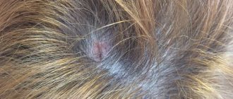
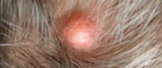
This type of tumor is common on the head. This is explained by its peculiar location, because it constantly appears near the hair follicles. Such a formation can only be treated surgically; other methods, conservative or non-traditional, lead to complications.
Such a formation cannot go away on its own, even if it opens, the pus comes out, and the size decreases, this cannot indicate the complete disappearance of such a problem. Over time, the sebaceous ducts will become clogged again, leading to new cyst formation.
Atheroma on the scalp is treated using methods such as:
- Planned removal of small tumors without inflammation is carried out using laser and surgical radio wave removal methods.
- If inflammatory processes occur, emergency opening, drainage, opening of the abscess, treatment of local inflammation using a symptomatic method, and removal with a scalpel are used.
After removing the cyst with capsule, a rehabilitation period begins.
Depending on the removal method used, it varies:
- If a small atheroma was removed and the wound was sutured, the sutures dissolve within a week or a week and a half, leaving no traces;
- With the help of laser and radio waves, the rehabilitation period is reduced to 5-7 days, because the size of the incision during such operations is minimal;
- Treatment of purulent atheroma requires a longer rehabilitation period; it often leaves a keloid scar.
The difficulty of treating a cyst on the scalp depends on the size and stage of inflammation. The sooner the patient seeks help, the fewer defects will remain after surgery. If gentle treatment methods are used, then there is no need to shave off the hair near the cyst, so there will be no appearance defects.
Diagnostics
Diagnosing atheroma is not a difficult task for experienced surgeons at the Shifa clinic. Already at the stage of the initial examination, our doctors palpate a round elastic neoplasm, which is a sebaceous gland cyst.
An analysis of the patient’s complaints and medical history remains mandatory in verifying the diagnosis. The localization and external characteristics of the tumor in 90% of cases make it possible to identify atheroma at the first request for help.
Differential diagnosis is carried out with soft tissue neoplasms (fibromas, lipomas, osteomas). To do this, specialists at the Shifa clinic use the following additional procedures:
- Ultrasound examination (ultrasound). The technique allows you to clarify the size, volume of the tumor and the presence of signs of inflammation.
- Diagnostic puncture.
- General blood analysis.
- Coagulogram to assess blood clotting.
- Blood test for infectious group (HIV, hepatitis B, C, syphilis).
These examinations help surgeons at the Shifa clinic assess the patient’s general health, identify possible contraindications and prevent complications. All diagnostic procedures can be completed in just a few days in our center without waiting in long lines.
Development of suppurating atheroma
Purulent atheroma occurs with acute microbial inflammation of the contents of the sebaceous gland. The glands are located under the layer of skin, tightly adjacent to the hair follicles. They are necessary to produce lubricant for the skin and hair.
If the glands are clogged, sacs are formed, which are gradually filled with sebaceous mass, which resembles gruel. If a cyst is not treated immediately after it occurs, an infection often gets inside, which leads to inflammation and suppuration.
The usual sizes of atheroma are within a few centimeters. It is not a real tumor, it only resembles it in appearance. When microbes enter the cyst cavity, they cause inflammation with the formation of pus.
If the atheroma is red, swollen, pain is felt when pressed, and the body temperature rises, then this indicates the beginning of purulent melting of the abscess contents. When the pus melts the skin, the contents come out.
It is dangerous if the pus does not come out, but spreads under the skin. This phenomenon leads to the development of widespread inflammation, which is called an abscess.
If suppuration appears, you should immediately consult a doctor. The only effective treatment is to remove the abscess through surgery. In the operating room, the surgeon removes the contents and the shell of the formation itself, then the bacteria are fought.
The purulent cavity is often treated with antiseptics, drainage is introduced through the wound, and the remaining pus is removed with its help. In case of severe inflammation and a large amount of pus, doctors prescribe antibiotics.
Problems often arise in removing the cyst along with the membrane in the case of purulent atheroma. If the specialist was unable to completely remove all remnants of the membrane, then a relapse will occur over time. Treatment of an abscess takes a long time, sometimes a person needs at least several weeks to fully recover.
What is lipoma?
A lipoma is a benign growth that usually appears as a “lump” under the skin that is made up of fatty tissue.
Lipoma is often called a “fat tumor,” but do not be alarmed—it has nothing to do with malignant tumors. Almost nothing is known about why lipomas occur. It is believed that this is due to the malfunction of certain enzymes and a hereditary predisposition.
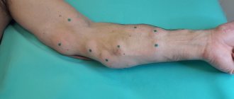
Most often, wen, oddly enough, appears where there is little fat under the skin: on the back and in the area of the shoulder girdle, on the outer side of the shoulder, thigh. This “bump” is soft and painless to the touch. If you press it with your finger, it easily “rolls” under the skin. Dimensions rarely exceed 5 cm.
There are also exotic localizations: around the nerve (in this case, the lipoma leads to pain), in the spinal canal, in the joints, in the thickness of the muscles.
Lipomas usually grow slowly and in most cases do not cause any problems other than cosmetic discomfort.
