| Idiopathic guttate hypomelanosis | |
| Other names | Symmetrical leukopathy progressive |
| Speciality | Dermatology |
Idiopathic guttate hypomelanosis
is a very common acquired disorder that affects women more often than men, presenting with skin lesions that occur primarily on sun-exposed areas of the skin, suggesting that sun exposure may be important.[1]:860
Reason for appearance.
Each group has separate symptoms and etiology of development. The most dangerous disorders are those that have internal prerequisites for development.
External reasons
External root causes are less dangerous.
https://www.youtube.com/watch?v=ydIbQGsf6nc
White pigment spots on the body are the result of:
- injuries received;
- increased sensitivity to external stimuli;
- constant interaction with chemicals;
- prolonged exposure to direct sunlight.
Probable causes also include abuse of solariums. Most often, damage to the integrity of the skin leads to disruption. The treatment approach directly depends on the cause of the disorder. Only a doctor can find out why the skin color has changed after a comprehensive diagnosis.
Depigmented areas occur in people when using aggressive household chemicals. People with hypersensitivity to chemicals are more likely to have a reaction.
Important! When influenced by external factors, the problem is not dangerous. This is a cosmetic defect.
To get rid of the problem, you need to minimize contact with the irritant.
Internal reasons
If white spots appear on the body due to internal reasons, treatment is required. It is necessary to eliminate the underlying disease.
Internal root causes include:
- autoimmune diseases;
- kidney diseases;
- gastrointestinal pathologies;
- hormonal imbalances;
- fungal diseases;
- avitaminosis.
White areas on the body are a symptom of the underlying disease. The internal factor acts as a trigger mechanism for the disorder.
The production of the hormone begins due to the enzyme tyrosinase, which triggers various chain reactions. This process is regulated by a combination of several genes. And when a violation or failure appears in them, white spots appear - hypomelanosis. This genetic disease is determined by the recessive type and is common among marriages between relatives.
Such failures are difficult to predict, and it is very difficult to influence them. It is possible to predict the appearance of spots in children whose relatives have freckles, light spots, or a clearly defined patch of gray hair. Conducted research has made it possible to find methods and drugs that can normalize melanin synthesis.
Causes of hypomelanosis: why white spots appear on the skin
The main cause of the disease must be sought in the human genetic apparatus. There is a connection between the development of hypomelanosis in parents and their children - the hereditary nature of skin pigmentation disorders. The disease is inherited in a recessive manner, and the carrier of the hypomelanosis gene can be identified by white spots on the skin, freckles and areas of gray hair on the head that have clear boundaries.
The formation of spots on new areas of the skin and the progression of hypopigmentation on already affected areas is provoked by excessive sun exposure.
Classification of hypomelanosis
There are several categories of hypomelanosis, and each type of disease has its own differences regarding symptoms of manifestation, time of occurrence, causes, etc. In total, the following types of disease can be distinguished:
- Hypomelanosis Ito. It is a genetic feature, rarely transmitted hereditarily, and more often has a sporadic path of origin. On the skin, the pathology appears in a wavy or zigzag pattern. Almost always, this category of disease is accompanied by problems in the neurological sphere, which is caused by disturbances in embryogenesis that occurs during pregnancy. Because of this, other defects in the child’s development are often present.
- Teardrop-shaped. This type of disease affects women who have a reduced amount of melanin in the blood. As a rule, these are representatives with light skin type. Changes develop when the cover is exposed to the sun or ultraviolet radiation for a long time. Due to the fact that the production of melanin occurs much more slowly than its restoration, hypomelanosis develops.
- Idiopathic. This category usually includes all those forms of the disease, the origin of which cannot be reliably identified.
- Progressive macular hypomelanosis. This type of pathology differs in that it is provoked by the activity of bacteria on the skin of the body, during which chemical components are produced that interfere with the normal production of melanin.
The skin of the legs and forearms is most often affected, but otherwise hypomelanosis can spread throughout the body. Rarely, light marks appear on the face.
Varieties.
Guttate hypomelanosis - the disease is characterized by the appearance of white, round spots with a diameter of up to 1 cm. Women aged 35-55 years are most susceptible to this type of disease. The number of melanocytes decreases several times. This disease is caused by genetic inheritance.
Hypomelanosis is a disease common to everyone and can be hereditary. It is characterized by a violation of cell development in the prenatal period, especially in the 2-3 trimester of pregnancy. Skin pigmentation is determined by the presence of areas of unusual shape. Children may have additional symptoms, such as mental impairment, autism, seizures, and microcephaly. Diseases of other organs may also occur.
Idiopathic hypomelanosis is characterized by a violation of melanin synthesis; areas of the skin in such cases do not have pigment, they can be called colorless. Under the influence of ultraviolet rays, the pathology will progress. The cause of the disease is a genetic malfunction, usually manifests itself in children, but can also be observed in adults.
Classic idiopathic hypomelanosis
This type of pathology includes all other forms of manifestation of the disease of unknown etiology.
It manifests itself from the formation of lighter areas on the skin of irregular shape. They can appear on any part of the skin, unlike teardrop-shaped lesions, the lesions can be large in size and merge together as the disease progresses. This form is characterized by only one symptom – the appearance of light spots. The spots do not become inflamed, do not itch or cause discomfort. Treatment is the same as in the case of guttate hypomelanosis.
Causes and exacerbations
The mechanisms of pathology development differ in each case.
- However, it is believed that most cases of the idiopathic form of the disease occur due to impaired melanocyte production processes. With guttate hypomelanosis, their rapid destruction occurs, but with Ito, problems with the migration of these cells in the 2-3 trimester of pregnancy are to blame.
- Some scientists have come to the conclusion that there are family cases of this disease, and the hereditary factor is to blame. One way or another, the undeniable fact is that it is problems with melanocytes that lead to the appearance of spots.
- In childhood, hypomelanosis can accompany various other hereditary pathologies. For example, local changes in the color of the hair and skin are detected in tumorous scleroderma and Vaanderburg syndrome.
- The disease can worsen with prolonged exposure to the sun.
general information
Human skin is the most extensive and sensitive organ; any neoplasm on it or a change in pigmentation indicates that some kind of malfunction is occurring in the body. Therefore, under no circumstances should any modification of the skin be ignored.
The appearance of any spots on the skin always causes inconvenience and a desire to get rid of them as quickly as possible. Before you sound the alarm, you need to find out the possible reasons for their appearance. Let's look at the most common pathologies that lead to the appearance of white spots.
Diagnostics.
A Wood's lamp is used to visualize the spots. A brain CT scan and histological examination may also be prescribed.
Treatment of the disease is characterized by the use of corticosteroids, retinoids, and cryomassage of the affected areas. Treatment methods also include phototherapy and bioresonance therapy.
Diagnosis of changes in the dermatological area helps to examine the skin using a Wood's lamp. If it is necessary to differentiate the pathology from vitiligo, albinism or another disease associated with loss of pigmentation, histological examination is prescribed. In this case, you will need to take the altered bodily area for a biopsy.
Additional diagnostic methods are prescribed, as a rule, for the idiopathic form, as well as for Ito hypomelanosis. In particular, an electroencephalogram, neurological examination, ultrasound and MRI are necessary.
Disruption of pigmentation processes can manifest itself in various forms. To verify the pathology, it is necessary, in addition to a visual examination, to use a study using a Wood's lamp. It is especially often used in the presence of light skin and unclearly manifested pathology.
Diagnosis of hypomelanosis is based on identifying clear boundaries of a hypopigmented lesion when illuminated by a lamp in a darkened room. Thanks to it, it becomes possible to detect a site and verify it.
Diagnosis of Ito hypomelanosis additionally includes a computed tomography scan of the brain, which reveals enlargement of the 3rd and lateral ventricles, unclear boundaries between the brain substance, and atrophy of the frontal lobes.
Histological examination makes it possible to detect an insufficient number of melanocytes in the area of hypopigmentation. In addition, with Ito hypomelanosis, other features may be present in the lesion, for example, vascular nevi, cocoa spots, Ott's nevus, or Mongoloid blue spots.
[14], [15], [16], [17]
Guttate hypomelanosis is a type of leukoderma, a skin disease associated with the loss of pigment in the body. With this pathology, dysfunction or absence of melanocytes, cells responsible for the production of melanin, a natural brown pigment that determines the shade of the skin, eyes, and hair, is observed in the epidermis.
First of all, the doctor conducts an external examination. This helps to establish a preliminary diagnosis. Afterwards the patient is referred for dermatoscopy. This is a tool that increases the examination area several times. Sometimes this is not enough to tell what these white spots are on the body.
A scraping is taken for diagnosis. This is necessary for a detailed study of damaged skin. Additionally, you need to donate blood. Diagnosis from photos is not possible.
If necessary, the patient is referred to see specialized specialists. This is usually necessary when traditional treatment has not given a positive result.
Hypomelanosis ITO
They belong to the group of neurocutaneous hereditary syndromes; in combination with changes in the skin, a malfunction of the functions of the central nervous system, internal organs and systems is observed. The main cause of the development of the disease is considered to be anomalies in the formation of the outer germ layer in the early stages of the perinatal period of the embryo.
Hypomelanosis ITO is a heterogeneous form of phakomatosis. It manifests itself in discoloration of individual areas of the skin, in combination with manifestations of neuralgia; defects in the development of the skeleton, teeth and muscle tissue are also possible. The disease is considered rare, occurring in one child out of 80 thousand. The disease has not yet been fully studied; in medical works it may be called neurocutaneous syndrome or achromatic pigment incontinence.
The cause of the violations causing this anomaly has not yet been determined.
There is a connection between chromosomal abnormalities such as mosaic trisomy, mutations in the X chromosome and other genetic aberrations. The individual gene responsible for the predisposition to achromatic pigment incontinence has not yet been identified, so it is not possible to identify the pathology using perinatal screenings.
Symptoms
Hypomelanosis ITO is manifested by a group of symptoms that includes characteristic changes in the skin, central nervous system disorders, abnormal development of the skeleton, muscles, poor vision or hearing.
ITO syndrome on the skin appears as white discolored spots that appear at an early age (2-3 years). The spots may be light in color or brownish and irregular in shape. Skin spots may be congenital, or they may appear for the first time months or years of a child’s life. The localization of areas of hypopigmentation can be very diverse.
The most commonly affected area is the torso. The lesions can be located symmetrically, with the affected area increasing in one direction. The lesions may take the form of stripes, coils, and multiple spots may resemble a world map. The disease is characterized by the loss or acquisition of excellent pigmentation in certain areas of the hairline; focal hair loss, the appearance of angiomas, ITO nevus, and thickening of the nails are not excluded.
Pathologies of skeletal development may include: bone deformation, kyphosis-scoliosis, joint hypoplasia, the presence of rudimentary processes, asymmetrical arrangement of limbs, anomalies in the structure of teeth.
In the complex, benign vascular tumors of internal organs, cardiac malformations, hernias, defects in the structure of the genital organs and gastrointestinal tract can be observed.
Impaired vision functions include lightening of the retina, which in turn causes clouding of the surface of the stratum corneum, and possible development of strabismus. In most cases, hearing loss is diagnosed with hypomelanosis ITO. In more than 10% of all cases, a severe form of hearing loss is determined.
Patients are also characterized by a number of neurological disorders: muscle weakness, retardation, epileptic seizures. It cannot be said that children with ITO hypomelanosis are mentally retarded, but in more than 50% of all cases, difficult learning and a low level of socialization (autistic behavior) are observed. Epilepsy seizures are observed in more than 50% of all sick children.
To differentiate the pathology, complex diagnostic methods are used: external examination, MRI of the whole body, head nomogram, and the child is also advised to visit an ophthalmologist and ENT doctor.
Prevention.
Since the disease has a genetic type of inheritance, the following methods have been developed for prevention; they reduce the risk of developing the disease. Patients at possible risk of developing diseases are advised to avoid exposure to direct UV rays, not to visit a solarium, and to cover light spots.
Before figuring out how to treat a defect, you need to determine the cause. Typically, skin formation is one of the symptoms of ongoing diseases. Each of the ailments has a separate set of symptomatic manifestations. It is almost impossible to independently determine the true cause of the disease.
Pityriasis versicolor
Pityriasis versicolor is the most commonly diagnosed diagnosis. Another name is lichen versicolor. A white spot on the back is the main sign. The number of rashes increases. The formations spread to other parts of the body.
New growths are white and scaly. The shape is round, oval or cylindrical. The boundaries of the formations are clear. Additionally, the person has itching. There is an enlargement of the lymph nodes.
The cause of the disease is a fungal infection. The disorder is contagious.
Pityriasis rosea
Pityriasis rosea is not contagious. This is the result of viral diseases. White and pink formations appear on the body. There are small light dots around them. The causative agent of the disease is a fungal infection. The treatment is complex.
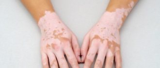
Vitiligo
White spots on the skin of the legs and arms – vitiligo. Have clear boundaries. Vitiligo occurs as a result of autoimmune disorders. Symptoms appear between 10 and 30 years of age. The disorder is hereditary.
Important! The disease cannot be cured. Therapy is aimed at reducing the severity of symptoms.
Vitiligo is a serious cosmetic problem.
White spots on the skin of the arms and torso are the result of delayed melanin formation. The disorder is hereditary. It is laid down at the genetic level. Activation of the disease occurs after exposure to ultraviolet radiation. Treatment is difficult. There is no single scheme. Therapy is aimed at combating skin manifestations.
Pigmentless nevus
This is a benign skin formation. May be congenital or acquired. There is no need to figure out how to treat the formation. These are ordinary moles with a deficiency of pigment.
All moles can degenerate into malignant formations. It is necessary to monitor their condition. The development of cancer is indicated by the growth of a mole.
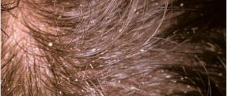
A white spot on the face may be a fungal infection. The affected areas rise above the epidermis. The surface begins to actively peel off. Lumpy areas appear. There is a risk of ulcers.
On the feet, the disease thins the skin. When it appears on the head, the hair becomes dull and lifeless. The most common fungal diseases are briefly described in the table.
| Disease | Description |
| Rubromycosis | Red and white spots appear on the body. Additionally, follicles and nodules appear. |
| Microsporia | The inflamed area is covered with scales. There is pronounced peeling. The lesions merge. |
| Trichophytosis | Affects exposed skin. The disease is transmitted through household objects. There is peeling on the skin. |
Fungal diseases also include seborrheic dermatitis, candidiasis and nail fungus. Almost all disorders are contagious. Requires urgent treatment. All personal hygiene products will need to be sanitized to prevent the spread of the disease.
Leprosy is a chronic infection. Affects skin and internal organs. White spots on the skin of the hands and body are different in size. The outlines are clear. Infection can occur through airborne droplets. There is also a risk of intrauterine infection.
Lumps and plaques may occur. The spots merge. Treatment requires consultation with highly specialized specialists.
Syphilis
Syphilis affects the entire body, including the skin. White spots on the skin of the legs and torso lead to enlarged lymph nodes. The main method of transmission of the disease is sexual.
The patient's body temperature rises. There is weakness and malaise. Without treatment, the disease progresses. The rash is characteristic of secondary syphilis.
Leucoderma
Leioderma is the result of the complete disappearance of pigment from skin cells.
The disease is a complication of other disorders:
- syphilis;
- pityriasis rosea;
- chronic dermatosis.
Melanin ceases to be produced completely. Leukoderma may have a false form. This is the result of the use of local cosmetics and medicines.
Hypomelanosis
A white spot on the face may indicate hypomelanosis.
The disease has several forms:
- guttate hypomelanosis;
- ito;
- idiopathic hypomelanosis.
All species have hereditary development factors. The disease may be accompanied by heart disease, disorders of the genitourinary system and disorders of the nervous system.
Pigmentless nevus Vitiligo Leucoderma Pityriasis rosea Hypomelanosis Ito Rash due to syphilis Vitiligo on the face Guttate hypomelanosis
Skin can be preserved without defects by following preventive measures.
Doctors recommend:
- tanning in doses;
- use sunscreen when tanning;
- refuse to visit the solarium;
- Healthy food;
- to refuse from bad habits;
- maintain personal hygiene;
- drink enough clean water.
It is important to monitor your body and consult a doctor promptly if unwanted symptoms appear. Prevention significantly reduces the likelihood of developing depigmented areas.
There is no effective practice regarding this disease, since its occurrence in a large proportion of cases is influenced by internal factors and genetics.
Only the guttate type of hypomelanosis can be prevented. To do this, you simply need to reduce the number and duration of sunbathing and refuse to visit the solarium. Since ultraviolet light penetrates the earth even on cloudy days, it is better to use sunscreen daily. You must be indoors until 4-5 pm.
State Budgetary Institution of Health Care of Sevastopol “Dermatovenerologic Dispensary”
Solar radiation and its effects on the skin
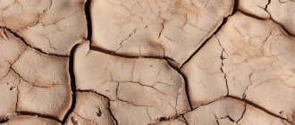
The main damaging effect on the skin, leading to the appearance of the first signs of aging and their progression, is ultraviolet (UV) radiation. It initiates a degenerative process in the skin, as a result of which it becomes drier and rougher, gradually loses its tone, and wrinkles and age spots appear on it. Changes caused by exposure to UV radiation are called photoaging. The source of ultraviolet radiation is the sun.
Depending on the length of electromagnetic waves, UV radiation is conventionally divided into UVA, UVB and UVC ranges. UVC rays, the rays with the shortest wavelength (200-290 nm), are the most dangerous because they have the highest energy. Fortunately, all UVC rays are blocked by the ozone layer of the earth's atmosphere.
UVB rays with a wavelength of 290 to 320 nm are only partially blocked by the ozone layer and reach the Earth's surface, although they practically do not penetrate glass. A significant part of them (more than 70%) is reflected by the stratum corneum of human skin, and yet some of the UVB rays penetrate into the epidermis, and about 10% even into the uppermost layers of the dermis. These rays have a strong damaging effect. They cause erythema (redness of the skin or sunburn) and tanning, and are responsible for many of the acute and chronic side effects associated with exposure to sunlight.
The wavelengths of UVA rays range from 320 to 400 nm. Of the entire UV spectrum, these rays have the lowest energy, but at the same time the highest penetrating ability. They are not retained by the ozone layer and pass through glass and the stratum corneum of the skin. In human skin, UVA rays reach the middle layers of the dermis and, although they do not cause sunburn, play a leading role in the development of actinic skin damage.
It has now been reliably established that both UV radiation spectra—both UVA and UVB—play an active role in the process of photoaging.
UV radiation triggers photochemical reactions in the skin, the products of which are free radicals and reactive oxygen species. By reacting with other biological molecules - protein, lipid or DNA molecules - they transfer their activity to them. Having thus turned into free and aggressive radicals, they, in turn, begin to react with the molecules surrounding them. In this case, proteins cross-link with each other, lipids of biological membranes are oxidized, and mutations occur in the genetic apparatus of the cell. Free radicals damage the connective tissue of the dermis, keratinocytes, have a negative effect on the vascular system of the skin, and enhance all manifestations of photoaging.
SIGNS OF PHOTOAGING
Premature skin aging begins with connective tissue, where collagen and elastin fibers are located. Collagen provides skin elasticity, and in young people its threads are constantly renewed. The enzyme collagenase destroys old threads, and new ones are synthesized in fibroblasts. Over time, under the influence of UV radiation, collagen synthesis in fibroblasts decreases and cross-linking of collagen filaments occurs. Collagen dimers are inaccessible to collagenase, so they gradually accumulate in the skin. Cross-linking of collagen fibers occurs after each free radical attack, while elastin synthesis increases, which together leads to a decrease in skin elasticity and the formation of wrinkles.
Signs of photoaging can be divided into physiological, clinical, anatomical and histological.
MAIN MANIFESTATIONS OF PHOTOAGING
With intense exposure to UV radiation, already at the age of 35-40 years, people develop signs of skin photoaging. It becomes drier, light wrinkles, pigment spots, and isolated telangiectasias appear. With age, these phenomena steadily progress. Degenerative changes, atrophy, keratosis begin to develop, spots of hypomelanosis (with partially lost pigment), yellow nodules and plaques appear. The listed clinical signs are combined into the concept of dermatoheliosis.
SOLAR ELASTOSIS
The main manifestation of dermatoheliosis is solar elastosis, which develops at the age of 40-50 years and clinically manifests itself as Milian lemon skin, characterized by reticular striations in the form of diamond-shaped slits and grooves against the background of thickened pale yellow skin). The process is usually localized on the face, neck, hands and forearms. As for other parts of the body, solar elastosis can only manifest itself as a yellowish tint to the skin. The disease is steadily progressive and, although not malignant in itself, serves as a marker for other more serious sun-induced skin lesions such as solar keratoses and squamous cell skin cancer.
Simultaneously with solar elastosis, other skin lesions develop as a result of UV irradiation, the number and nature of which may vary depending on the location of the affected area on the body. Such skin lesions include:
- • diamond-shaped skin of the neck;
- • Favre-Rakucho disease;
- • diffuse Dubreuil elastoma;
- • senile hyperplasia of the sebaceous glands;
- • Bateman's solar purpura;
- • O'Brien's actinic granuloma;
- • actinic comedonal plaques;
- • venous lakes;
- • lentigo solar;
- • idiopathic guttate hypomelanosis.
DIAMONDS NECK SKIN
Diamond-shaped skin of the neck (Cutis rhomhoidalis nuchae) clinically manifests itself as thickened, deeply diamond-shaped skin on the back of the neck, as well as yellow plaques. When stretched, this skin is no different from the lemon Milian skin on the face, that is, such a lesion is essentially a pronounced manifestation of solar elastosis in the neck area. Diamond neck skin occurs predominantly in men because women's neck skin is usually protected by hair.
FAVRE-RACOUCHO DISEASE
Intense insolation and wind contribute to the development of Favre-Racouchot disease in men over 50 years of age, which affects the skin of the face, especially the temporal areas and around the ears, as well as the back of the neck. In the lesions, against the background of thickened and compacted skin of a yellowish-red hue, collected in rough folds (phenomena of pronounced solar elastosis), nodular elements with a diameter of 0.5-1.0 cm, cysts with whitish contents with a diameter of 0.1-0.5 are visible cm and comedones, located in the central part of most nodules and cysts. In severe cases of the disease, cysts can merge, forming thickened yellowish plaques with many large comedones.
DIFFUSE ELASTOMA OF DUBREUL
A milder variant of Favre-Racouchot disease, diffuse Dubreuil elastoma, is characterized by the presence of well-defined yellowish plaques in the forehead, cheeks and lateral neck. Sometimes the disease manifests itself as a solitary papule or plaque in the area of the nose or back of the head, consisting of papule-like elements that resemble cobblestones. The skin in the area of the plaque is yellowish, wrinkled, soft, and gradually undergoes atrophy.
SENILE HYPERPLASIA OF THE SEBABY GLANDS
After prolonged intense sun exposure, senile hyperplasia of the sebaceous glands develops, characterized by the appearance of small light yellow papules in the forehead, lower eyelids, cheeks, nose and forearms, which over time acquire a darker yellow tint, a dome-shaped shape and umbilical depressions in the center. When the elements are squeezed, droplets of sebaceous secretion are released from them.
BATEMAN'S SOLAR PURPLE
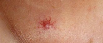
Long-term exposure to sunlight, which damages fibroblasts around blood vessels and leads to loss of collagen and vascular fragility, is a major predisposing factor in the development of solar Bateman's purpura in people over 60 years of age. The immediate cause of purpura is mechanical trauma, leading to rupture of dilated vessels of the subpapillary venous plexus. Clinically, the disease is characterized by the sudden appearance of hemorrhagic spots of red or purple color with a diameter of 1.0-5.0 cm with clear rugged contours on the back of the hands, extensor surfaces of the shoulders and forearms, and very rarely on the lower extremities or face. The surrounding skin is thin and has reduced elasticity. Due to the presence of hemosiderin in the elements and a decrease in the activity of macrophages, the lesions slowly evolve into pigment spots.
O'BRIEN'S ACTINIC GRANULOMA
O'Brien's actinic granuloma is formed as a result of an inflammatory reaction with a giant cell infiltrate that develops in the dermis in areas of solar elastosis. It most often occurs on the extensor surface of the forearms and upper back in people with fair skin color and the presence of freckles. It begins with the appearance of translucent reddish-brown papules, which gradually enlarge, lighten and turn into ring-shaped elements with a diameter of 1.0-3.0 cm. The skin over them is compacted and smooth. The center of the element is atrophic and depigmented. The elements are usually asymptomatic, but sunburn can cause severe erythema and irritation.
ACTINIC COMEDONA PLACLES
Actinic comedo plaques are single or multiple nodules or plaques localized on the extensor surface of the forearms and face, especially around the eyes and on the upper cheeks. The color of the plaques varies from pink to blue, and comedone-like elements may form against their background.
VENOUS LAKES
Venous lakes are clinically expressed in the appearance of soft blue or purple papules with a diameter of 0.5 cm, disappearing with diascopy. Single or multiple elements are usually located on the lower lip or in the helix area of the auricle, less often on other areas of the skin exposed to sunlight.
LENTIGO SOLAR
Lentigo solaris is the result of local proliferation of melanocytes in the area of the dermoepidermal junction. Clinically, it manifests itself as multiple pigment spots of a uniform light or dark brown, and sometimes black color, located on open areas of the skin and especially noticeable on the face (temples, forehead), the back of the hands and the extensor surface of the forearms. The spots are usually quite large - more than 1.0 cm in diameter, have a round shape and unclear boundaries. Although solar lentigo is the result of cumulative sun exposure, it, unlike freckles, does not darken with exposure to the sun.
According to some authors, the focus of solar lentigo, when basaloid cells spread into the lower layers of the dermis, can turn into a plaque of solar keratosis of the reticular type. In addition, lesions of solar lentigo can become inflamed, becoming the precursors of benign lichenoid keratosis. In very rare cases, long-standing solar lentigo progresses to Dubreuil's limited melanosis, a disease that transforms into melanoma.
TEAR-shaped IDIOPATHIC HYPOMELANOSIS
Guttate idiopathic hypomelanosis is characterized by the appearance of hundreds of asymptomatic depigmented porcelain-white spots with clear edges of a round or star-shaped shape measuring 0.2-0.5 cm, scattered like confetti on the anterior surface of the legs and forearms. The development of the disease is associated with prolonged UV irradiation, leading to a decrease in the number of melanosomes and melanin granules.
One of the dangerous consequences of photodamage to the skin is photocarcinogenesis—malignant degeneration of cells that occurs under the influence of UV radiation. Under the influence of UV radiation, the expression of genes encoding both suppressor proteins and proteins that promote tumor development is disrupted. UV radiation stimulates the production of the protein c-fos, which stimulates cell proliferation. At the same time, constant long-term UV irradiation can cause mutations in the gene encoding the tumor suppressor protein p53. As a result, a discrepancy between cell proliferation, differentiation and apoptosis occurs. With age, the imbalance of these processes worsens, which gradually leads to the development of skin tumors.
MODERN WAYS TO COMBAT PHOTO-AGING OF SKIN
The initial manifestations of photoaging are reversible and with skillful handling of the sun and rational skin care, their regression can be achieved.
To eliminate the phenomena of dermatoheliosis, more active therapeutic measures are needed (the use of systemic and topical aromatic retinoids, peelings, cryotherapy, laser therapy, dermabrasion, surgical excision). Treatment is usually lengthy and in some cases traumatic. In addition, it requires certain financial costs. Unfortunately, especially with already widespread processes, it is not always possible to achieve the desired result. At the same time, with the right and rational approach to insolation, photoaging can be prevented, prolonging the youth of the skin for many years.
The most effective way to combat photoaging in the modern world is its active prevention .
First of all, it is necessary to inform the general public about scientific data confirming the damaging effects of UV radiation on the skin and the body as a whole, and to form a critical attitude towards excessive insolation and the fashion for tanning. In order to prevent photoaging, a set of measures should be used aimed at protecting the skin from damaging solar radiation. To do this, you should, if possible, avoid direct sunlight , use clothing that protects your skin from the sun, and sunbathe only during the period of minimal summer sun activity.
It must be remembered that not only UV radiation from direct sunlight has a damaging effect, but also UV radiation reflected from the ground and surrounding objects, passing through clouds, water and even light clothing. In this regard, the leading element of prevention is the use of sunscreens that prevent skin damage from sunlight.
We invite you to watch a video about vitiligo treatment
Scientists have not yet been able to develop a pathogenetic treatment. Therefore, treatment is carried out only at the symptomatic level. The main task is generalization and reduction of manifestations.
For treatment, corticosteroid medications are used directly on the lesion. They also talk about the effectiveness of topical retinoids, pimecrolimus and cryomassage.
Sometimes photochemotherapy is used, as well as methods of replacing melagenin with the drug.
If necessary, bioresonance therapy is used, which should restore normal functioning of the nervous system and improve immunity.
The treatment is complex. Selected by a doctor. Therapy is selected individually. It is necessary to take into account the diagnosis and the degree of neglect of the pathology.
If treatment is ineffective, the selected therapy should be reconsidered.
Drug treatment is most effective. Medicines are selected taking into account the cause that caused the disorder. Additionally, you need to take vitamin supplements to generally maintain your general condition.
Therapeutic therapy can be supplemented with any physiotherapeutic techniques that improve local metabolic processes.
- Some of the best results are shown by cryomassage procedures.
- Photochemotherapy also significantly helps restore melanin formation.
Hypomelanosis idiopathic guttate (photo)
There is no developed and effective medicine that can affect the mechanisms of origin of hypomelanosis and prevent the occurrence of new spots. Mainly because the patient usually has a disease at the genetic level. However, treatment is still prescribed. It consists of prescribing medications that can reduce the severity of skin manifestations and prevent the development of spots.
As for medications, the following are prescribed:
- Corticosteroids. This category of medications is selected and prescribed strictly on an individual basis. Injections are made directly into the pigmented area.
- Pimecrolimus-based ointments (Elidel).
- Products with local retinoids (Differin, Retasol, Retinoic ointment).
- The drug Melagenin. Used for replacement therapy.
For macular hypomelanosis, the basis of treatment is Erythromycin ointment and other local antibacterial agents. Additionally, the skin should be constantly moisturized.
As a rule, symptomatic therapy is used in the treatment of hypomelanosis. Due to the fact that depigmentation is caused by genetic disorders, medical scientists have not yet found an effective way to treat the disease.
There are currently two methods used to get rid of white patches on the skin.
Therapeutic. Various physiotherapeutic procedures are used to improve metabolism in the epidermis. Cryomassage and hardware phototherapy are common.
- Treatment of guttate hypomelanosis involves injections of corticosteroid drugs. The drug is injected directly into the white spot. The medication is selected by the doctor individually for each case.
- Use of liniments containing pimecrolimus.
- The use of retinoid ointments and creams.
- Treatment with a solution to restore pigment – Melagenin with placenta extract for external use.
- Prescription of antibacterial liniments for progressive macular hypomelanosis.
Forecast
A patient with hypomelanosis is always given a favorable prognosis. The only exception is the form of Ito disease, when congenital disorders play a significant role in determining the prognosis of life.
If you try to avoid constant sun exposure, you will be able to avoid the spread of the disease to healthy tissue.
All iLive content is reviewed by medical experts to ensure it is as accurate and factual as possible.
We have strict sourcing guidelines and only link to reputable sites, academic research institutions and, where possible, proven medical studies. Please note that the numbers in parentheses ([1], [2], etc.) are clickable links to such studies.
If you believe that any of our content is inaccurate, out of date, or otherwise questionable, please select it and press Ctrl Enter.
Hypomelanosis is a pathology of the formation of pigmentation of the skin against the background of any disease. The development of hypomelanosis is based on a violation of the production of melanin by melanocytes located in the thickness of the skin. This pathological condition can manifest itself in the form of leukoderma, a reduced amount of melanin, as well as its complete absence.
The triggering point in the development of hypomelanosis is damage to one or more links in the production and transformation of melanin. This may be the absence of melanocytes in the skin, a violation of the formation of full-fledged melanosomes and their transportation to keratinocytes.
The main clinical manifestation of the pathology is considered to be white spots that arise as a result of a previous disease with superficial dyschromia of the skin. Hypomelanosis is often observed among children, occurring shortly after illness.
To make the correct diagnosis, histological examination is often used. Indeed, in its absence, hypomelanosis can be missed, which leads to developmental delays in childhood. Therapeutic goals for pathology are aimed at peeling procedures and the use of retinoids.
[1], [2], [3], [4], [5], [6]
When the first symptoms appear, you should consult a doctor who will prescribe the necessary therapy to help reduce the risk of relapses.
Other possible causes of white spots on the body
One of the most common causes of white spots in adults and children is fungal infections. This disease is characterized by the appearance of white or pink spots on the skin.
Pityriasis versicolor - at an early stage of the disease it is easy to confuse with hypomelanosis. The infection can develop against the background of excessive sweating, immunodeficiency conditions, chronic endocrine diseases, as well as due to taking a number of aggressive medications. In people with good immunity, the fungus may not cause any symptoms for several weeks.
At the first stage of the disease, the spots are white, but after that they acquire a reddish or brown tint. The spots are localized in the shoulder girdle and neck area and can be observed in different age groups of the population. The incubation period for the infection can last up to a month, and the disease itself, from the moment the first symptoms appear, can last up to six months. Despite the fungal etiology (the causative agent is the fungus Pityrosporum Orbiculare), the disease does not bring much discomfort. The spots do not flake, itch or become inflamed. For treatment, antifungal ointments are used in combination with antiseptics.
Hypomelanosis in a child
An insufficient amount of pigment produced in babies may indicate the presence of Wardeburg syndrome, which is transmitted genetically in a dominant manner. Its main clinical manifestations are considered to be white strands of hair, areas of hypopigmentation on the skin, different colors of the iris and eye levels, as well as a wide bridge of the nose and congenital deafness.
In addition, hypomelanosis in a child is observed with tumorous sclerosis, which is characterized by the appearance of white spots, up to 3 cm in size and localized on the body, as well as nodules on the forehead, arms and legs. In addition to skin manifestations, mental retardation, epilepsy, retinal phakomatosis, cyst-like formations in the kidneys, lungs, bones and cardiac rhabdomyomas are observed.
Hypomelanosis in a child is observed with Ito hypomelanosis with the appearance of hypopigmented areas of the skin of various shapes in the form of waves and stripes. Such symptoms may disappear on their own with age.
Vitiligo is also a defect in pigment synthesis, which is characterized by the appearance of white areas of skin with a clear contour. Localization is possible on the face, genitals, feet, hands, and joints.
Hypomelanosis
Hypomelanosis is a pathology characterized by a zoned decrease in the level of melanin pigment. The appearance of foci of hypopigmentation can be provoked by either endogenous or exogenous factors.
The main sign of the disease is the appearance of white spots. These areas of discoloration can have different shapes and be localized on any area of the skin. Only a qualified dermatologist can accurately determine the disease through examination under a special fluorescent lamp and scraping from the lesion. It is believed that this disease cannot be cured.
The treatment is symptomatic and cosmetic in nature; the patient is also given recommendations, adherence to which helps to avoid the appearance of new spots.
Etiology of hypomelanosis
This disease is one of the manifestations of leukoderma. Leukoderma is a group of pathological conditions in which natural pigment is destroyed. There are several forms of manifestations of hypomelanosis; the main factor influencing the development and course of this pathology is genetic predisposition.
Some forms of the disease may manifest themselves due to the influence of UV rays, cold, or taking certain groups of medications. The only manifestation of hypomelanosis is white spots of varying sizes on the skin; as a rule, the pathology does not affect internal organs and systems. In rare cases, when pathology develops against the background of dysembryogenesis, dysfunction of the central nervous system, abnormalities in bone tissue formation, heart defects and reproductive system can be observed as concomitant symptoms. It is difficult to provide accurate data on the prevalence of the pathology, since in most cases its manifestations do not cause much discomfort to the patient, and he does not go to the doctor.
Causes of the disease
Depending on the typology, the process of development of the disease and the causes that provoke it may differ. The main reason, undoubtedly, is a dysfunction in the production of melancites or a high degree of their destruction, however. As a result, light spots with a reduced pigment content appear on the skin. Foci of hypomelanosis can have different shapes, with clearly defined contours.
It cannot be said that the reasons for the development of this pathology are completely known to doctors.
In most cases, spots are hereditary in nature, and the tendency to their appearance can be transmitted through a generation or several.
Hypomelanosis in a child deserves special attention, since in most of its manifestations it indicates concomitant hereditary pathologies or chromosomal syndromes. Most often, Vaanderburg disease occurs in children (hereditary pathology is accompanied by the appearance of a light patch of hair on the head). Tumorous scleroderma, also most often observed in childhood, is manifested by small lightened spots of different localization.
One of the diseases that belongs to the group of hypomelanosis is vitiligo, a hereditary disease that manifests itself in the complete disappearance of melanin from large areas of the skin. The true etiology of vitiligo has not yet been studied. It is known that the first symptoms are observed in childhood. First, small lesions appear, which eventually merge into larger ones. The localization of lesions can be almost symmetrical and observed in those areas that are most susceptible to triggers (sunlight, friction, sweat, etc.). Doctors consider this pathology to be autoimmune.
Typology of the disease
photo - white spots appeared on the skin
Varieties of hypomelanosis are characterized by differences from one another in clinical course, age at which the first signs appeared, triggers, and a number of other factors. The typology includes only independent diseases, and not those that arise against the background of existing pathologies.
Guttate hypomelanosis
This form of pathology can most often be observed in female representatives of the population aged 35-55 years. Women who have a light skin tone and who spend long periods of time in direct sunlight are most susceptible to hypomelanosis.
As a result, in the affected areas there is a decrease in the number of melanocytes by almost 2 times. In addition, there are opinions that guttate hypomelanosis is associated with HLA-DR8.
Genetic factors play an important role in the development of this disease, especially if it is observed in close relatives.
Clinical manifestations of hypomelanosis are characterized by the appearance of spots on the skin that are white and round in shape. The diameter of such altered areas reaches up to 1 cm.
Guttate hypomelanosis first appears on the lower leg (extensor surface) and then spreads to the forearms, upper back and chest. This pathological condition is not characterized by damage to the skin of the face.
Progression of the process is observed with age, as well as with excessive exposure to direct sunlight.
Features of the treatment of hypomelanosis: an effective treatment regimen
Hypomelanosis is a pathology that spreads at the genetic level, so there is currently no pathogenetic treatment for the disease. Symptomatic therapy is used:
- Correct and active sun protection
- Clothing that covers exposed skin
- Corticosteroids are also used locally in the form of ointments.
- Retinoids: local and systemic use
- Elidel 1% cream 2 times a day - until hypopigmentation decreases or completely disappears.
The development of hypomelanosis is provoked by excessive sun exposure, so the best prevention of pathology is to avoid direct sunlight on unprotected skin.
Read more articles on skin diseases in the “Dermatology” section on estet-portal.com. Thank you for staying with us!
Share: