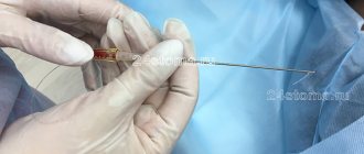
Biorevitalization is a cosmetic procedure in which hyaluronic acid is injected under the skin using a syringe with a thin needle. The injection is performed in a papular manner, that is, with the formation of subcutaneous blisters. In the process of assimilation of the composition, they are absorbed without the need for any external influence.
Papules after biorevitalization: what is it?
Papules are small elevations above the skin level that contain a gel-like composition with hyaluronic acid. They appear due to the fact that the drug is injected to a shallow depth. The formation of papules is a feature of biorevitalization, in contrast to similar cosmetic procedures. This is a natural skin reaction that is not a pathology, although it scares many.
If there are no papules after biorevitalization, this may indicate:
- disrupted technology (the drug was injected very deeply);
- low density of the substance (HA is diluted with other components);
- excessive dryness of facial skin, imbalance of water balance;
- large thickness of the dermis.
The size of the resulting papules depends on the angle of the needle entering the skin, the composition of the hyaluronic acid-based product and the area of treatment. For example, in the décolleté area the diameter of the tubercles is usually 3 mm, while in the periorbital area it is no more than 1 mm.
Treatment of papules
The appearance of papules (nodules) should be a reason to make an appointment with a doctor. Treatment should begin by identifying the cause that causes the pathology. In accordance with the examination results, experienced specialists at our medical center will select the most effective drugs that do not cause side effects.
Prevention is of great importance in preventing further damage. In our center, drug treatment is supported by the development of recommendations regarding personal hygiene, facial skin care, and diet. If necessary, an appointment with related specialists may be recommended.
How many days will it take for the papules to resolve?
Within a few hours after the procedure, the papular tubercles noticeably increase (up to 30%), and then the drug begins to gradually be absorbed with their smoothing. It is impossible to say exactly how long papules last after biorevitalization, because everything depends on the characteristics of the body. The average period is 2-3 days, the maximum is 10 days. But with good skin condition and a high ability to regenerate, the tubercles become barely noticeable or disappear completely already on the 3rd day after the procedure. Those with sensitive skin should immediately prepare for a longer recovery period.
The cosmetologist can give an approximate time taking into account the following factors:
- skin density at the treatment site;
- thickness of the surface layer of the epidermis;
- degree of hydration of dermal cells;
- circulatory and lymph flow activity.
Differential diagnosis of papules for skin pathologies other than syphilis
Parapsoriasis or Broca's disease
Parapsoriasis is a rare, non-contagious skin disease that belongs to lymphoproliferative dermatoses.
It occurs predominantly in adult men of middle age.
Today there is no precise definition of this disorder.
Due to its variable clinical presentation and lack of specific histopathology findings.
However, the lesions usually appear as pink and scaly patches.
They vary in size from 1 to 5 cm.
The rashes are often chronic and resistant to medication.
There are two types of parapsoriasis: small-globular parapsoriasis (in English terminology SPP) and plaque parapsoriasis (LPP).
Of the two types, LPP is considered a premalignant dermatosis that can develop into T-cell lymphomas.
Whereas SPP is a chronic benign condition.
Again, because this disorder is difficult to diagnose, there are no specific epidemiological statistics.
In 10% of those who have plaque-type parapsoriasis, the disease progresses to a malignant form.
The cause of parapsoriasis is unknown; research on this topic is still underway.
It is conventionally accepted that parapsoriasis develops against the background of infectious processes and immune disorders.
Fine-grained psoriasis is usually asymptomatic.
There is no deterioration in general health, but rashes develop, ranging from pink to yellow-brown.
Round or oval spots (macula) ranging in size from 2 to 5 cm.
Maculae are mainly located on the abdomen, sides, back or lower extremities.
When examined under a microscope, there are very nonspecific data: mild spongiosis, parakeratosis, exocytosis of small lymphocytes.
There is also a variant of the disease called digital dermatosis.
In this case, the rashes look like elongated spots rather than round ones.
To make a definitive diagnosis, an epidermal biopsy is recommended.
It is advisable to perform several procedures on different areas of the skin.
Treatment begins with moderate-to-high potency corticosteroids for 8 to 12 weeks.
Wilson's ringworm
Lichen planus is an acquired, chronic, immune system-mediated disease.
Appears as polygonal, purple, itchy, papules or plaques on the skin and similar lesions on mucous membranes, such as the oral cavity.
Lichen planus is thought to be an immunologically mediated disorder.
This appears to be a response of CD8 cells to antigens in the basal cell layer and dermoepidermal junction.
Mainly in keratinocyte cells.
The reason for this phenomenon is still unknown.
There is only some speculation about the effects of certain viruses (eg, hepatitis C and hepatitis B) or drug factors.
Characteristic skin lesions of the lichen plan are polygonal, purple, itchy, flat papules.
They can stick together to form plaques.
Lesions may be close together or widely separated from each other.
They may have white spots or lines called Wickham's furrows, and they may also be hyperpigmented.
The lesions are usually distributed symmetrically on the upper/lower extremities, especially around the elbows and wrists.
However, they can be found anywhere on the body, including the glans penis and vulva.
Papules tend to disappear after some time, leaving signs of hyperpigmentation of the skin.
Psoriasis vulgaris (vulgar psoriasis)
Psoriasis vulgaris or scaly lichen is the most common form of psoriatic skin lesions.
Occurs in almost 80% of patients.
The disease is characterized by thickening of the epidermis, development of an inflammatory reaction on its surface, redness and silvery-white scales.
Skin lesions may be accompanied by itching, burning and pain on palpation.
Typical, inflammatory, erythematous, pruritic, and silvery-scaly patches of skin occur in psoriasis vulgaris on the extensor sides of the elbows and knees.
Also on the scalp, behind the ears.
However, other parts of the body may also be affected: the skin of the genitals, perineum, mammary glands, palms of the hands.
In turn, scaly lichen is divided into two subtypes:
- Type I: The disease usually occurs in patients under 40 years of age and is therefore called the early type. Other family members may also suffer from this form of psoriasis. Often, patients’ blood contains genetic markers of susceptibility to the disease.
- Type II. Called late type, it develops after 40 years. There is usually no family predisposition; DNA markers in the patient’s blood are significantly lower than in the first subtype. This type of psoriasis is milder and responds more effectively to treatment.
Miliary papular syphilide
Rapulosa milliaris nigra is a benign skin disease.
Often affects patients with secondary syphilis.
The symptom is accompanied by the appearance of soft, dermal papules on the skin.
The color is practically no different from the natural shade of the skin.
Often localized in the area of hair follicles, on the face, around the eyes, and back.
Papules are harmless, but may be accompanied by severe itching and irritation.
The disease is quite rare and tends to occur in patients with dark skin, more often in African Americans.
The pathology is quite difficult to respond to drug therapy and practically does not respond to medications.
There are several methods for removing and treating rapulosa milliaris: curettage, electrocoagulation, electrosurgery, cryotherapy and electrotherapy.
Each treatment carries a risk of scar tissue formation, decreased pigmentation, and keloid formation.
It is important to differentiate miliary syphilide from lichen scrofulous.
In this case, the rashes are grouped and are papules in the form of perifollicular nodules that affect children or adults with tuberculosis infection.
Yellowish-reddish-brown papules usually develop on the back, chest, and abdomen.
Not accompanied by the formation of scar tissue.
The pathology can be found under synonyms - Lichen Scrofulosorum, Tuberculosis Cutis Lichenoides.
Reasons for the long resorption of papules
If the papules do not disappear within 10 days or increase with the formation of edema, you should consult a cosmetologist. This skin reaction is usually observed due to the following reasons:
- allergies to drug components or excipients;
- presence of contraindications to biorevitalization;
- connective tissue density in treatment areas;
- low quality of the drug or violation of its storage regime;
- ignoring the cosmetologist’s recommendations for aftercare;
- failure to maintain distance between injections;
- incorrect injections;
- violations of hygiene standards.
Other types of syphilitic rashes
The above-described rashes occur more often with secondary syphilis.
The following types of rash are less common:
- Weeping erosive papular syphilide. Typically, the lesion develops on the skin between the toes. Weeping erosions occur as a result of constant friction and injury to papules.
- Monetoid (numular) papular syphilide. The papules are quite impressive in size and tend to merge into large lesions. The shape resembles hemispherical formations of dark red color.
- Vegetating papules or condylomas lata. This phenomenon is characteristic only of secondary type syphilis, in case of recurrence. Most often, the localization of formations is observed in the abdomen, between the fingers. Gradually, the papules grow, becoming lumpy. They are not malignant, but they are quite easy to injure and provoke an infectious process.
General phenomena in secondary syphilis:
- Painless wounds in the genital area, perineum
- Acne all over the body
- Enlarged, swollen lymph nodes
- Rashes on mucous membranes
- Headache, fatigue, fever
- Weight loss, sometimes hair loss
- Joint pain
- Forgetfulness and mental problems
- Blindness or hearing problems
The development of morphological elements in the form of rashes is characteristic of the second stage of the infectious process.
It is called secondary syphilis.
In the second stage of infection, skin rashes are visible, including on the mucous membrane of the throat.
The skin rash usually occurs on the palms and soles of the feet, but can occur anywhere on the skin.
Often, patients do not notice such rashes or mistake them for an allergic reaction, etc.
If a patient has syphilis and does not seek treatment, the infection can lead to serious, life-threatening conditions.
Diseases such as nervous disorders, brain damage, mental disorders, blindness, aortic aneurysm, damage to blood vessels and heart vessels may develop.
Long-term absence of adequate treatment can lead to death.
Ways to quickly eliminate papules
In order for the rehabilitation process to proceed as quickly as possible, it is prohibited:
- touch, comb, knead the bumps;
- sleep on your stomach, cover your head with a blanket;
- use decorative cosmetics;
- wash your face with tap water (in the first 2 days - only filtered water);
- go outside without UV protection cream, be exposed to the wind and frost for a long time;
- visit the pool, gym, sauna (for 2 weeks);
- smoke, drink alcohol.
Sometimes pharmaceutical ointments to relieve swelling and inflammation help reduce the duration of resorption of papules. Before using them, you should consult with a cosmetologist at the Bionics clinic. For 2-3 days you can apply a soothing mask.
Risk factors
The main non-modifiable risk factor for developing basal cell carcinoma is age.
.
Approximately 80% of patients are actually over 60 years of age
, and rarely - under 20 years of age. Men are more susceptible to BCC than women and are 30% more likely to experience a recurrence, especially within six months of the first lesion being identified and/or if they are over 65 years of age.
Other factors that increase the risk of developing basal cell carcinoma:
- Etiological
:- prolonged exposure to direct sunlight, in the mountains or southern regions;
- frequent exposure to artificial UV rays in a solarium.
- Exogenous
:- X-ray and radioactive radiation;
- exposure to carcinogens on the body (skin contact with arsenic, harmful working conditions);
- reduced immunity due to organ transplantation or HIV infection;
- the presence of severe sunburn in childhood, wounds, scars.
- Genetic
:- Phototypes I and II according to the Fitzpatrick scale
; - nevus of the sebaceous glands of Jadassohn;
- basal cell nevus syndrome, or Gorlin syndrome (leads to multiple BCCs);
- xeroderma pigmentosum;
- Bazex syndrome (can lead to early development of multiple BCCs);
- generalized basaloid follicular hamartoma syndrome;
- Rombo syndrome;
- Happle-Tinschert syndrome.
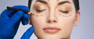
Causes of inflammation of the eyelids
Inflammation of the eyelids can be caused by the following reasons:
- pathological activity of mites living in the skin (a common phenomenon when the immune system is weakened);
- allergic reactions to certain types of medications, plant pollen, food;
- viral and microbial infections in the body;
- chemical, thermal and mechanical eye injuries;
- disruption of the cardiovascular and nervous systems;
- endocrine and hormonal disruptions.
1 Diseases of the skin around the eyes and inflammation of the eyelids. Diagnosis and treatment
2 Diseases of the skin around the eyes and inflammation of the eyelids. Diagnosis and treatment
3 Diseases of the skin around the eyes and inflammation of the eyelids. Diagnosis and treatment
Diagnosis of basal cell carcinoma
The diagnosis is made based on histological examination
. Differential diagnosis of basalioma is carried out with:
- molluscum contagiosum,
- hyperplasia of the sebaceous glands,
- amelanoma
, _ - intradermal melanocytic nevus,
- Merkel cell carcinoma,
- trichoepithelioma,
- squamous cell carcinoma
- keratoacanthoma,
- inflammatory dermatoses (psoriasis and eczema),
- actinic keratosis,
- Bowen's disease
- morphea (focal scleroderma),
- dermatofibrosarcoma,
- scar.