Ringworm in humans has a different nature; it can be either an infectious or non-infectious disease. It manifests itself as spots of different colors, swelling, itching, and painful sensations. Requires immediate treatment to avoid negative consequences. At the same time, contacting a dermatologist is mandatory, since there are 7 types of lichen, and it is almost impossible to make a diagnosis without a visit to a specialist.
What is ringworm?
Ringworm is an infectious skin disease caused by the parasitic fungi Microsporum canis and Trichophyton tonsurans, it is part of the group of ringworms that primarily affect the skin, hair and nails. This is an infectious dermatological pathology caused by dermatophyte fungi. It is also called dermatomycosis, scab, micro- and dermatophytosis.
Doctors distinguish several types of ringworm, depending on the location, nature of the course and the causative agent.
The main types of the disease are:
- chronic or ringworm of smooth skin;
- surface;
- ringworm of the nail plate;
- infiltrative-suppurative.
Since ringworm is caused by two types of fungi, in medicine it is diagnosed as microsporia and trichophytosis. These fungi have persistent pathogenicity, pronounced virulence and often cause acute diseases with varied clinical manifestations.
A disease caused by fungi of the genus Microsporum canis is microsporia. Depending on the location, there is microsporia of smooth skin and microsporia of the scalp.
Trichophytosis is caused by the activity of the dermatophyte Trichophyton tonsurans, which, through the anthroponotic route of infection, causes inflammation of the scalp and smooth skin, and through the zootroponotic route, inflammation in the deep layers of the dermis, accompanied by suppuration.
The presence of fungal spores does not always provoke damage to the skin and its appendages. The transition of the fungus from an inactive state to a pathogenic state is facilitated by metabolic and endocrine disorders, immune diseases, hypovitaminosis or vegetative-vascular dystonia. This increase in the pathogenicity of dermatophytes provokes an exacerbation of inflammatory processes, the spread of the fungus through the blood and lymph flow.
The disease is weakly expressed during the development of a latent infection and when localized in previously affected areas, and the high virulence of the fungus is formed during a relapse, gradually decreasing during the course of the infection.
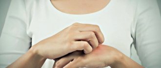
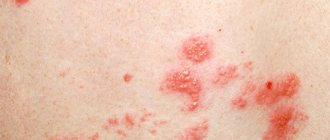
Symptoms of ringworm: At the end of the incubation period, itchy ring-shaped spots of red-pink color appear on the skin, on the surface of which bubbles appear, after which a crust and peeling remain. The hair at the site of the lesion breaks and falls out.
Normally, the immune system is able to cope with the pathogen and prevent its activity, but there are a number of factors that increase the risk of ringworm, namely:
- skin microtraumas;
- lack of vitamins and microelements;
- prolonged contact with the source of infection;
- high humidity and air temperature;
- exacerbation of chronic diseases, etc.
In a situation where there is an adult or child with lichen at home, it is necessary to take preventive measures to reduce the likelihood of its spread, namely:
- use separate towels and bed linen;
- wash your hands thoroughly with soap, especially if you touched the patient’s things;
- use gloves when touching areas of disease;
- undergo examination and take tests to identify pathogens, etc.
The main method for diagnosing ringworm is the use of a Wood's lamp, under the light of which the affected areas acquire a characteristic greenish glow. Microscopic examination and culture of skin and hair scales are effective to confirm the diagnosis.
Timely diagnosis and a properly selected treatment regimen can effectively eliminate ringworm in children and adults, preventing its spread. The pathology is prone to relapse, since the presence of spores of viable fungi can cause repeated damage. It is extremely important to complete the course of treatment and follow all doctor’s recommendations.
Why does lichen appear?
It is difficult to name with 100% certainty the exact causes of infection and the method of formation of the lichen patch on the skin. However, there are a number of factors that provoke the appearance of the disease:
- low level of immunity;
- constant exposure to stressful situations;
- chronic emotional and physical fatigue;
- infections;
- genetic predisposition to skin diseases.
Even the gender and age category of the patient can influence the form of the disease.
The causes of lichen also depend on its type.
Shingles appears due to contact with the Herpes zoster virus. The causative agent of the disease is the Herpesviridae virus. The disease is activated when there is a herpetic infection in the patient’s body.
Pityriasis or “pink lichen” has an infectious and allergic cause.
Lichen versicolor or pityriasis versicolor is located in the stratum corneum of the skin. A person can become infected with it through close contact with a carrier of the disease, as well as through the use of household items (towels, bed linen, dishes) of a sick person. The main causes of this type of disease are excessive sweating, hot weather and inflammatory processes on the skin. The causative agent of lichen versicolor is a fungus.
Flat red fungus is directly related to disturbances in the patient’s body’s metabolic processes and low level of immune properties. Contact with certain medications and chemicals can become an irritant. This type of lichen has a genetic predisposition, that is, if close relatives often suffer from lichen planus, then the risk of the disease in this case is maximum.
Trichophytosis is one of the most common types of disease and affects children and adults who come into contact with a sick animal or household items carrying fungal spores.
How is ringworm transmitted?
Ringworm is transmitted in two ways:
- from a sick animal – zoonotic with an incubation period of one week;
- from an infected person – anthroponotic with an incubation period of up to 6 weeks.
Children are at risk because they most often come into contact with the carriers of the pathogens of this disease - cats and dogs. Children's skin is characterized by low density of the stratum corneum and weak protective properties of the water-lipid mantle. The stratum corneum prevents the penetration of the fungus into the layers of the skin, since its spores are located between the horny scales, and the water-lipid mantle forms a barrier to the penetration of pathogens.
The infection is also transmitted by non-compliance with personal hygiene standards and the use of other people's personal belongings containing fungal spores (hats, combs, bed linen, etc.).
Pathogens and carriers of lichen
The content of the article
Dermatologists distinguish several types of lichen, caused by different pathogens.
Most often, the appearance of lichen on human skin is initiated by fungi:
- zooanthropophilic - transmitted to humans from an infected animal, including from pets;
- anthropophilic - live only on human skin;
- geophilic - live in the soil and come into contact with human skin upon contact with the ground.
In addition to fungi, viruses are often the causative agent of lichen. Their cunning lies in the fact that they can live asymptomatically in the human body for a number of years and become more active when the immune system is weakened. This is exactly how, for example, those familiar to many with herpes or shingles behave.
How does ringworm begin?
Favorable conditions contribute to the introduction and reproduction of a pathogenic fungus in the skin, triggering the infectious process. In the initial stages, this period is asymptomatic. After which the development of the pathology acquires clinical manifestations, the signs of which depend on the stage of the pathology.
The stages of the infectious process during the development of ringworm are:
- incubation period;
- period of mushroom growth;
- refractory stage (period of insensitivity, rest);
- regression.
In the skin, the fungus forms branched mycelium, which gradually invades new areas of the skin, while old lesions become the location of its spores. This period of fungal growth is determined by the rapid increase in the colony of the pathogen, active division of skin cells and the high rate of exfoliation of the affected epidermis.
Since most often the growth of the colony outstrips the rate of change of the stratum corneum, the infectious process spreads. An intense immune reaction (inflammation) in the lesion is displayed in the form of a red ring, which, in the chronic course of mycosis, becomes the permanent residence of the pathogen.
What is pityriasis versicolor
Pityriasis versicolor, or pityriasis versicolor, is a chronic skin disease of a fungal nature. It appears in the form of specifically pigmented spots of various sizes and shades, without signs of inflammation. Most often, the areas affected by pityriasis versicolor are located on the body (back, abdomen, neck, shoulders) and scalp. Representatives of both sexes are most susceptible to the disease at a young age, and children under 7 years of age are the least susceptible.
External manifestations of the disease usually do not cause concern, so they are often perceived as harmless cosmetic defects. For this reason, the pathology becomes chronic: spots periodically appear, then disappear, and after some time, under the influence of provoking factors, the disease worsens again.
What does ringworm look like?
Clinical manifestations of ringworm depend on the type of pathogen, course and stage of the disease. Dermatophytes secrete enzymes and toxins that destroy keratin protein (they feed on it), so the severity of symptoms directly depends on the ability of fungi to produce these substances.
Common symptoms of ringworm on the scalp are:
- hair thinning;
- the appearance of localized peeling of the skin;
- redness of the epidermis;
- hair breaking off at the root;
- the appearance of small bubbles with cloudy contents along the edges of bald patches, after opening which yellow crusts form.
Ringworm of smooth skin includes the following symptoms:
- pronounced itching;
- the appearance of round red spots;
- light epidermis at the site of damage, surrounded by gray scales;
- the edges of the damaged area consist of pink-red bubbles;
- growth of the lesion in diameter.
The chronic form of the disease is accompanied by the presence of microscopic blisters on the affected areas, the formation of scars after their opening and severe itching. In a situation where the site of the lesion is the nail plate, its separation, discoloration and thickening are observed.
Ringworm of the infiltrative-suppurative form is characterized by the appearance of large bright red spots up to 10 cm in diameter, the outer surface of which is lumpy and uneven, and purulent follicles form along the edges. Pain, swelling and hyperemia of the affected areas are also observed.
The appearance of the first signs of the disease requires a mandatory visit to a dermatologist. The lack of qualified treatment can lead to the rapid spread of infection throughout the body and cause a number of complications: tissue scarring, death of hair follicles, allergic reactions and the addition of a secondary infection.
Treatment options for lichen depending on its type
Just like the causes and symptoms, treatment regimens differ depending on the type of fungus.
If shingles fungus is diagnosed at the very beginning of the disease, the doctor may prescribe the following medications: Acyclovir, Zovirax. According to individual indicators, drugs such as “Curantil” and “Furosemide” can be prescribed. If the immune system is particularly weakened, intravenous drips with immunoglobulin may be prescribed.
The treatment regimen for advanced shingles consists of taking the following medications:
- analgesics;
- antipyretic drugs in the presence of high fever;
- B vitamins, which help strengthen the body’s protective functions;
- sedatives for insomnia;
- prescribing diuresis when signs of intoxication appear;
- the skin at the site of the lesion can be treated with a solution of brilliant green, metacil ointment, "Solcoseryl".
Pityriasis in most cases goes away on its own and does not require treatment. The exception is severe itching, for which you can use antihistamines and hormonal creams.
During the course of the disease it is recommended:
- wear underwear made from natural fabrics;
- limit bathing and interaction of the affected area with water;
- exclude the use of cosmetics.
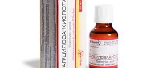
Treatment of versicolor versicolor is possible using the following medications: 5% salicylic ointment, Cycloperox, 5% sulfur ointment, Bifazol, Miconazole, Lamisil. If foci of the disease have spread throughout the body, then it is recommended to take Orungal or Itraconazole.
The flat red fungus causes significant discomfort to the patient due to severe itching and burning sensation. Suprastin, Tavegil and Zyrtec will help cope with itching. In addition, corticosteroids and antimalarial medications may be prescribed.
If lichen planus affects the mucous membrane, then treatment with Solcoseryl is necessary. If this type of disease recurs, antibiotics are prescribed.
If trichophytosis affects areas of the skin without the presence of hair, then the area can be treated with iodine and antifungal ointment (Exoderil, Microspor).
If there is hair in the area of the focus of trichophytosis, it is better to shave it off. In advanced cases, it is necessary to take antifungal drugs orally (for example, Irunin).
The course of the disease must be accompanied by taking a vitamin complex to maintain immunity.
Diet for fungal diseases
When the skin is affected by fungal diseases, it is important to maintain proper nutrition. In this case, it is useful to use the following products:
- breakfast with cereal porridge;
- dairy products;
- greens and green vegetables;
- honey;
- still mineral water;
- foods containing iron.
If you have pityriasis, you need to switch to dairy and plant foods. For other types of fungus, you should include in your menu:
- rose hips, viburnum and sea buckthorn;
- peanuts, hazelnuts and nuts;
- squid and salmon;
- seeds and vegetable oil;
- apples, apricots, cherries, blueberries, cherries;
- prunes and raisins;
- green bell peppers and carrots.
Along with the list of healthy foods, there is also a prohibited food group:
- dishes using spices and hot additives;
- alcoholic products;
- tangerines.
The following categories of drinks and food are subject to restrictions:
- coffee, tea and hot chocolate;
- cheeses that are too salty;
- broths cooked with meat and mushrooms;
- confectionery sweets with butter creams;
- canned food;
- fatty fish.
How is ringworm treated?
In each case, treatment for ringworm should be selected individually, since its composition and duration have their own specifics and are determined by the type, location and stage of the pathology.
Only prescriptions from a dermatologist are effective; they take into account the current clinical picture, the characteristics of the patient’s physical health and his age.
Therapy is aimed not only at relieving the symptoms of the pathology, but also at inactivating the pathogen. The chronic form of the disease also provides support for the immune system, normalization of metabolism and hormonal levels.
The following groups of drugs are used to treat ringworm:
- systemic and local antimycotics;
- antiseptic solutions;
- multivitamin complexes.
Symptoms of pityriasis rosea
The patient has a large pink spot. Its diameter is 20 mm or more. After a few days, the plaque becomes pale and yellow in color. The skin begins to peel off in the infected area. After the formation of spots, 3-5 days later they are observed on the body, arms, their diameter is 1 cm. The spots are pink, round or cylindrical in shape. The rashes begin to dry out and scales appear in the form of folds.
The patient's health worsens. Weakness, fever, headache, drowsiness, myalgia, redness of the throat, and poor appetite are observed. Based on the symptoms, pityriasis can be mistaken for an allergy. In this case, self-medication will lead to a deterioration in the patient’s health. The symptoms of the pathology are similar to other dermatological diseases.
The spots cover the back, thighs, arms and cause itching. They form within 3 weeks, then slowly disappear. The total duration of the pathology is up to 2 months. Dermatologists at the medical clinic treat patients with severe and initial forms of the disease.
How long does it take to treat ringworm?
The treatment process for ringworm is lengthy, usually taking at least six weeks, with systemic therapy lasting mainly 15-25 days.
The effectiveness of therapy is assessed using laboratory tests. In a situation where tests indicate a significant decrease in the number of fungal spores after 2 weeks of a medication course, it is necessary to continue the chosen treatment. In some cases, fungal shedding can last for several months.
After the signs of pathology disappear, you need to scrape the epidermis three times in previously damaged areas:
- immediately after completion of therapy;
- 7 days after therapy;
- after 2-3 months.
The result of therapy is considered successful if all three tests show negative results.
Treatment
Therapy for deprivation is specific, requiring time and some effort. “If you find spots on your skin, you must go to a skin and venous dispensary. There they will take a scraping from the skin at the site of mushroom formation. If it is positive, take another culture. If the result is positive and the type of rash is determined, treatment is prescribed. They are discharged only after three negative test results.
Antifungal tablets are used as treatment, local treatment is the same ointments. Everything is very serious, it is difficult to treat, so you should prepare yourself for a long process,” warns the specialist.
At the same time, you should be wary of complications. “If left untreated and the spots touched or scratched, there is a risk of infection. Moreover, it will spread throughout the skin. But it’s mainly an external influence, it doesn’t have a systemic impact,” says Tatyana Egorova.
How to treat ringworm in children?
Treatment of ringworm in a child is the responsibility of a pediatric dermatologist, since the characteristics of the child’s body require special doses and composition of medications.
Hygiene and child care are also important elements of pathology therapy:
- boiling and ironing clothes in contact with affected areas of the skin;
- treating household surfaces with disinfectants to prevent the spread of spores;
- frequent change of bed linen;
- washing in the shower rather than the bath, etc.
Types of lichen
In order to cure lichen, it is not enough to make a general diagnosis. It is equally important to promptly determine the type of lichen, since each is characterized by its own symptoms and requires its own treatment.
Dermatologists distinguish the following types of lichen:
- shearer;
- encircling;
- pityriasis;
- pink;
- red flat;
- tubular;
- solar;
- scaly.
Only a competent dermatologist can draw a line between them and correctly prescribe a set of therapeutic measures. Therefore, under no circumstances should you self-medicate. The surest way is to go to the clinic at the first sign of deprivation.
Now about what changes in the skin should alert a person.
Can I go to work or school with ringworm?
The presence of this diagnosis requires isolation of the patient from the team for at least two weeks. In schools and other children's institutions, it is mandatory to notify parents for timely detection of the disease in other children.
The disappearance of ringworm symptoms and 3 negative tests for the presence of fungus are a reason to return to school or work.
If a dermatomycosis infection has been detected in a team, then to prevent pathology, you can use antifungal shampoos for some time, carry out wet cleaning with the addition of antiseptic solutions, and strictly observe personal hygiene.
- Treatment of lichen in Kharkov;
- treatment of lichen in Uzhgorod;
- treatment of deprivation in Sumy;
- treatment of lichen in Poltava;
- treatment of lichen in Odessa;
- treatment of deprivation in Nikolaev;
- treatment of lichen in Mariupol;
- treatment of deprivation in the Dnieper.
3
1
1
Article rating:
3.8 out of 5 based on 5 ratings
Author: Mangusheva Victoria Yurievna
Dermatovenerologist, trichologist. Candidate of Medical Sciences, doctor of the highest category. Work experience more than 10 years.
Ringworm on the skin: treatment
Treatment of the disease is especially effective at an early stage. Ringworm can be cured in all patients, regardless of age. In this case, you need to consult a doctor even with minor manifestations. For diagnostic purposes, a dermatologist conducts a visual or instrumental examination (using a special lamp), prescribes a series of tests - studying urine, blood, as well as immunological studies and skin scrapings.
After establishing the exact cause and type of lichen, treatment is prescribed. Most often it is associated with the use of special ointments externally (antifungal drugs). The course of therapy lasts from 2 weeks to 3-4 months.
Main areas of treatment:
- Etiotropic – the use of drugs that eliminate the cause of the disease. These can be fungicides or antiviral agents (based on Acyclovir).
- Elimination of itching, spots and other unpleasant symptoms.
- Carrying out procedures - UV therapy, physiotherapy, increasing resistance (strengthening the immune system), maintaining personal hygiene.
- In rare cases, it is necessary to disinfect the premises, especially linen, furniture, and personal belongings.
It is not recommended to treat lichen yourself at home. This is dangerous because you can waste time and also infect other people. In addition, the patient often cannot determine which drug to treat the pathology with. For example, a person “prescribes” an antimicrobial ointment while he develops herpetic (viral) lichen. In rare cases, lost time can result in severe consequences such as blindness, arthritis, and other complications.
Ringworm scaly
The causes and treatment of this lichen, better known as psoriasis, are not fully understood. However, it is noted that it appears more often in people suffering from diseases of the endocrine and nervous systems and prone to alcoholism.
Psoriasis is a chronic disease, often occurring latently, i.e. is hidden. First, small reddish spots appear on the skin - most often the bends of the legs and arms, lower back and buttocks, causing severe itching due to dryness. Gradually, the spots grow and turn into a lesion that affects a large area of the skin. The peculiarity of psoriasis is that when the plaque is removed, the lichen begins to bleed.
It is necessary to treat scaly lichen under the supervision of a doctor, who will select the necessary topical medications (when applying them, you must strictly follow the instructions), and, if necessary, prescribe antibiotics.
Are all types of lichen contagious?
Many people believe that absolutely all types of lichen are dangerous to others. Meanwhile, this is not entirely true. So, with pink, ringworm or shingles, the patient will really need to be isolated. But, for example, pityriasis or scaly ones are not dangerous for others.
And this is another reason why any change in the skin should be the reason for going to a specialist - only he will be able to correctly determine the type of lichen, assess the degree of its danger, give recommendations for treatment, and finally select a set of medications and therapeutic measures aimed at including general strengthening of the body.
Tinea tubularis
Malasezzia is the name of the fungus that causes tubular lichen. This fungus can live in the human sebaceous glands for a long time without revealing itself in any way. Under certain conditions: for example, a decrease in immunity or an imbalance in the acid balance of the skin, severe hypothermia or excessive sweating, endocrine disorders or the development of certain chronic diseases - it begins to multiply intensively.
As a result, small flaky spots appear on the skin, which gradually grow and merge into one large spot. And although the victim practically does not feel itching or pain, he should not postpone a visit to the doctor.
After diagnosing the disease (methods used - scraping test, iodine test, use of a Wood's lamp, etc.), the dermatologist prescribes local or complex antifungal drugs.
A modern view of the problem of lichen planus
Lichen planus (LP) is one of the most common chronic recurrent diseases of the skin and oral mucosa. In 1859, Hebra first described lichen ruber acuminatus. The English dermatologist E. Wilson in 1869 first gave a clinical description of this disease, which differs from lichen acuminata by flatter papular elements. The first report on LP in the domestic literature was made by V. M. Bekhterev and A. G. Polotebnov in 1881. In the general structure of dermatological morbidity, LP is 1.2% and reaches up to 35% among diseases of the oral mucosa; in children the disease is diagnosed in 1–10% of cases. In recent years, the number of patients with LP has increased significantly; rare and difficult to diagnose forms have begun to be registered. In patients with LP of the oral mucosa, the disease develops with manifestations in the skin area in 15% of cases and in the genital area in 25%. In 1–13%, isolated damage to the nail plates is observed [1–3]. The incidence of malignant transformation varies from 0.4% to more than 5% over a follow-up period of 0.5 to 20 years, with almost all patients with the atrophic and erosive form of the disease developing cancer. Erosive-ulcerative forms of LP in 4–5% of cases are considered pre-cancerosis [4]. Over the last period of time, there has also been a noticeable increase in the number of patients with atypical, infiltrative and severe forms of this pathology, which have the greatest tendency to malignancy in 0.07–3.2% of cases. LP appears at any age, but most cases occur in the age group from 30 to 60 years. The disease develops in women more than twice as often as in men, mainly in perimenopausal women [3, 5].
LLP is characterized by the frequency of combination with various somatic diseases: chronic gastritis, gastric and duodenal ulcers, biliary cirrhosis of the liver, diabetes mellitus, etc. In addition, lichenoid lesions of the esophagus, stomach, intestines, bladder, endometrium may occur, which suggests a multisystem nature pathological process in LP [2]. Of particular importance in the occurrence of LP are dysfunctions of the liver and digestive tract. Important initiating factors are infections (in particular, hepatitis B and especially hepatitis C). Many authors believe that factors that cause antigenic stimulation of keratinocytes have a damaging effect on hepatocytes, and emphasize the connection between LP and primary biliary cirrhosis, paying attention to the erosive-ulcerative form of dermatosis, which may be a risk factor in the development of hepatitis or cirrhosis [ 6–8]. Metabolic changes in the body associated with changes in liver function, dyslipidemia, metabolic syndrome, disorders of carbohydrate metabolism can lead not only to cardiovascular diseases, but also to inadequate immune responses, therefore the combination of LLP with chronic hepatitis, biliary cirrhosis, xanthomatosis, diabetes mellitus, necrobiosis lipoidica, and amyloidosis are described quite often in the literature. [4, 9–11]. A number of authors identify a group of dermatoses (psoriasis, keratoses, keratoacanthomas, squamous cell skin cancer, vitiligo, discoid lupus erythematosus, limited scleroderma, lichen sclerosus, pemphigus vulgaris and bullous pemphigoid), which have in their pathogenesis general disorders of keratinization, immune response, metabolism, endothelial function and occurring in combination with LP [4]. Currently, data on hereditary predisposition to LP have been accumulated. In seventy described cases of familial disease with this dermatosis, it was noted that relatives in the second and third generations were mostly affected. According to a number of authors, in patients with common forms of dermatosis, histocompatibility antigens are more often recorded - the HLA systems: A3, B5, B8, B35, and HLA-B8 and HLA-B5 were found in erosive-ulcerative and verrucous varieties. There is also a significant increase in the fixation of haplotypes HLA-A3, B35 and B7 [7, 12, 13].
In accordance with modern ideas about the skin as an organ of immunity and in connection with the appearance of lichenoid rashes, LLP is characterized in many inflammatory processes by the inferiority of the regulation of immunity and metabolism, which explains the pathological inadequate reaction to injuries, drugs, chemicals, viruses, impaired enzymatic activity with a decrease glucose-6-phosphate dehydrogenase and other factors. The immunoallergic course of the disease involves the complex participation of neurovegetative, vascular, metabolic disorders, infectious, viral, intoxicating, hereditary and other factors and allows us to trace the initial stages of the formation of pathological changes in the skin. The role of changes in the cellular component of immunity in the pathogenesis of LP is due to an increase in the CD4+ content in the active phase of the disease, an increase in the T-helper/T-suppressor ratio, and activation of CD8+ cytotoxic lymphocytes. There is a direct interaction of the major histocompatibility complex type I - MHC I with the T-cell receptor of CD8+ lymphocytes, or the interaction of MHC II with the T-cell receptor of CD4+ lymphocytes and their subsequent differentiation into T-helper type I, producing a number of cytokines, including IL- 2 and interferon-γ. Cytokines interact with MHC I activated CD8+ lymphocytes, induce their proliferation and are an additional confirmatory factor that triggers cytotoxic mechanisms. The main mechanism of cell death in LP is apoptosis, induced by the interaction of tumor necrosis factor family cytokines produced by CD8+ lymphocytes, such as TNF-α and FasL, with the corresponding receptors on the surface of keratinocytes [14, 15].
LLP is characterized by a chronic relapsing course, the duration of which varies from 5 to 40 years. The onset of the disease occurs with rashes, itching, malaise, nervous stress, and weakness. Often elements of CP manifest themselves acutely. Clinical signs for classic cases of lichen planus are characterized by a dermoepidermal papule with a diameter of 1-3 mm, having a polygonal outline, an umbilical central recess, no tendency to peripheral growth, the presence of the so-called Wickham grid, visible in the depths of the papules after applying water or glycerin to the surface. Rashes of papules have a bluish-red or lilac color with a pearlescent tint and a polished sheen when illuminated from the side. Usually, having reached a size of approximately 3–4 mm, the papular elements subsequently stop increasing, but have a pronounced tendency to merge with each other, forming larger lesions in the form of plaques, various figures, and rings. During this period of development of dermatosis, a Wickham network is formed on the surface of the plaques in the form of small whitish grains and lines caused by unevenly expressed hypergranulosis. Lichenoid papules are located symmetrically on the flexor surfaces of the forearms, lateral surfaces of the torso, on the abdomen, mucous membranes of the oral cavity and genitals. Lesions in LP can be localized or generalized, acquiring the character of erythroderma. Despite the therapy, relapses of the disease can occur 1–5 times a year. The most torpid course of LP occurs in patients with verrucous, hypertrophic and erosive-ulcerative forms and in combination with diabetes mellitus, arterial hypertension and damage to the mucous membranes (Grinshpan-Wilapol syndrome) [3, 7, 16].
Manifestations of LLP on the skin are quite variable, and they are divided into forms: typical (classical); atypical; hypertrophic; pemphigoid; follicular; pigment; erythematous; ring-shaped. There are also erosive-ulcerative forms. There are frequent combinations of the erosive-ulcerative form of LLP with hypertension and diabetes mellitus (Grinshpan's symptom), hair loss and atrophic lesions of the scalp of a scarring nature (Little-Lassuer syndrome), a combination of lesions of the scalp with lichen planus and lupus erythematosus (overlap syndrome) [3, 17].
In annular LLP, the nodules are located in the form of circles of different sizes. Bullous LP is a rare variant in which blistering elements (vesicles and blisters) appear on papules or healthy skin. In acute LP, there are pointed papules that resemble skittles. Hyperkeratosis is pronounced, the skin looks like a grater on palpation. Localized predominantly in the neck, shoulder blades, and lower extremities, the highest form of manifestation of this variety is verrucous LLP. In the pigmented form of dermatosis, hyperpigmented spots or dark brown nodules appear. The erosive-ulcerative form of lichen planus is localized in the oral cavity, less often on the genitals. Women aged 40–60 years are most often affected. With the actinic variety of LLP, the rashes are located on open areas of the body (the dorsum of the hands and forearms). In some cases, one patient experiences a combination of types of the disease. To establish a diagnosis, at least one typical element of LP must be found. The mucous membranes are very often involved in the pathological process; they can be isolated or combined with skin lesions. Isolated damage to the oral mucosa often occurs in the presence of metal dental crowns, especially if they are made of different metals. According to the clinical course, varieties are distinguished: typical; exudative-hyperemic; bullous; hyperkeratotic. White papules are visible on the mucous membrane of the cheeks, some are finger-shaped and resemble fern leaves. This is the most frequently involved area of the oral cavity. On the tongue there are also white-violet plaque-like elements in the form of spilled milk (mainly in the exanthematic form). The bullous form of tongue damage is characterized by severe pain, which forces patients to refrain from eating. In the erosive-ulcerative form, shallow erosions covered with fibrinous films are visible, located on the tongue and mucous membrane of the cheeks; there are also bright red painful erosions on the gums (desquamative gingivitis). Non-erosive manifestations do not cause any sensations and are often unexpectedly revealed to patients during a doctor’s examination. With the so-called overlap syndrome, the lesions are usually located on the scalp and resemble lupus erythematosus and LP not only clinically, but also histologically. Little-Lassuer syndrome presents difficulties in diagnosis and treatment. This disease belongs to cicatricial alopecia. Most researchers consider it a manifestation of lichen planus. This syndrome is characterized by scarring alopecia of the scalp, non-scarring alopecia of the armpits and pubis, follicular bright red papules on the lateral surface of the shoulders, thighs, and buttocks, resembling thorns. In such patients, typical LP rashes, pointed rashes such as lichen spinosum on the skin of the trunk and extremities, and areas of pseudopellas on the scalp are simultaneously detected, and in all lesions the LP pattern is histologically detected. The initial signs of the disease are itching and rashes of lichenoid elements. In some patients, the nodules are initially localized only on the torso, and only after a few years do areas of scarred baldness appear on the scalp. Follicular pointed nodules on the shoulders, elbows, wrists, hips, flesh-colored, with horny spines at the top, without signs of inflammation. The grater symptom is pronounced. At the same time, you can find typical shiny papules of LLP. On the scalp (at the crown, back of the head, at the temples) there are round or oval bald patches that merge and have scalloped edges. The skin on these lesions is tense, shiny, atrophic, as if retracted. Their color is normal, sometimes pink-bluish. There may be islands of intact hair. As already mentioned, these lesions are accompanied by non-scarring alopecia or thinning of pubic and axillary hair. Sometimes there are papules on the mucous membranes and nail dystrophy. The presence of all three signs is not necessary for diagnosis.
Nail damage in LLP is characterized by the destruction of the nail fold and nail bed, the appearance of longitudinal striations, which have the appearance of a sail or yard. A frequent manifestation of the disease is rashes that clinically and histologically resemble LP, but are not such, but are called lichenoid tissue reaction. Most often, it is caused by taking various antibiotics (streptomycin, tetracycline), diuretics, angiotensin-converting enzyme inhibitors (captopril, enalapril), complexing agents (penicillamine, cuprenil), oral hypoglycemic agents, antimalarials (quinine, chloroquine), anti-tuberculosis drugs (aminosalicylic acid ), drugs for the treatment of arthritis (gold preparations). If the manifestations are similar, evidence of a lichenoid tissue reaction is improvement after discontinuation of the intended drug [15, 17].
The histomorphological features of typical elements of LP make it possible to diagnose the disease according to the characteristic histological pattern, guided by pathohistomorphological examination. The main ones are: unevenly expressed acanthosis; hyperkeratosis with areas of parakeratosis; increase in rows of cells of the granular layer (granulosis); vacuolar degeneration of basal cells of the epidermis; diffuse arcade-shaped, strip-like infiltrate [3].
In typical cases, the diagnosis of lichen planus is made based on the clinical picture. The classic (typical) form of LLP is distinguished from limited neurodermatitis, in which matte papules are formed, densely located from the periphery of the lesion to the center with the formation of lichenification of the skin, accompanied by intense itching and the presence of scratching in typical places. And also from syphilis, characterized by the presence of erosive, ulcerative or condylomatous rashes on the genitals, regional lymphadenitis or polyadenitis, roseolous-papular-pustular elements on the skin of the body, papules on the palms and soles, treponema pallidum in scrapings, positive serological reactions. Lichenoid parapsoriasis does not affect the mucous membranes and is characterized by a torpid course and the presence of the wafer phenomenon. Lichenoid tuberculosis of the skin is distinguished by the presence of an epithelioid cell infiltrate, which is absent in LLP. In lichen planus, the rashes are predominantly localized on the penis; the histomorphological specimen reveals perivascular granulomas of epithelioid cells. The hypertrophic, verrucous form of LP is distinguished from amyloid and myxedematous lichen, warty skin tuberculosis, chromomycosis, Hyde's nodular pruritus, warty form of neurodermatitis and chronic psoriasis. The atrophic form of LP must be distinguished only from the primary lichen alba of Tsumbusch (guttate scleroderma). Usually this does not cause difficulties: atrophy in LP develops secondarily, within the existing primary elements. The follicular form of LP must be differentiated from Darier's disease, pityriasis rubra pilaris (lichen acuminata or Devergie's disease), Kirle's disease, and lichen styloid. And Little-Lassuer syndrome - from follicular mucinosis, Siemens syndrome and Lutz syndrome. In some cases, it is difficult to distinguish discoid lupus erythematosus from foci of cicatricial atrophy on the scalp in Little-Lassuer syndrome. Histological changes in these dermatoses have much in common: follicular hyperkeratosis, exocytosis of infiltrate cells in the hair follicles, degeneration of cells in the basal layer of the epidermis, fibrin deposition on the basement membrane, the presence of hyaline bodies in the epidermis due to the death of epidermocytes. deposition of IgG, IgM, IgA in the area of the basement membrane, etc. The erythematous form of LP should be distinguished from toxidermia. Both diseases can be provoked by gold preparations, antibiotics, and antimalarial drugs. Establishing the correct diagnosis is helped by the results of histological and immunomorphological studies, detection of LLP elements on the mucous membranes of the oral cavity or genital organs. When LP is combined with discoid lupus erythematosus, the distinctive features may be areas of atrophy, localization of lesions on the ears and exacerbation of the process under the influence of insolation, which is more typical for lupus erythematosus. LLP is characterized by the presence of elements typical for this disease on the skin or mucous membranes [3, 7, 18, 19].
LLP of the mucous membranes only should be differentiated from leukoplakia, syphilitic papules, pemphigus vulgaris, lichenoid reaction of the oral mucosa, Keir's disease, plasmacytic Zon balanitis, bowenoid papulosis.
Isolated nail lesions in LP should be differentiated from nail lesions in psoriasis, eczema, Devergie's disease, Darier's follicular dyskeratosis, infectious and fungal diseases [7, 12, 20].
The choice of treatment method for patients with LP depends on the severity of clinical manifestations, duration of the disease, and information about the effectiveness of previously administered therapy. Each clinical type of the disease requires an individual approach to therapy. It is necessary to clarify the duration of the disease, the relationship of its occurrence with neuropsychic stress or past infections, previous treatment, and the presence of concomitant diseases. If the patient has applied for the first time and has not been previously examined, it is necessary to conduct an in-depth examination before starting treatment to determine the state of the nervous system, digestive tract, including the state of liver function, and also make sure that there is no hidden or overt diabetes mellitus. If only the oral mucosa is affected, it is necessary to consult the patient with a dentist to exclude developmental anomalies or the presence of artifacts that create problems in the mouth, including those of a traumatic nature. It is necessary to clarify the role of stress in the development of LP. It has been established that stress, through a system of neurohumoral factors, has a general effect on the body of a patient with LP, affecting the adaptive structures of the central nervous system, psycho-emotional status, state of immunity, aggravating the clinical course and clearly worsening the prognosis.
In the presence of limited rashes, treatment begins with the use of topical glucocorticosteroid drugs. For external therapy of patients with LP, glucocorticosteroid drugs of medium and high activity are used. In the presence of widespread rashes throughout the skin, systemic drug therapy and phototherapy are prescribed. Considering the positive results from the use of corticosteroid and antimalarial drugs prescribed in combination orally, it is recommended to add drugs from these groups to patients with LP. In the treatment of patients with LP, tablets or injections of systemic glucocorticosteroid drugs are used. To treat the common form of LP patients, retinoids are used for 3–4 weeks. In the erosive-ulcerative form, a cytostatic agent can be used for 2–3 weeks. As the disease progresses, detoxification therapy is used. To relieve itching, first-generation antihistamines are prescribed for 7–10 days, both orally and in injectable forms. Regardless of the clinical characteristics, medications are prescribed that calm the nervous system, normalize sleep, and reduce itching. One of the methods of treating patients with LP with typical manifestations, with the prevalence of the process and progression phenomena, is phototherapy (PUVA, selective phototherapy). PUVA therapy is carried out according to the usual method in a total dose of 15–20 procedures. Unfortunately, patients with LP often have contraindications to this type of treatment (hypertension, obesity, dysfunction of the thyroid gland, cardiovascular system, etc.).
Another active and effective method for this form of the disease is the combined use of corticosteroids and antimalarial drugs. It has been noted that the effectiveness of simultaneous administration of these medications is much higher than each one separately. For the treatment of patients with the hypertrophic (verrucous) form of LP, delagil and simultaneous intralesional administration of Diprospan can be used. The latter should be injected strictly intradermally so as not to cause atrophy. With these manifestations of the disease, the destruction of the most pronounced elements should be carried out, for which a destructive laser or the method of radio wave surgery can be used. If in most cases of patients with LP, external treatment is not carried out, then in this case it is advisable to prescribe active steroids, including those containing salicylic acid, under an occlusive dressing. In recent years, for erosive and ulcerative LLP of the mucous membranes of the oral cavity and genitals, along with corticosteroids and antimalarial drugs, metronidozole has been successfully used for 2–3 weeks. In ordinary forms of the disease, the use of external agents is not required. In rare cases, corticosteroid ointments can be prescribed for individual lesions. There are few publications on the use of cyclosporine, neotigazone, and cyclophosphamide in the treatment of patients with LP [3, 17].
During the period of exacerbation of the disease, patients are recommended to take a gentle regimen with limited physical and psycho-emotional stress. The diet should limit salty, smoked, fried foods. In patients with damage to the oral mucosa, it is necessary to exclude irritating and rough foods. Of the physiotherapeutic methods of therapy, phototherapy (suberythematic doses of ultraviolet irradiation) deserves attention. It should be emphasized that in all cases, treatment of patients with LP should be comprehensive and individual. It is necessary to provide for the prescription of drugs aimed at treating concomitant diseases, which often complicate the course of this dermatosis. Lichen planus, currently considered an immune-dependent disease, may indicate concomitant somatic pathology and systemicity of the pathological process. In this regard, a patient with LP requires certain management tactics associated with a thorough examination and consultation of many specialists. In order to prevent possible malignancy of long-existing hypertrophic and erosive-ulcerative lesions, patients should be under clinical observation. Persons with frequent relapses of the disease are also subject to it [3, 4, 17].
One of the main tasks in the prevention of LP is the fight against relapse of the disease. In this regard, it is important to sanitize foci of focal infection, timely treatment of identified concomitant diseases, prevention of taking medications that can provoke the development of the disease, general health measures, hardening of the body, prevention of nervous strain, and sanatorium-resort treatment. The length of the period of active disease depends on the prevalence and location of LP. The prognosis for the patient’s life is generally favorable, however, palmoplantar LP is characterized by a longer, recurrent course; on the mucous membranes, without treatment, manifestations of LP can persist for decades and transform into cancer.
Literature
- Dovzhansky S.I., Slesarenko N.A. Clinic, immunopathogenesis and therapy of lichen planus // Russian Medical Journal. 1998. No. 6. pp. 348–350.
- Lomonosov KM Lichen planus // Attending Physician. 2003. No. 9. pp. 35–39.
- Yusupova L. A., Ilyasova E. I. Lichen planus: modern pathogenetic aspects and methods of therapy // Practical medicine. 2013. No. 3. pp. 13–17.
- Slesarenko N. A., Utz S. R., Artemina E. M., Shtoda Yu. M., Karpova E. N. Comorbidity in lichen planus // Clinical dermatology and venereology. 2014. No. 5. P. 4–10.
- Lykova S. G., Larionova M. V. Benign and malignant neoplasms of internal organs as a factor complicating the course of dermatoses // Ros. magazine leather and veins diseases. 2003. No. 5. pp. 20–22.
- Ilyasova E. I., Yusupova L. A. Modern clinical and diagnostic aspects of lichen planus. Collection of materials of the All-Russian scientific and practical conference “Kazan dermatological readings: synthesis of science and practice.” pp. 43–48
- Butov Yu. S., Vasenova V. Yu., Anisimova T. V. Licheny // Clinical dermatovenerology. 2009. T. 2. pp. 184–205.
- Butareva M. M., Zhilova M. B. Lichen planus associated with viral hepatitis C, features of therapy // Vestn. dermatol. and venerol. 2010. No. 1. P. 105–108.
- Zakrzewska JM, Chan ES, Thornhill MH A systematic review of placebo-controlled randomized clinical trials of treatments used in oral lichen planus // Br J Dermatol. 2005. No. 153 (2). R. 336–341.
- Shengyuan L., Songpo Y., Wen W., Wenjing T. et al. Hepatitis C virus and lichen planus: a reciprocal association determined by a meta-analysis // Arch Dermatol. 2009. No. 145 (9). R. 1040–1047.
- Harman M., Akdeniz S., Dursun M., Akpolat N., Atmaca S. Lichen planus and hepatitis C virus infection: an epidemiologic study // Int J Clin Pract. 2004. No. 58 (12). R. 1118–1119.
- Ilyasova E.I., Yusupova L.A. Lichen planus. Collection of scientific articles and abstracts of the scientific-practical conference “Sexually transmitted infections and reproductive health of the population. Modern methods of diagnosis and treatment of dermatoses.” pp. 109–116.
- Cevasco NC, Bergfeld WF, Remzi BK, de Knott HR A case-series of 29 patients with lichen planopilaris: the Cleveland Clinic Foundation experience on evaluation, diagnosis, and treatment // J Am Acad Dermatol. 2007. No. 57 (1). R. 47–53.
- Fedotova K. Yu., Zhukova O. V., Kruglova L. S., Ptashchinsky R. I. Lichen planus: etiogy, pathogenesis, clinical forms, histological picture and basic principles of treatment // Clinical dermatology and venereology. 2014. No. 6. pp. 9–19.
- Garayeva Z. Sh., Yusupova L. A., Mavlyutova G. I. et al. Lichen planus. Modern aspects of differential diagnosis. Collection of materials of the All-Russian scientific and practical conference “Kazan dermatological readings: synthesis of science and practice.” pp. 17–23
- Molochkov V. A., Prokofiev A. A., Bobrov M. A., Pereverzeva O. E. Clinical features of various forms of lichen planus // Ros. magazine leather and Venus. bol. 2011. No. 1. P. 30–36.
- Chistyakova I. A. Lichen planus // Consilium Medicum. Dermatology. 2006. No. 1. pp. 39–41.
- Tsvetkova G. M., Mordovtseva V. V., Vavilov A. M., Mordovtsev V. N. Pathomorphology of skin diseases. M.: Medicine, 2003. 496 p.
- Barbinov D.V., Ravodin R.A. Criteria for histological diagnosis of lichen planus // St. Petersburg dermatological readings: mat. IV Ross, sc. pract. conf. St. Petersburg, 2010. P. 18.
- Brauns B., Stahl M., Schon MP et al. Intralesional steroid injection alleviates nail lichen planus // Int. J. Dermatol. 2011. No. 50 (5). P. 626–627.
L. A. Yusupova*, 1, Doctor of Medical Sciences, Professor K. F. Khairetdinova**
* GBOU DPO KSMA Ministry of Health of the Russian Federation, Kazan ** GAUZ RKVD Ministry of Health of the Republic of Tatarstan, Kazan
1 Contact information