Kluban Olesya Anatolevna
Neonatologist
April 29, 2017
Toxic erythema of newborns is a problem that 40 to 70% of mothers face on the 2nd-3rd day of a baby’s life. “Terrible pimples” are usually very scary for new mothers, and the diagnosis “Erythema toxicum” sounds so scary that some parents find themselves on the verge of panic.
But should we be afraid? Olesya Anatolyevna Kluban , a neonatologist at the ISIDA clinic, answers this question
Erythema toxicum, despite its scary name, is a benign non-infectious process that occurs during the adaptation period of newborns. It appears, as a rule, 48-72 hours after the birth of the baby, sometimes (rarely) immediately after birth and even less often - on the 10-14th day of life. This problem occurs equally among all races and both sexes and is more common in babies born at term weighing more than 2500 g.
Table of contents
- Etiology and pathogenesis
- Post-traumatic vascular dyschromia
- Principles of treatment
Post-traumatic and post-inflammatory vascular dyschromia are acquired pigmentation disorders, the pathogenesis of which involves the vascular component of the skin against the background of acute or chronic inflammation or injury.
In our company you can purchase the following equipment for the treatment of post-traumatic and post-inflammatory vascular dyschromia:
- M22 (Lumenis)
Why does toxic erythema occur?
“The causes of toxic erythema in infants remain unknown ,” says Olesya Anatolyevna Kluban, neonatologist at the ISIDA clinic. – The most common hypotheses for the occurrence of toxic erythema are as follows :
- activation of the newborn’s immune system in response to stress (childbirth, hypoxia, cooling);
- inflammatory response to skin colonization by normal bacterial flora at birth;
- acute hypergic skin reaction caused by maternal lymphocytes transferred shortly before or during childbirth;
- allergic reaction of the body to external and internal irritants;
- reaction of adaptation mechanisms of newborn skin in relation to mechanical or thermal stimulation.”
Etiology and pathogenesis
Blood and lymphatic vessels of the skin are normal
Arterioles and venules form two plexuses in the dermis ( Fig. 1 ):
- The upper horizontal one is located in the papillary dermis, from which the capillary loops of the dermal papillae emerge.
- The lower horizontal one is located on the border of the dermis and subcutaneous fat. It is formed by vessels coming from the muscles and subcutaneous fat.
The arterioles and venules of the lower horizontal plexus connect with the upper horizontal plexus and also form lateral outflows that supply blood to the hair follicles and sweat glands.
The external diameter of the arterial vessels of the papillary dermis varies from 17 to 26 µm - they are called terminal arterioles. Their wall consists of a continuous monolayer of endothelial cells with a basement membrane, which are externally covered with pericytes ( Fig. 2 ). Under normal conditions, endothelial cells suppress blood coagulation and prevent plasma proteins from entering the surrounding tissue.
The lymphatic vessels of the skin form two plexuses, similar to the upper and lower horizontal plexus of blood vessels and located near them. Branches from the superior lymphatic plexus pass into the dermal papillae and drain into larger lymphatic vessels in the deep dermis and upper part of the pancreas.
Unlike endothelial cells of blood vessels, endothelial cells of lymphatic vessels are layered on each other, having almost no tight junctions and possessing a rudimentary basement membrane (or no basement membrane at all). In addition, the lumen of the lymphatic vessels is much wider, and the wall is thinner - this allows tissue fluid to freely enter them ( Fig. 3 ).
Rice. 1. Superior ( papillary plexus ) and inferior ( cutaneous plexus ) horizontal choroid plexuses in the skin (Elsevier)
https://image.slidesharecdn.com
Rice. 2. Skin biopsy of the terminal arteriole ( left ) and venule ( right ) of the skin. The arteriole has a thicker wall and a fragmented elastic plate (dark blue dots) (Saara T. et al. Diagnosing vascular dementia by skin biopsy - Uniqueness of CADASIL. Skin Biopsy - Perspectives 2011)
Blood and lymphatic vessels of the skin during inflammation
Blood and lymphatic vessels make a significant contribution to the pathogenesis of the inflammatory process and its clinical manifestations. Dilated arterial and venous capillaries with increased blood flow underlie rubor and calor - redness and local increase in temperature. Excess exudate, caused by an increase in the permeability of blood vessels and the drainage capacity of lymphatic vessels, leads to tumor - edema. Finally, dolor and functio laesa (pain and dysfunction) occur after the influx of leukocytes into the area of inflammation.
Activation of the endothelium by inflammatory mediators (VEGF, TNF-α, IL-6, IL-1β, etc.) increases its sensitivity to substances that ensure interaction with leukocytes: E-selectin, intercellular adhesion molecule 1 (ICAM-1), adhesion molecule vascular cell 1 (VCAM-1) ( Fig. 3 ). In chronic inflammation, the network of blood vessels remains activated and highly permeable for a long time, which supports the accumulation of fluid in the tissues. Under its influence, the endothelial cells of the lymphatic capillaries diverge, and fluid from the tissues begins to leak into the lymphatic network. The mechanisms that control the enlargement of the lymphatic lumen are currently unknown, just as the function of dilated lymphatic capillaries has not been studied. They are believed to help suppress the inflammatory process.
Inflammatory diseases and conditions that involve the vascular component of the skin (Huggenberger R., Detmar M.. The cutaneous vascular system in chronic skin inflammation. J Investig Dermatol Symp Proc 2011; 15(1): 24–32):
- Rosacea
- Atopic dermatitis
- Alopecia areata
- Bullous pemphigoid
- Dermatitis herpetiformis
- Exudative erythema
- Systemic sclerosis
It should also be noted here that atherosclerosis, bronchial asthma, rheumatoid arthritis and inflammatory bowel diseases. Below we will describe diseases and conditions of the skin in the pathogenesis of which the participation of the vascular component of the skin is best studied.
Rice. 3. Modern ideas about the role of blood and lymphatic vessels in chronic skin inflammation (Huggenberger R., Detmar M. The cutaneous vascular system in chronic skin inflammation. J Investig Dermatol Symp Proc 2011; 15(1): 24–32)
Skin blood vessels ( right ) are composed of a monolayer of endothelial cells ( red ) with a continuous basement membrane ( gray ), which are externally covered by pericytes ( blue ). Lymphatic endothelial cells ( green, left ) do not have tight junctions, overlap each other, and have a rudimentary basement membrane. They are connected to the extracellular matrix through fibrillin filaments ( green threads ). The lumen of lymphatic vessels is much wider, and the wall is thinner than that of blood vessels.
Endothelial cells of blood vessels express receptors for vascular endothelial growth factor ( VEGF ) - VEGFR-1 and VEGFR-2 . At the same time, endothelial cells of lymphatic vessels express VEGFR-2 and VEGFR-3. A key “player” of chronic inflammation, VEGF-A , can bind to VEGFR-1 and VEGFR-2 , i.e. influence both blood and lymphatic vessels. Long-term stimulation of VEGF-A and VEGF-C leads to vascular remodeling, increased vascular permeability, increased expression of adhesion molecules and chronic skin inflammation. Against this background, the lymphatic vessels dilate, which probably suppresses inflammation. Its inhibition is also facilitated by the binding of chemokines to the D6 .
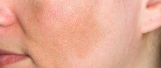
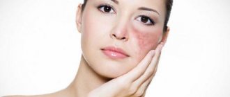
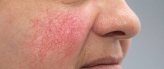
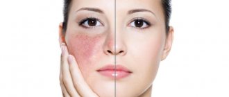
Post-inflammatory vascular dyschromia using the example of rosacea
Rosacea is characterized by pronounced vascular changes in the skin. All the mechanisms of this pathology have not yet been studied, but it is believed that the following are involved in its pathogenesis:
- innate immunity;
- reactive oxygen species;
- ultraviolet radiation;
- skin microbiome;
- vascular changes.
Locally, blood flow increases in the skin, which, together with dilatation of the choroid plexuses in the dermis, leads to typical clinical manifestations of rosacea (see below). It has been reported that vascular endothelial growth factor A (VEGF-A) levels, angiogenesis and lymphangiogenesis are increased in patients with rosacea. Clinically, this is confirmed by episodes of hot flashes and erythema. Ultraviolet irradiation worsens rosacea, probably by stimulating keratinocytes to produce even more VEGF-A.
Interestingly, the role of lymphatic vessels in the pathogenesis of rosacea is currently unknown. Some patients experience skin swelling resembling lymphedema, and with phymatous changes, severe skin lymphedema may occur. This indicates an important role for lymphatic dysfunction in the pathogenesis of rosacea, but what role it plays is still unclear.
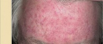
The clinical manifestations of rosacea consist of a spectrum of symptoms and signs. Erythema with telangiectasias occasionally appears on the cheeks and forehead of patients, usually after certain triggers. Inflammatory papules and pustules may be observed on the nose, forehead and cheeks ( Fig. 4 ). Unlike acne, patients with rosacea do not report increased oiliness; instead, they experience dryness and flaking. The absence of comedones also speaks in favor of rosacea in the differential diagnosis with acne vulgaris. Finally, rosacea almost never causes scarring on the skin.
In the final stages, patients may experience rhinophyma, a thickened and deformed nose. Although in some cases rhinophyma appears in isolation, without other clinical signs of rosacea.
Rice. 4. Rosacea (Danish national service on dermato-venereology)
Post-traumatic vascular dyschromia
Post-traumatic vascular dyschromia is preceded by some kind of injury - a bruise, skin injection, incision, etc. As a result, blood enters the soft tissues, where oxidation of hemoglobin and ferritin occurs with subsequent accumulation of hemosiderin. Predisposing factors:
- swelling of the lower extremities;
- diabetes;
- diseases of the cardiovascular system;
- high blood pressure;
- venous hypertension;
- chronic venous insufficiency;
- lipodermatosclerosis.
Post-traumatic vascular dyschromia manifests itself as spots of any size and shape, ranging in color from yellow to brown or black, appearing in any area of the body, but more often on the lower extremities ( Fig. 5 ).
Rice. 5. Post-traumatic hyperpigmentation on the legs due to vascular damage and hemosiderin accumulation under the skin (Photo: MavCure)
https://i0.wp.com/www.healthline.com
Clinical manifestations
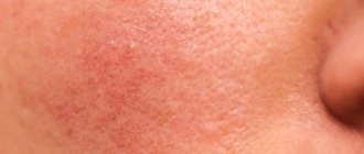
Rashes with toxic erythema look like red spots from 0.1 to 2 cm in diameter, which can merge with each other and be accompanied by slight swelling. In the center of the spot there may be a papule of dense yellowish consistency. Sometimes instead of papules, pustules with white contents are formed. Often, due to their external similarity, the rash is compared to a rash caused by hives or a mosquito bite.
The rash can be in the form of single elements, but it can also spread abundantly throughout the body. It does not occur on the palms and soles. More often it begins on the face and torso, then appears on the extremities. Individual elements can appear and disappear within a few hours. On average, the duration of the rash is 3-5 days. But sometimes toxic erythema lasts up to 10-14 days.
As a rule, the general condition of the baby is not affected when toxic erythema occurs. Sometimes, with a profuse rash, itching may occur and, as a result, the child may experience general anxiety.
Principles of treatment
Treatment of post-traumatic and post-inflammatory vascular dyschromia depends on the specific disease or skin condition. For example, for patients with rosacea , avoiding trigger factors is important. Each person’s set of such factors is individual and is determined experimentally. As a preventative measure, sunscreen cosmetics with UVA and UVB filters are recommended. Patients with rosacea better tolerate physical filters - zinc oxide and titanium dioxide.
For post-traumatic vascular dyschromia , topical corticosteroids, griseofulvin, cyclosporine A, vitamin C and bioflavonoids may provide some benefit.
An effective method for treating post-inflammatory and post-traumatic vascular dyschromia is IPL therapy - the use of intense pulsed light. The IPL module is included in the universal M22 platform device (Lumenis). Its positive effect is based on selective photothermolysis - the absorption of light of a certain wavelength by blood hemoglobin. IPL parameters are selected in such a way that the hemoglobin located in the dilated vessels accumulates maximum light energy. As a result, local heating and destruction of pathological capillaries that feed the area of dyschromia occurs.
A significant advantage of IPL over lasers is the impact area - intense pulsed light has a wide beam, which allows you to quickly and efficiently treat large areas of vascular networks, malformations, hemangiomas, telangiectasia, varicose capillaries, etc.
A certain difficulty for IPL is melanin, which accumulates in the skin over the area of dyschromia and acts as a screen, making it difficult for light to pass through to the hemoglobin in the blood. To solve this problem, the IPL module of the M22 platform device has several light filters that “cut off” excess light radiation in order to selectively heat only hemoglobin in dilated vessels. To increase the effectiveness of the impact, several passes with the IPL module are also used.
Questions from our users:
- skin dyschromia classification
- dyschromia types, methods of correction
- dyschromia in cosmetology
- causes of dyschromia in a child