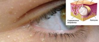
Vesiculopustulosis is a fairly common infectious skin disease among newborns that affects the openings of the child’s sweat glands. Vesiculopustulosis appears in newborns within a few hours after birth. Multiple blistering rashes with transparent contents begin to appear on the child’s skin, which subsequently becomes cloudy. In some cases, children are born with congenital signs of this disease. However, regardless of whether the disease is congenital or acquired, it begins to develop in the womb, due to the presence of some kind of chronic inflammatory process.
What causes the disease?
In medical practice, vesiculopustulosis is a disease characterized by damage to the mouths of the sweat glands by pathogenic agents. The most common causative agent is staphylococcal infection. The pathology got its name due to the fact that rashes on the body appear in the form of vesicles and pustules. These are pustular formations on the skin of an inflammatory nature.
With timely and correct treatment, vesiculopustulosis goes away quickly and does not cause complications. Among the factors that provoke the disease are the following:
- Damage to the skin of a newborn.
- Impaired functioning of the sweat glands.
- Diaper rash, prickly heat.
- Scratches, abrasions.
- Contact with a carrier of infection.
- The baby can become infected from the mother while in the womb or while passing through the birth canal.
For example, if a woman has foci of inflammation provoked by certain types of pathogens, the child may well become infected from the mother.
This is why it is so important to treat acute and chronic infectious diseases during pregnancy planning.
What causes vesiculopustulosis in newborns?
The main causes of vesiculopustulosis are damage to the integrity of the child’s skin. Also, the penetration of infection is possible in the prenatal period from mother to fetus if there are bacterial foci in the woman’s body.
Cost of consultation with a pediatrician in Moscow clinics:
- The first clinic of Izmailovo, Dr. Bandurina
1700 ₽
- Family Clinic
1950 ₽
- SM_clinic
2100 ₽
- Make an appointment by phone:
(499) 685-47-75
Another 159 clinics
Pathogens
From an etiological point of view, vesiculopustulosis is one of the types of pyoderma. This is an inflammatory lesion of the dermis of a purulent nature. Among the causative agents of infection, the following pathogens are distinguished:
- Staphylococci.
- Streptococci.
- E. coli.
- Klebsiella.
- Mushrooms, in particular Candida.
Most often, Staphylococcus aureus causes pathology in newborns. In a person with normal immunity, these bacteria do not manifest themselves in any way. It is believed that the majority of the world's population are carriers of these pathogens.
After birth, the baby exchanges microflora with the mother, which leads to the colonization of staphylococcus in his body. With skin lesions or weakened immunity, the infection manifests itself aggressively. This leads to the development of vesiculopustulosis.
Maybe
Conditions for increasing risk factors
Newborn babies have very thin and vulnerable skin. Despite this, it already has the ability to suppress the activity of some pathogens. If the baby is not properly cared for, the likelihood of damage to the dermis by pustular rashes increases. This occurs under the influence of such provoking factors:
- Overheating of the baby as a result of strong wrapping.
- Increased sweating when the indoor air is too dry and hot.
- Swaddling in diapers that have not been ironed beforehand.
- Damage to the dermis. If nails are not trimmed in a timely manner, the newborn may scratch himself.
- Increased sweating in an infant.
- Immune diseases in the newborn, in particular HIV.
- Premature birth, lack of weight.
- Diseases in the mother caused by staphylococcal infection.
- Lack of hygiene.
To prevent the development of the disease, it is necessary to properly care for the newborn, bathe him, change his diaper in a timely manner, iron his clothes and diapers.
In addition, you need to maintain the correct temperature and humidity in the room where the baby is.
Classification of the disease
Doctors divide vesiculopustulosis into two groups, depending on the time of occurrence of the pathology and its cause. Let's look at these groups in a little more detail.
Congenital
This type of disease in infants develops and is diagnosed from the first days after birth. Infection occurs from the mother inside the womb or during birth. If a woman has acute or chronic illnesses caused by staphylococcus, it is unlikely that it will be possible to avoid infection of the baby.
Acquired
If pathology in a newborn develops within a few weeks after birth, vesiculopustulosis is considered acquired. Here, the disease usually appears due to overheating, prickly heat, skin damage or poor hygiene.
The baby can also become infected from surrounding people. For example, if the health workers involved in his care do not follow sufficient preventive measures.
comparison table
If we compare both forms of the disease, these data can be indicated in a table.
| Type of disease | Development time | Route of infection |
| Congenital form | The first days after birth | Intrauterine infection or infection during passage through the birth canal |
| Acquired form | Within a few weeks of birth | Poor hygiene, skin damage, overheating |
Having discovered the first signs of vesiculopustulosis in a baby, it is necessary to begin treatment as quickly as possible. Competent and timely therapy will help prevent dangerous complications and quickly get rid of the pathology.
Symptoms in a child
The clinical picture of the disease largely depends on the individual characteristics of the child’s body. In newborns with normal immunity, signs of general body intoxication are usually absent. Manifestations such as weakness, drowsiness, loss of appetite, and apathy appear in underweight babies and in children born prematurely. Most often, these symptoms are absent, and the symptoms consist of the appearance of skin rashes.
Clinical manifestations of vesiculopustulosis in newborns:
- Formation of pink spots of small diameter in the area of skin folds and places with the greatest localization of sweat glands.
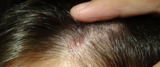
- The highest concentration of rashes is observed in the groin folds, in the area of the armpits, on the neck, in the buttocks, face and hairline.
- The rash tends to merge and form larger lesions.
- Red spots quickly degenerate into pustules - vesicles.
- After a few days, the blisters fill with purulent contents.
- The vesicles dry out, a crust forms in their place, and later peeling and healing are observed.
Healing of the dermis, as a rule, is not accompanied by the appearance of scars and cicatrices, since the damage to the skin during the disease is shallow.
Clinic for complicated course
In severe cases of vesiculopustulosis, the symptoms expand. Severe forms of the disease are most often observed in weakened children or with improper treatment of the disease in its early stages. In this case, the signs are as follows:
- Increased body temperature, sometimes up to 38-39°C.
- The child becomes capricious, cries, and refuses to eat.
- The rash affects large areas of the body. The rash combines into large lesions.
- Due to an increase in body temperature, refusal to eat and drink, digestion is disrupted, constipation or, conversely, diarrhea may occur.
- Dryness of the oral mucosa is observed.
If such symptoms develop, you should never self-medicate. The newborn must be shown to the pediatrician immediately.
Often the cause of vesiculopustulosis is overheating of the child. Read about what diaper rash is and how to deal with it in one of our articles.
Symptoms and clinical picture of vesiculopustulosis in newborns
The most characteristic manifestations of vesiculopustulosis are the formation of small blisters filled with clear liquid, spreading throughout the body and scalp. After a few days, the blisters transform into a pustule with purulent contents, after which crusts begin to form.
Symptoms of vesiculopustulosis:
- formation of blisters with serous contents and redness around;
- temperature increase to 38-39 degrees;
- constant anxiety and tearfulness;
- refusal to feed;
- dry lips and mucous membranes of the oral cavity.
When adequate therapy is prescribed, the signs of vesiculopustulosis completely disappear.
Diagnostics
The disease is diagnosed according to the following indicators:
- Visual inspection of the child's skin.
- Anamnesis collection.
- Identification of markers of the inflammatory process using a blood test.
- Carrying out bacteriological culture to identify the causative agent of infection.
In uncomplicated cases, therapy is carried out at home.
Parents must strictly follow the doctor’s recommendations and carry out the prescribed treatment measures. If the patient is in serious condition, he has an elevated body temperature, general intoxication of the body, rashes affect large areas of the skin, therapy is carried out exclusively in a hospital setting.
Diagnostic methods
To make a diagnosis, the attending physician conducts a thorough examination of the child, interviews the parents, and prescribes the necessary laboratory tests for vesiculopustulosis to identify the causative agent of the infectious process.
The main methods for diagnosing vesiculopustulosis include:
- physical diagnosis (examination, palpation, auscultation);
- general blood test (increased ESR, leukocytosis);
- bacterial culture of serous contents (determination of the pathogen).
The severity of the pathology
Treatment tactics are selected by a specialist depending on the severity of the disease.
Mild course
If the patient has an uncomplicated course, treatment can be carried out at home. The doctor examines the newborn and assesses the degree of skin damage and laboratory test data. After this, he prescribes medications and gives parents the necessary recommendations.
Severe course
In severe cases of the disease, it is recommended that the newborn stay in a hospital setting.
Let us remind you that a pathology is considered severe if the baby has extensive damage to the skin, body temperature rises, and signs of general intoxication of the body are expressed.
Complicated course
The most dangerous is the complicated course of vesiculopustulosis. This form of pathology, as a rule, occurs in children with weak immunity and in the presence of other diseases. In this case, the infection affects not only the superficial layers of the skin, but also the deep tissues. As a result, abscesses and cellulitis may develop.
Methods of treating the disease
Among the main types of therapy for vesiculopustulosis in a newborn, various treatment methods are used. The following measures are used:
- Treatment of rashes with antiseptic agents.
- Use of antipyretics. The most commonly used is Paracetamol.
- Adjusting the air temperature in the room and maintaining air humidity.
- Taking antibacterial drugs.
- Treatment of crusts and ulcers is carried out using herbal baths.
- Use of physiotherapy.
During therapy, you should never try to open the blisters yourself.
Reviews from experts indicate that this can lead to the infection spreading to adjacent areas of the skin and the course of the disease becoming much more complicated.
Features of general therapy in children
Due to the fact that vesiculopustulosis in newborns is a consequence of the proliferation of staphylococcal infections, treatment of the disease is usually carried out with the help of antibacterial drugs. Second generation cephalosporins are most often prescribed. In addition, the following groups of drugs are prescribed:
- Antipyretic medications. Among babies in the first months of life, Paracetamol, Panadol, Nurofen are used. In this case, forms such as rectal suppositories and syrup are used.
- If antibiotics are ineffective, the patient is prescribed treatment with antistaphylococcal plasma and gamma globulin.
- It is possible to increase the body's defenses through vitamin therapy.
- If complications such as an abscess or phlegmon develop, surgery is performed.
Vesiculopustulosis in young children and adults, as a rule, occurs in a mild form, except in cases where the disease is advanced or complicated. In addition to systemic drugs, local therapy also gives good results in treatment.
Using Local Remedies
Vesiculopustulosis has such a characteristic clinical sign as skin lesions. In this regard, therapy is carried out using local disinfectants and wound healing agents.
The following treatment methods are used:
- Treatment of diseased areas with antiseptics (diamond green, Fukortsin).
- Lubricating the blisters with ointments with an antibacterial effect. Drugs such as Erythromycin or Zinc ointment work well.
- Bathing a child in decoctions of medicinal herbs. Plants such as chamomile, calendula, string, and nettle are suitable for this.
- If there are large vesicles on the body, they can be removed using a cotton swab generously soaked in an alcohol solution.
- In severe cases of pathology, the doctor may prescribe therapy using ultraviolet treatment.
During illness, it is important to properly care for the baby, regularly change diapers, iron diapers and clothes, ventilate the room more often and not wrap the newborn up.
If vesiculopustulosis is mild, local treatment is sufficient. In complicated cases, systemic therapy is used.
Skin of newborns: differential diagnosis of pathological conditions, features of care
I.A. GORLANOV
, Doctor of Medical Sciences, Professor,
L.M.
LEINA , Ph.D.,
I.R.
MILYAVSKAYA , Candidate of Medical Sciences,
St. Petersburg State Pediatric Medical University Issues of caring for the skin of a child in the first years of life remain relevant to this day.
Due to the anatomical and physiological characteristics of children, the protective function of the skin, protecting against adverse external influences, is significantly reduced in them. The influence of various damaging factors, improper use of care products - all this can lead to disruption of the normal condition of the skin. The most common skin lesions in children of the first year of life include diaper (contact) dermatitis, the prevalence of which can be up to 50%. The importance of proper baby skin care as the main element in the prevention of dermatitis is undoubted. Features of the skin of infants and young children
The skin of a newborn baby has its own characteristics, which is associated with its transition at birth from an aquatic intrauterine environment. At birth, the skin is covered with vernix caseosa, which is 80% water, as well as lipids and proteins [1]. “Virginic lubricant” lipids are formed in the epidermis and sebaceous glands. As studies by Marchini G. et al. have shown, the original lubricant contains antibacterial peptides that play a protective role both before birth and in the first days of life [2].
The skin of newborns and infants is soft, velvety, pink. It is much thinner than the skin of an adult. In newborns, the structure of the epidermis at this age differs in a number of features. Their epidermis is much thinner. The basal cells are small in size, and the number of desmosomes at the junctions of cells is also reduced. The stratum spinosum has 2–5 rows of cells. The granular layer is poorly developed and consists of 1–2 rows of cells. Unlike the skin of adults, in infants the shiny layer is practically absent even on the palms and soles. The connection between the cells of the stratum corneum is weak, the horny plates are easily rejected (physiological parakeratosis) [1, 3].
By the time of birth, two layers are distinguished in the dermis, although not very clearly: the upper one is papillary and the lower one is reticular. The border between the epidermis and dermis is smoothed. In newborns and infants, the dermis is thinner than in older children and differs in structure. With the exception of the palms and soles, the papillary layer is not yet fully formed. The skin of newborns is highly hydrophilic. The subcutaneous fatty tissue of newborns and infants also has a number of features. At birth, the mass of subcutaneous fat is 4-5 times greater than in adults. In newborns, brown adipose tissue predominates, which is replaced with white with age. In terms of its chemical composition, subcutaneous fatty tissue also differs from that of adults. It is dominated by saturated fatty acids.
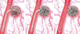
Skin appendages are imperfect at birth. There are 12 times more eccrine sweat glands in newborns than in adults. Their secretory section is represented by 6-12 loops of epithelial tubes, and the excretory duct is located in the dermis and epidermis. Unlike adults, the excretory ducts of the sweat glands in the epidermis of infants are usually straight and not corkscrew-shaped. The result of their immaturity is frequent blockage of the eccrine sweat glands with keratin plugs. With age, the sweat glands become more formed, larger, and the intensity of sweating after 2-3 months. after birth it returns to normal [1].
Newborns are characterized by hyperplasia of the sebaceous glands. Their number per 1 cm of facial skin in newborns is 4–8 times greater than in adults. After 40 weeks During pregnancy, the skin of a mature newborn is almost fully developed anatomically. Its weight is 10−13% of a newborn's body weight, compared to 3% in an adult. Anatomical maturity only partially correlates with functional maturity, and this discrepancy concerns both barrier function and thermoregulation. In a newborn child, the connection between the epidermis and the dermis is weak, while the dermal-epidermal junction plays an important role in the skin’s ability to withstand mechanical stress, therefore, in the neonatal period, injuries associated with detachment of the epidermis from the dermis can occur [2–4].
The characteristics of the skin of newborns and infants contribute to the easy occurrence of skin lesions with a pronounced exudative reaction and generalization of inflammatory processes. Dermatoses that occur at an early age differ significantly from diseases in adults and require specific care and treatment.
Violation of the skin barrier function is manifested by an increase in transepidermal water loss and the risk of percutaneous intoxication from external agents. For example, transdermal absorption occurs directly through the stratum corneum, hair follicles and sweat glands. And in premature babies, the barrier function of the skin is formed by the 4th week. life. The risk of percutaneous absorption in newborns, according to Eichenfield LF, Frieden IJ, Esterly NB, is presented in table [5].
Physiological and pathological conditions unique to the skin of newborns and infants are explained by its structural and functional properties. Features of proliferation and differentiation of epidermal and dermal cells, lipid composition and structure of connective tissue fibers determine the elasticity and velvety of the skin. The abundance of blood vessels and the constantly dilating capillary network give the skin of a newborn a peculiar bright pink color. A large number of functionally labile lymphatic tubules forming “lymphatic lakes” form pastosity and easily occurring edema.
Due to anatomical and physiological characteristics, various manifestations may appear on the skin during the neonatal period, including physiological or borderline conditions that do not require active intervention, as well as severe diseases [6–8].
Physiological conditions include physiological desquamation, erythema of the newborn, telangiectasia, blue (Mongolian) spots, sebaceous ichthyosis (Ichthyosis sebacea), milia.
Borderline changes in the skin of newborns can be considered miliaria and toxic erythema, which may require medical intervention.
Diseases requiring treatment include vesiculopustulosis, pyoderma of various etiologies, limited skin defects (Aplazia cutis), subcutaneous adiponecrosis, diaper dermatitis of varying severity.
Physiological peeling. Peeling of the skin (desquamation of the skin of newborns) is observed 24-36 hours after birth, more often in post-term infants and lasts up to 3 weeks. If desquamation is visible immediately after birth, congenital ichthyosis must be excluded. Bepanten cream can be used as a treatment for physiological peeling.
Erythema of newborns (physiological catarrh of the skin) is characterized by hyperemia of the skin due to dilation of capillaries in response to exposure to an ambient temperature lower than intrauterine. This erythema usually persists for 2-3 days and then decreases until it disappears. Redness of the skin is replaced by more or less pronounced pityriasis-like or lamellar peeling.
Telangiectasias are densely located dilated capillaries of the skin in the form of spots, most often located in the area of intrauterine presentation of the head - in the back of the head or on the forehead, sometimes in the area of the eyebrows and eyelids (nevus of Unna). The spots disappear when pressed and intensify when the child cries. It is believed that this is not a capillary nevus, but a transient vasodilation. Such telangiectasias disappear on their own after 1-1.5 years. No treatment required. According to Kanada KN et al., the incidence of telangiectasias is observed in 83% of newborns [9].
Blue, or Mongolian, spots are found only in representatives of the Mongoloid race and children with dark skin. Usually on the skin of the lumbosacral region, less often on the shoulder blades or buttocks, a bluish-purple spot of irregular shape of various sizes is found. When pressed, the stain does not disappear, the skin in the lesion is not changed. Usually this is a single formation, but there are also multiple ones. Histological examination reveals spindle-shaped melanocytes in the dermis (normally they are not found there). This appears to be the result of incomplete migration of melanocytes into the epidermis from the neural crest. Mongolian spots usually disappear spontaneously by 5-6 years.
Sebaceous ichthyosis (Ichthyosis sebacea) occurs due to increased secretion of quickly drying sebum by the sebaceous glands. The skin of a newborn baby becomes dry, rough to the touch, and acquires a brownish tint. Superficial cracks appear on the surface of the sebaceous crust. After a week, the process ends with abundant pityriasis or lamellar peeling. If a child has embryonic hair (lanugo) preserved on his skin, then when it is glued together with sebaceous secretion, the so-called hair is formed. seta. After the crust is rejected on the 6th–8th day, the skin takes on a normal appearance. The child’s condition with sebaceous ichthyosis is not affected and no treatment is required. Skin care is carried out by holding warm baths followed by lubricating it with lanolin cream or dexpanthenol. Sebaceous ichthyosis is differentiated from congenital.
Milia are small epidermal cysts that develop from the sebaceous glands of the hair vellus. Occurs in more than 50% of mature newborns. Milia appear from the moment of birth and are whitish-yellow papules the size of a pinhead or millet grain (milium). They are usually localized in the forehead, on the nose and cheeks, and in premature babies even on the trunk and limbs. These elements are epidermal retention cysts containing, in addition to thick fat, horny scales. Within 3−4 weeks. milia disappear. No treatment is required [3].
Miliaria is a reaction of the child’s body to overheating, associated with blockage of the sweat glands due to their functional immaturity. Miliaria occurs in approximately 15% of newborns. It is caused by a temporary blockage of the excretory ducts of the eccrine sweat glands. The appearance of prickly heat is promoted by high temperature and humidity of the rooms in which newborns are located. There are crystalline and miliaria rubra. With crystalline miliaria, blockage of the sweat gland ducts occurs at the level of the stratum corneum. Small blisters without an inflammatory component appear on the skin, filled with transparent contents. Bubbles sizes up to 1 mm. With prickly heat, the level of blockage of the sweat gland ducts is deeper. The rashes are represented by small erythematous papules and papulovesicles. The localization of rashes is any, but more often they appear on areas of the skin covered by clothing (torso, axillary and groin areas). With severe hyperhidrosis, prickly heat appears on the face, neck and scalp. Being inherently a physiological condition, prickly heat is often complicated by the addition of a secondary pyococcal infection, resulting in the development of vesiculopustulosis or even abscesses of the sweat glands.
When treating prickly heat, baths with potassium permanganate and drying agents in the form of powders are usually used. Prevention consists of ensuring temperature control, daily bathing, and air baths [1, 3, 6].
Newborn acne (Acne neonatorum) occurs in newborns at 2–3 weeks. life, are more common in boys than in girls. The cause of this condition is not completely clear; their appearance is usually associated with hormonal stimulation of the sebaceous glands by maternal androgens. Acne appears more often and lasts longer in breastfed children. The rashes are usually located on the face in the cheek area, on the forehead and on the nose in the form of small papules and pustules, surrounded by a halo of hyperemia. Less commonly, similar elements appear on the chest and shoulders. Breastfed children get sick. The process on the skin resembles that of teenage acne vulgaris and is associated with postpartum hormonal changes in the mother’s body. Acneiform rashes in newborns can be associated with the saprophyte Malassezia furfur and Malassezia sympodialis and are referred to as neonatal cephalic pustulosis [6, 7, 9, 10].
For single pustular elements, treatment consists of treating the skin with disinfectant 30−40% alcohol solutions of 1−2% salicylic acid, powder containing neomycin and bacitramine, and salicylic-zinc paste. It is possible to use external antifungal agents. The mother is prescribed a dairy-vegetable diet and increased fluid volume. In case of a common form of acne with severe pustulization, antibacterial therapy is prescribed taking into account the sensitivity of the pathogenic flora and specific bacteriophages.
Toxic erythema of newborns is observed in almost half of newborns on the 2nd–5th day of life. It is rare in premature babies. This condition is considered borderline between normal and pathological. Modern research confirms the hypothesis of an excessive reaction of the immune system to the first colonization of the skin by non-pathogenic microorganisms [2, 3]. In addition, it has been established that the mother’s consumption of large amounts of foods that are obligate allergens (chocolate, condensed milk, fish, citrus fruits, etc.) after childbirth can play a role in the formation of toxic erythema. Most often, this condition is considered as the first manifestations of an allergy (as well as dry skin, flushed cheeks, etc.).
In the clinical picture of the disease, limited and generalized forms are distinguished. With limited toxic erythema, single small (miliary and lenticular) spotted and urticarial elements of a bright pink color appear on the skin of the extensor surfaces of the limbs, trunk, and buttocks; a vesicle can be seen in the center of only a few. The child's condition is not affected. In the generalized form, the rash is abundant, polymorphic with a predominance of papulovesicles, urticaria and even pustules, prone to rapid spread and fusion (Fig. 1). The general condition of the child, as a rule, is not disturbed. Eosinophilia is present in the blood in 20% of cases. A generalized form is often treated with antihistamines. Treatment of toxic erythema involves prescribing a hypoallergenic diet to the mother. Externally, vesicular and pustular elements are treated with a 1% solution of brilliant green. The rash usually lasts 2-4 days, with a generalized form up to 7-10.
Limited congenital defects of the skin and subcutaneous tissue (Aplazia cutis) can occur when the amniotic membranes of the fetus are disrupted, at the site of intrauterine healing of hemangiomas, or during an attempted abortion. It is possible that such a disease is preceded by viral or bacterial infections at the beginning of pregnancy or occupational hazards of the mother (radiation, chemical agents, etc.). The frequency of skin aplasia is 1: 5 thousand. Changes on the skin are visible immediately at the birth of the child. More often, defects in the skin, subcutaneous tissue, and sometimes deeper defects are visible on the scalp, as well as on the torso or limbs. The lesions take the form of granulating ulcers or scars of a round or oval shape ranging in size from a few millimeters to 5-6 cm in diameter. Sometimes aplasia cutis is combined with such congenital anomalies as cleft upper lip and hard palate, malformations of the limbs, etc. The differential diagnosis of Aplazia cutis is carried out with a nevus of the sebaceous glands, which can also be seen from birth in the form of a lesion with no yellowish hair protruding above the surface skin. If aplasia is localized in the midline of the skull, it is advisable to conduct an ultrasound examination to exclude encephalocele.
The disease should be differentiated from possible injuries during childbirth, congenital syphilis, and congenital epidermolysis bullosa. Treatment depends on the child's condition. In case of numerous injuries, antibacterial therapy and agents that improve microcirculation and epithelization are prescribed. Ulcerative defects are treated with disinfectant solutions (hydrogen peroxide, furatsilin, chlorhexidine), ointments containing antibiotics, solcoseryl, actovegin. There is no treatment for scars [1, 3].
Subcutaneous adiponecrosis appears in the 1st–2nd week. life and is an essential disease of young adipose tissue with a disorder of lipid metabolism due to the peculiar ratio of fatty acids (the predominance of saturated acids over polyunsaturated ones). Histologically, extensive necrosis of subcutaneous fatty tissue with the presence of reactive inflammation, giant and epithelioid cells is determined. The pathological process occurs at the sites of injury during obstetric manipulations (application of forceps, turning the fetus, resuscitation measures). Large, full-term newborns are usually affected.
Clinically, sharply demarcated nodes or infiltrates appear in the area of the back, shoulders, and buttocks, up to the size of the child’s palm. The skin over the infiltrates is initially cyanotic or violet, then becomes pale. The nodes are usually painless and the general condition of the child is practically not affected. Treatment consists of prescribing thermal procedures (dry warming bandages, Sollux), vasodilators and antioxidant drugs [1].
Diaper dermatitis
One of the most common skin changes is diaper dermatitis (ND), the incidence of which ranges from 35 to 50% [24]. At the same time, premature babies are high-risk patients. PD is more often observed in girls. A greater predisposition to PD was noted in children with increased sensitivity to allergens, which is explained by the frequent development of exudative phenomena and dyspepsia. The disease belongs to the group of region-specific dermatoses, in which an acute inflammatory reaction of the skin occurs in the area covered by diapers. In recent years, thanks to the widespread use of industrial diapers with good hygroscopic properties, the number of diaper dermatitis has decreased. The disease is multifactorial in nature and is initiated by a combination of a number of factors: physical, chemical and biological (interaction of the skin with urine and feces). The main condition for the occurrence of diaper dermatitis is skin occlusion due to the use of diapers. The surface in contact with diapers is excessively moistened, the stratum corneum is loosened, as a result of which the barrier functions of the epidermis are reduced and the skin becomes more sensitive and vulnerable. Such changes contribute to the colonization of yeast-like fungi Candida albicans and gram-positive bacteria, in particular Staphylococcus aureus and streptococci, on the surface of the skin. Enzymes secreted by microorganisms break down uric acid, promote the synthesis of ammonia and create an alkaline skin reaction, which in turn activates the enzymatic activity of feces [11–13].
Most often, contact dermatitis develops in those areas that come into direct contact with the diaper. The buttocks and genitals are most affected. First, spots appear with erythema of varying intensity, then the skin in the affected areas becomes swollen, sometimes peels, and later papular rashes may appear. Children who spend a long time in diapers with poor absorption at night may develop erosions and sometimes even ulcers on the surface of the skin. Diaper dermatitis, a type of simple contact dermatitis, is more typical for children in the first month of life, which is explained by the anatomical and physiological characteristics of the skin (Fig. 2).
The second most common after simple diaper dermatitis is candidiasis diaper dermatitis. It is represented by different-sized bright, clearly defined erythematous foci of irregular shape, spotted and papular in nature. Characterized by a fairly rapid growth of lesions along the periphery, a tendency to the appearance of fresh rashes in folds and on contacting surfaces. Along the periphery of the lesions one can often see “screenings” of miliary papules and small flaccid superficial pustules, after the opening of which erosions with fragments of epidermis of a regular round shape remain (Fig. 3). There is a tendency to increase the area of eroded surfaces, which have a bizarre shape with clearly defined boundaries of scalloped outlines. The likelihood of secondary candidal infection is very high with any form of diaper dermatitis that lasts more than three days. In infants, candidiasis in the anogenital area is often combined with thrush in the oral cavity, where there is a white cheesy coating and cracks in the corners of the mouth. These patients may also have intestinal candidiasis. The diagnosis is based on clinical data and microscopy - detection of hyphae and pseudomycelium [14, 15].
PD of bacterial etiology is most often caused by b-hemolytic streptococci, and is called papulo-erosive streptodermia. Predisposing factors to the occurrence of this form of dermatitis are the formation of ammonia in wet diapers, excretion of antibiotics in the urine, and washing clothes with synthetic scented detergents. On the skin of the buttocks, the back of the thighs, and in boys, on the scrotum, on an erythematous, somewhat edematous background, miliary and lenticular papules of a pinkish-bluish color appear slightly protruding above the skin, on the surface of which erosions quickly appear.
Prevention and treatment of PD
The key to treating PD is good skin care to prevent irritation and damage to the epidermis. This is achieved by cleansing the skin, protecting it from adverse effects, in particular contact with secretions [25]. Skin care principles
1. It is necessary to change diapers in a timely manner. In particular, the shift should be carried out early in the morning (immediately after the child wakes up), immediately before the baby goes to bed, after each feeding, if the child shows signs of anxiety; in any case, at least every 3-4 hours. 2. Clean and dry the skin regularly and thoroughly, especially in the anus and genital area. Due to the low threshold of irritability, it is advisable to use detergents sparingly. Soap can irritate the skin due to alkaline components, and synthetic detergents (bath bubbles, shampoos) due to the degreasing effect they produce. Therefore, it is necessary to bathe a baby using detergents no more than 2-3 times a week, while washing should be done regularly, and after bowel movements it is mandatory. 3. Air baths should be performed every time you change diapers. 4. To prevent skin contact with secretions (urine and feces), it is advisable to use modern disposable diapers, the inner cellulose layer of which contains a gel-forming material with high moisture-absorbing ability. 5. Applying barrier cream at every diaper change. When erosions occur, treatment with a 1% aqueous solution of aniline dyes (brilliant green or eosin) is necessary.
During PD, the use of creams and ointments is recommended to treat affected areas of the skin. In dermatological practice, for the purpose of healing, improving trophism and tissue regeneration, topical products containing vitamin B5 (pantothenic acid), which is not without reason called the antidermatitis factor, are used. In the body, provitamin pantothenic acid (synonymous with vitamin B5) is an integral part of coenzyme A, which is involved in many enzymatic reactions of aerobic cell metabolism. Firstly, coenzyme A, acting as an intermediate carrier of acyl groups, promotes the formation of acetyl-CoA with subsequent initiation effective oxidative breakdown of carbohydrates and fatty acids, resulting in the formation of ATP. Thus, vitamin B5 is one of the essential components that allows the complex system of cellular bioenergetics and metabolism to function normally. Pantothenic acid stimulates the production of glucocorticoids, which partly explains the effectiveness of vitamin B5 in the treatment of allergies. The most important property of this vitamin is its participation in tissue regeneration and healing of damaged epithelium.
The drugs of choice, which have proven their effectiveness and safety for the prevention and treatment of PD, are the Bepanten line. The active substance of Bepanten is dexpanthenol (synthetic provitamin pantothenic acid - B5). which helps prevent the development or speedy healing of already formed microcracks. The provitamin dexpanthenol, which is part of the drug, is quickly converted into pantothenic acid in skin cells, exerting its therapeutic effect, enhancing the metabolic activity of dermatocytes and stimulating regeneration processes
The drug is available in the form of a 5% ointment, as well as in the form of a 5% cream and a cream with antiseptic properties containing chlorhexidine - Bepanten plus.
In pediatric practice, the form of Bepanten is actively used in the form of an ointment, which, in addition to the active ingredient - dexpanthenol, includes lanolin, almond oil, and liquid paraffin. The main inactive substance of Bepanten, lanolin, is close in lipid composition to the secretions of the sebaceous glands. Being semi-permeable, lanolin provides a pronounced protective effect without interfering with gas exchange. Lanolin also penetrates the stratum corneum of the skin, providing its hydration, and reduces friction, one of the main causes of diaper dermatitis.
Since Bepanten ointment does not stain the skin, does not contain dyes or preservatives, is practically odorless, and the fatty components of the ointment are well absorbed by the skin, it is deservedly popular among both pediatricians and parents. When using Bepanten ointment or cream, it is also necessary to take into account its lack of antibacterial and antiviral effects.
Bepanten ointment should be applied daily in a thin layer to the affected areas of the baby's skin during swaddling until the symptoms of diaper dermatitis disappear. For preventive care, the ointment is applied to the skin of the buttocks and groin area with each diaper change. Bepanten ointment is especially effective for the prevention of PD, prickly heat, and milk scabs on the skin of infants.
It is advisable to use Bepanten cream as a care product for dry, irritable skin, as well as as a preventative for healthy skin.
When identifying skin abrasions, products that also have disinfectant properties are used as a topical agent. Thus, Bepanten Plus cream, which contains 0.5% chlorhexidine, provides, in addition to the regenerative effects of dexpanthenol, antiseptic and disinfectant. An important advantage of the cream for use in pediatric practice is its ability to not cause pain when applied to a wound surface.
The use of Bepanten Plus cream is effective for the treatment of diaper rash accompanied by weeping.
The high clinical effectiveness of Bepanten ointments and creams has been confirmed by a number of clinical studies. [2−3, 19−20]. According to foreign and domestic researchers, when assessing the effectiveness of using Bepanten ointment in the treatment of infected skin lesions in newborns, the following data were obtained in 58% of children - complete disappearance of dermatitis, in 48% - a significant decrease in skin symptoms [21−23].
If candidiasis of the skin is suspected, pastes, creams and ointments from the group of imidazoles (clotrimazole, ketoconazole) are used externally. External therapy for diaper dermatitis of bacterial etiology includes the use of fusidic acid in the form of a cream or paste. Systemic therapy is usually not required [16]. Conclusion
Compliance with hygiene and skin care rules prevents the development of PD and secondary (associated) skin infections in young children. Treatment and observation of children with uncomplicated PD is carried out by pediatric neonatologists or general pediatricians. In the case of a complicated or prolonged course of PD, differential diagnosis requires consultation with a pediatric dermatologist.
Bibliography
1. Zverkova F.A. Skin diseases of young children. St. Petersburg: Sotis, 1994:236. 2. Ivanova N.A., Kostrakina L.N. Experience with the use of bepanten and bepanten plus in the treatment of atopic dermatitis in children. Consilium medicum. Pediatrics 2005;1:39-41. 3. Yatsyk G.V., Akoev Yu.S. Clinical effectiveness of various newborn skin care products based on D-panthenol. Cons. Med. Pediatrics 2004;6(2):41-3. 4. Marchini G, Lindow S, Brismar H, Stabi B, et al. The newborn infant is protected by an innate antimicrobial barrier: peptide antibiotics are present in the skin and vernix caseosa. Br. J of dermat., 2002;147:1127-1134. 5. Peter G. Heger Pediatric Dermatology M., 2013.633. 6. Schmid-Wendtner MH, Korting HC. The pH of the skin surface and its impact on the barrier function. Skin Pharmacol. Physiol., 2006;19(6):296-302. 7. Eichenfield LF, Frieden IJ, Esterly NB. Textbook of neonatal dermatology. 2001. 8. Darmstadt GL, Dinlos JG. Neonatal Skin Care. Pediatric Dermatology 2000;47(4):757-8. 9. Color Textbook of Pediatric Dermatology. William L. Weston, Alfred T. Lane, Joseph G. Morelli. Mosby, fourth edition, 2007:466. 10. Hurwitz. Clinical pediatric dermatology. Fourth edition, Elsevier Saunders, 2011:624. 11. Canada RN, Merin MR, Munden A, Friedlander SF A prospective study of cutaneus findings in newborns in the United States: correlation with race, ethnicity, and gestational status using updated classification and nomenclature. The J. of Pediatr., 2012;161(2):240-5. 12. Zuniga R, Nguyen T Skin conditions: common skin rashes in infants. FP Essent 2013;407:31-41. 13. Marty O. Visscher Recent Advances in Diaper Dermatitis. Etiology and Treatment Pediatr Health., 2009;3(1):81-98. 14. Visscher MO, Chatterjee R, Munson KA, Pickens WL, Hoath SB: Changes in diapered and nondiapered infant skin over the first month of life. Pediatr. Dermatol., 2000;17(1):45-51. 15. Wolf R, Wolf D, Tuzun B, Tuzun Y. Diaper dermatitis. Clin. Dermatol., 2000;18(6):657-660. 16. Scheinfeld N. Diaper dermatitis: a review and brief survey of eruptions of the diaper area. Am. J. of clinical. Dermatol., 2005;6(5):273-81. 17. Marty O. Visscher. Recent Advances in Diaper Dermatitis: Etiology and Treatment. Pediatr. Health. 2009;3(1):81-98. 18. Concannon P, Gisoldi E, Phillips S, Grossman R. Diaper dermatitis: a therapeutic dilemma. Results of a doubleblind placebo controlled trial of miconazole nitrate 0.25%. Pediatric. Dermatology. 2001;18(2):149-55. 19. Ebner F, Heller A, Rippke F, Tausch I. Topical use of dexpanthenol in skin disorders. Am. J. Clin. Dermatol., 2002;3(6):427-433. 20. Tahiliani AG, Beinlich CJ. Pantothenic acid in health and disease. Vitam. Horm., 1991;46:165-228. 21. Gillman T. The dermis. An Introduction to the Biology of the Skin, Philadelphia. 1970. 22. Pulet G. Effect of Bepanten. Realitis Pediatricas. 52, 2000. 23. Revyakina V.A. Modern technologies for skin care in children with atopic dermatitis. Attending Physician 2004;3. 24. Jordan WE, Lawson K, Berg R, Fromxman J. Diaper dermatitis: frequency and severity among a general infant population. Pediatr. Dermatolog., 1986;3:198-207. 25. Liou LW, Janniger CK. Skin care of the normal newborn. Cutis., 1997;59(4):171-174.
Traditional treatment
Experts insist that it is absolutely impossible to use traditional therapy as the main method of treatment for pyoderma in a newborn. Traditional recipes can act exclusively as an auxiliary therapy. It is necessary to take into account that many herbs can provoke a serious allergic reaction in a newborn.
The mistake many mothers make is that they start using medicinal raw materials in the form of herbs without any control. This is incorrect and can provoke the development of not only an allergic reaction, but also more dangerous complications, such as pulmonary edema. To exclude such an outcome of the situation, treatment should be carried out in strict compliance with the recommendations of a specialist.
The effects of herbal baths
With vesiculopustulosis, the main goal of alternative treatment is to improve the condition of the child’s skin, disinfection, eliminate the inflammatory process, and relieve symptoms. For this, it is recommended to use plants such as chamomile, string, yarrow, calendula, St. John's wort, nettle and others. Therapeutic baths have the following effect in case of illness:
- Remove the inflammatory process.
- They have a wound healing effect.
- They help disinfect and dry wounds.
- They have an antimicrobial effect.
- Simulate local immunity.
In addition, herbs soothe the affected areas of the dermis and heal wounds and injuries. With proper and regular use of medicinal baths, it is possible to achieve good results and prevent dangerous complications in the baby.
Preparing a decoction and bathing a newborn
To treat a child with vesiculopustulosis, it is recommended to take a bath using a decoction of herbs once a day. It is better if the procedure is carried out before bedtime. This will help not only fight infection, but also speed up falling asleep and normalize sleep.
The procedure is carried out as follows:
- Preparation of the decoction. For a liter of boiling water you need to take two tablespoons of herbs. The product is brought to a boil and left in a water bath for another 15 minutes. After this, remove the broth from the heat, cool and strain.
- The resulting decoction is added to the bathtub. The water temperature should be 37°C. To control the temperature, you can use a special thermometer or do it with your elbow. The temperature should not be felt.
- Bath the newborn for 10-15 minutes.
- After the procedure, pat the baby’s body dry with a soft, clean towel and lubricate the affected areas with an antiseptic.
The described method of treatment cannot be used if the child has an elevated body temperature, or if the baby is crying or being capricious. In such a situation, it is better to postpone swimming.
4.Treatment
If possible, they strive to eliminate the factors described above that contribute to the further development of the infectious-inflammatory process. The skin is wiped with antiseptic solutions (for example, a solution of aniline dye in water or alcohol), antibacterial and anti-inflammatory (including hormone-containing) ointments are applied, and nutrition and thermal conditions are normalized. Ultraviolet irradiation is effective. During the period of active therapy, bathing the baby is prohibited. Systemic administration of antibiotics (orally) is a last resort, which should be resorted to only in the most severe cases.
Complications
According to medical statistics, vesiculopustulosis in children is treated quite quickly. Complications arise if therapy is delayed or carried out incorrectly. In addition, the likelihood of complications increases in the presence of the following provoking factors:
- Premature birth.
- Weight deficiency in a child.
- Formula feeding.
- Immune diseases in the newborn, in particular HIV infection.
- Presence of birth injuries or congenital malformations.
If the disease develops in children at risk, more careful attention is required on the part of medical workers and parents.
Complications of vesiculopustulosis:
- Osteomyelitis.
- Development of sepsis.
- Abscess.
- Phlegmon.
- Inflammation of the sweat glands.
- Omphalitis is inflammation of the umbilical wound.
Treatment tactics for the occurrence of certain complications in children are selected depending on the type of pathology, the severity of its course and symptoms in the newborn.
If any complications develop, the child must stay in the hospital under close medical supervision.
1.General information
Vesiculopustulosis occurs to one degree or another, if not in every newborn, then, in any case, very often, and is no longer perceived by many parents as a disease. Meanwhile, this is precisely a pathology, it has specific causes and a specific pathogen, and further development can vary, to put it mildly, within wide limits - up to very serious complications (see below).
Vesiculopustulosis (colloquially “pemphigus”) belongs to the group of pyodermas, i.e. purulent skin lesions, although in most cases it does not look as scary as it is formulated here. We are talking about yellowish-white multiple dots, which in the first days of a newborn’s life can be observed throughout the body or in some local area. With the right attitude and approach, in one or two, maximum three or four weeks, you can forget about the problem.
A must read! Help with treatment and hospitalization!
Rules for disease prevention
Many mothers who have attended courses for pregnant women at the antenatal clinic become familiar with the necessary recommendations for caring for a newborn even before the birth of the child. The leading gynecologist often talks about how to care for a baby.
Let's highlight some important tips that will help prevent vesiculopustulosis in a newborn:
- Before picking up the baby, be sure to wash them with soap and warm water or treat them with an antiseptic.
- Chronic or acute diseases caused by various infectious agents should be treated before pregnancy and childbirth.
- In the first weeks of life, the baby should be limited from contact with strangers and animals. It is important to understand that even an older child who is already in the house can be a carrier of Staphylococcus aureus.
- A newborn's clothes and diapers should be washed frequently and ironed thoroughly.
- The diaper must be changed as it gets full.
- The baby should be dressed according to the air temperature. The baby should not overheat and sweat.
- You should not skip routine checkups with your pediatrician.
- When the first symptoms of the disease appear, you should consult a doctor as soon as possible.
Vesiculopustulosis in an infant has a congenital or acquired form. The baby can catch the infection in utero or from surrounding people. In most cases, the disease is mild. Complications develop with untimely or incorrect treatment. Complicated forms of pathology are treated in a hospital setting.
All information in this article is provided for informational purposes. It should not be used as a call for self-medication!