Some of the cute spots on the baby’s skin are there from birth, others appear as they grow older. Whether to be happy with them or not is an ambiguous question. In any case, you need to be attentive to the child and his “pattern” on the skin.
Tatyana Levanovich TATYANA LEVANovich Dermatovenereologist at SM-Clinic, doctor of the highest category
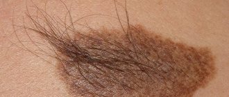
The age of appearance, number and pattern of birthmarks and moles is passed on from mothers and fathers; this is a kind of DNA-programmed connection with relatives. Due to hormonal changes, moles can appear or disappear suddenly. Therefore, the picture that parents see in a five-year-old child will definitely become different by adolescence. But regardless of the number and location, each mole must be treated with care: do not pull out hair, do not comb, do not scratch - irreversible consequences are possible.
Linear nevi - symptoms
Linear epidermal nevus was first described by Baerensprung in 1863 as naevus unius lateralis. These are congenital changes, most often present at birth, but sometimes they may not appear until childhood.
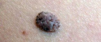
At the first stage of the disease, they look like limited areas of excessive pigmentation that do not protrude above the surface of the skin, smooth spots or papules that match the color of the skin. Later in the disease they take on a papillary form. The discoloration is associated with mild hyperkeratosis.
Epidermal moles can be found on any area of the skin, including the anus and urogenital areas. They are often located on the face and neck, scalp, trunk or limbs. The most common changes are linear and unilateral, along Blaschko's line, not extending beyond the midline of the body.
They can be:
- single, clinically resembling seborrheic keratosis;
- multiple warts, sometimes generalized, affecting large areas of the skin and persisting throughout life.
Sometimes they have a linear, vortex, striped system, often generalized, covering large areas of the body.
Moles appear in early childhood, grow with the patient and do not regress. Sometimes they can flare up and disappear, especially in the case of inflammatory linear papillary epidermal nevus, which can be accompanied by pain, burning and erosions, but never completely goes away.
Cases of the development of a malignant neoplasm in an epidermal nevus are rare, but not excluded. Isolated cases of development at the site of the lesion have been described:
- keratoacanthomas;
- basal cell carcinoma;
- squamous cell carcinoma.
Such moles may be associated with other developmental defects of the central nervous system, skeletal system, vision, as well as with some developmental disorders, for example, Proteus syndrome, McCune-Albright syndrome, Klippel-Trenaune syndrome.
What is a nevus (mole)?
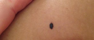
A mole is a local accumulation of melanocytes in a certain area of the skin. As a rule, this is a round or oval spot with a diameter of up to 6 mm in all shades of brown. Nevi can be congenital or acquired. The latter appear due to:
- hormonal fluctuations. Most nevi appear during adolescence and pregnancy;
- chronic allergic or infectious dermatological diseases (dermatitis, acne, lupus, psoriasis);
- prolonged exposure to ultraviolet radiation;
- genetic disorders.
Diagnosis - histopathological features of linear nevi
There is considerable variability in histopathological studies. The most common manifestations:
- hyperkeratosis;
- acanthosis;
- papillomatosis.
Some moles may exhibit acantholysis and dyskeratosis, resembling Darier's disease.
Sometimes you can find a picture of epidermolysis with hyperkeratosis, reminiscent of congenital bullous erythroderma ichthyosis. In these cases, mutations in the keratin 1 or 10 genes are found.
Diagnosis of nevus
Types of epidermal nevi
There are several clinical types of epidermal nevi. This classification is not definitive, since epidermal moles can be hard, linear and inflamed at the same time.
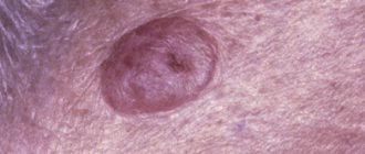
Soft papillary nevus of the epidermis . This is most often a small flesh-colored or gray formation with a soft, velvety surface. Individual papules forming clusters resemble senile warts.
Hard papillary nevus.
Unilateral, segmented, most often located on the trunk and limbs. These are tall or flat, raised discolorations of normal skin, light or dark brown, with a papillary surface.
Inflammatory linear verrucous epidermal nevus.
This is a special form of epidermal nevus with inflammation and severe itching. ILVEN may worsen and disappear, especially in the form of psoriasis-like psoriasis.
According to the conclusions of the study. Altman and Mehregan, diagnostic criteria for such moles include a number of features:
- early onset is noted - in 75% of cases before 5 years,
- more common in females (4 times more often);
- frequent localization on the left lower limb and buttocks;
- itching is usually present;
- histopathological appearance resembling psoriasis - a characteristic pattern of parakeratotic areas with visible psoriasis-like epidermal hyperplasia, mild spongy edema and exocytosis of lymphocytes;
- Such nevi have no tendency to spontaneous regression and resistance to treatment.
The lesions are linear, pruritic, erythematous lesions with a flaking surface and may resemble psoriasis lesions with a linear pattern. They are distinguished from psoriasis by the absence of Munroe microcirculation and the lack of epidermal involucrin expression in ILVEN. The connection with psoriasis is confirmed by the participation of the cytokines IL-6, IL-1, TNF-alpha, ICAM-1 in the pathogenesis.
Horny nevus (nevus corniculatus)
. This is the rarest form of epidermal nevus. The clinical picture shows very intense keratosis with the formation of thick papillary growths and filamentous formations. Histopathological examination shows foci of acantholysis with depressions in the epidermis filled with dense keratin.
Pediatric epidermal nevus.
This variety is characterized by congenital hemidysplasia, unilateral ichthyosis and limb defects. All of these disorders can occur, but it can only be the cutaneous form. The lesions are located unilaterally and do not extend beyond the midline of the body, with the most common lesions being the flexor folds. They have the character of erythematous lesions covered with yellow hyperkeratotic scales and sometimes with horny plugs. This type of mole occurs only in women because the gene responsible for the condition is located on the X chromosome.
Epidermal nevus syndrome, which is a combination of epidermal nevus with other developmental defects, has been clinically identified.
There are 4 clinical variants of the syndrome (Michalovsky classification):
- four-symptom form - lesions of the skin, bones, nerves and eyes.
- three-symptomatic form - skin, bone and nervous changes.
- two-symptomatic form - skin and bone changes.
- monosymptomatic form - the presence of a linear papillary epidermal nevus.
Why is this happening
According to some studies, 75–80% of the total dose of solar radiation that a person receives in his entire life occurs within a period of up to 20 years.
To maintain your health, doctors recommend following these simple rules: do not visit a solarium under the age of 25 or over 55; do not ignore sunscreen, especially in areas with intense ultraviolet exposure; Do not spend a lot of time in the sun if your skin is not prone to tanning. Ultraviolet radiation increases risk factors if a child or adult:
- light skin type,
- nevi (moles) on areas of the body unprotected from the sun,
- genetic predisposition,
- growing moles;
- sunburn with blisters (in a child under 5 years of age, the risk of developing melanoma increases 80 times!).
- By the way, a large number of moles on a baby’s body does not mean anything bad. On the contrary, there is a belief that the more there are, the happier fate will be. According to statistics, malignant neoplasm occurs in 70% of cases on “clean” skin and only in 30% develops from a congenital nevus.
Differential diagnosis
To make an accurate diagnosis, a dermatologist must conduct a series of tests, since the symptoms of linear moles are similar to those of other skin diseases.
At issue are:
- warty neurodermatitis
- characterized by lycosis and itching; - lichen planus papillary
- characterized by itching and its occurrence at a later age, determined by the presence of typical lesions of lichen planus in other places and histological examination; - lichen striata - characterized by inflammatory papular rashes and spontaneous resolution within a few months.
How to prevent melanomas
- Minimizing the influence of ultraviolet radiation during active hours. Dermatologists do not recommend opening areas with moles between 11 a.m. and 4 p.m. Active sun rays can trigger the degeneration of ordinary pigmented areas into melanoma. If you need to go outside at this time, cover your skin with nevi with light, light fabrics and hats.
- Use of sunscreens. For people of the first and second color types, it is mandatory to use cosmetics with spf 30. Creams, mousses, and sprays create a protective film against ultraviolet radiation for 30-60 minutes.
- Avoid traumatizing moles. If the nevus is located in such an area that it is constantly injured (rubbed by a shirt collar, scratched when shaving, pressed by straps and underwear straps), then it is better to have it removed by a dermatologist.
- Get your moles examined annually by a dermatologist. If you have fair skin with multiple nevi, then dermatologists advise undergoing a dermatological examination every six months. An unscheduled visit to a specialist is needed when the nevus begins to bleed or hurt.
For your peace of mind, a dermatologist will diagnose the mole, talk about the causes, symptoms, methods of treatment and prevention of melanoma-dangerous nevi. Sign up for a consultation by phone: 483-21-78
Treatment of epidermal moles
Epidermal moles persist throughout life, do not tend to disappear spontaneously and are primarily a cosmetic problem, especially on the skin of the face and neck. These are benign formations, although there are risks of developing cancer. A case of squamous cell carcinoma inside a nevus has been described in the literature. Treatment of epidermal moles is necessary for cosmetic reasons.
Treatment uses many different methods, varying in effectiveness and availability. When treating epidermal nevi, the following is used:
- surgical removal of the lesion;
- electrocoagulation;
- cryotherapy;
- dermabrasion;
- CO 2 laser;
- dye laser;
- tretinoin;
- 5-fluorouracil;
- acitretin;
- calcipotriol;
- dithranol;
- podophyllin;
- keratolytic drugs.
Conservative methods fail to achieve complete recovery, and scars remain after surgical removal of the lesion. The mechanism of action of individual methods is not always fully understood. For example, calcipotriol can affect the production and function of cytokines and normalize the differentiation of epidermal cells, 5-fluorouracil has an antiproliferative effect.
Some of the treatments that were used in the past are now only of historical significance.
According to many scientists, the most effective method of treating linear lesions in a small area is to remove the nevus by surgical excision; however, when completely removing large lesions, it is necessary to cover the defect with a free skin graft, which is often associated with the formation of an unaesthetic scar.
The effect of laser on moles
How are nevi (moles) formed and what is their danger?
Nevi appear from pigment cells (melanocytes) at the border between the dermis and the epithelial layer of the skin. Due to external factors (ultraviolet) or internal (hormones), melanin accumulates in a certain area, forming a flat or convex brown neoplasm - a mole.
Nevi themselves are not dangerous. But if the neoplasm is constantly injured (for example, rubbed by clothing), ultraviolet light or chemicals come into contact with it, then there is a risk of the mole degenerating into melanoma (skin cancer).
Laser removal of warts
The use of lasers gives promising results. Various types of lasers have been used to treat moles, including: CO 2 lasers, dye laser. Landthalter et al. Dermatologists have successfully used argon laser in patients with localized papillary epidermal nevus. Also, very good results are achieved using the argon laser in most patients with superficial nevus, while satisfactory results have not been obtained in patients with papillary nevus.
Another method of laser treatment of warts is the removal of epidermal nevus with a pulsed Er: YAG laser operating at a wavelength of 2940 nm and having the properties of high absorption of water contained in the tissue, as well as CO 2 laser removal.
The pulse energy is aimed at ablation, and the area of thermal damage is small, which minimizes the possibility of scarring and guarantees a relatively rapid healing process. This type of laser can remove superficial changes with very good cosmetic results.
In the case of large papillary moles of the epidermis, the best method is CO2 laser treatment.
Uch. Michel et al have used the CO 2 laser in the treatment of papillary epidermal moles with good cosmetic results when the appropriate specifications are met. The small lesions have completely disappeared, leaving only a discolored scar. Relapses were observed with too gentle procedures.
Based on these results, scientists claim that CO 2 laser treatment is more effective than other lasers, but at the same time has a higher risk of scarring. Laser treatment is a relatively new, but very promising method of treating papillary nevi of the epidermis.
Sources
- Du Vivier A.: Atlas of Clinical Dermatology, 2002.
- Gur E, Zucker R: Complex facial nevi: a surgical algorithm, 2000.
- Rosen T: Keratoacanthomas arising within a linear epidermal nevus. J. Dermatol. 1982.
- Mikhalovsky R.: Dermatological syndromatology, 1984.
- Goldberg HS: Basal cell epitheliomas developing in a localized epidermal nevus. 1980.
- Ichikawa T. et al.: Squamous cell carcinoma arising in a verrucous epidermal nevus, Dermatology, 1996.
- Molin L, Sarhammar G: Perivulvar inflammatory linear verrucous epidermal nevus treated with CO2 laser, 1999.
- Michel JL et al: CO 2 laser dermabrasion in the treatment of linear papillary epidermal nevi, Dermatologica, 2001.
- Pavlacik M. et al.: Epidermal nevus syndrome - a two-symptomatic form, 1996.
- Zvulunov A. et al.: Calcipotriol for topical use for the treatment of inflammatory linear verrucous epidermal nevi: 1997.
- Pearson IC, Parland CC: Treatment of epidermal nevi with pulsed erbium laser, 2005.
- Boyce S, Alster TS: CO 2 laser treatment of epidermal nevi long-term success. 2002.
What is melanoma and what are they?
Melanoma (skin cancer) is a malignant disease. Unlike other types of cancer, melanoma is very aggressive and quickly metastasizes to internal organs. Dermatologists are concerned that the annual number of reported cases of melanoma is increasing by 4%. Although, with early diagnosis, a patient with melanoma 0.8 mm thick has a 100% chance of recovery.
Skin cancer especially likes to appear on the lower leg, face, and forearm. Less commonly affects mucous membranes. Women get sick twice as often as men.
The diagnosis of melanoma is made by dermatological oncologists after cytological and histological examination of nevus cells.
Doctors identify the following types of skin cancer:
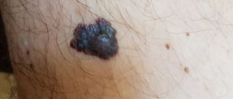
- Superficial melanoma. Outwardly it resembles a black plaque with a shiny surface. It grows quickly along the periphery, but with early diagnosis it can be easily cured
- Palmoplantar melanoma. It looks like a raised dark-colored mole with a characteristic lighter circle around it. An aggressive form of cancer.
- Subungual melanoma. It resembles a black stripe on the nail plate and can be mistaken for a phalanx injury. But if such a line appears suddenly, then it is worth visiting a dermatologist.
- Melanoma of the mucous membrane. They occur both in open areas (mucosa of the lips, eyelids) and in hard-to-reach places (mucosa of the anus, vagina). Any change in the color or structure of the mucous membrane is a reason to visit a dermatologist.
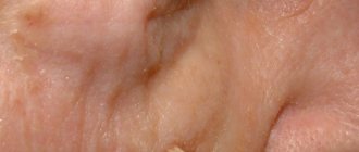
- Melanoma type lentigo maligna. The tumor appears at the site of sunburn. It can occur even years after the burn. That's why it's so important not to let children go out into the sun without sunscreen and to protect their skin as much as possible.
- Nodular melanoma. This type of cancer prefers men over 50 years of age. Outwardly, it looks like a blue or purple raised area on the skin that can hurt and bleed periodically.