Choroidal melanoma is a malignant pigmented tumor of the choroid of the eye (choroid). This tumor is one of the most common intraocular neoplasms. Choroidal melanoma is the main cause of mortality and disability in cancer patients with damage to the organ of vision. The disease can be practically asymptomatic, but at the same time has a high tendency to metastasize, so the problem of identifying it in the initial stages is extremely relevant.
- Etiological factors
- Clinical picture
- Stages of development of choroidal melanoma
- Types of eye melanoma
- Diagnostics
- Treatment options for choroidal melanoma
- Prevention and follow-up
- Prognosis for life with choroidal melanoma
Etiological factors
Most cases of choroidal melanoma are sporadic, that is, caused by one or another mutation of the melanocytic precursor cell, which can give rise to a pathological tumor clone. In addition, there is an assumption about the hereditary cause of this disease. The influence of such a typical provoking factor for skin melanoma as increased insolation for this tumor is also not excluded.
Elderly people are at risk (the average age of tumor manifestation is 60 years). Men get sick a little more often. Those with fair skin and hair, nevi and freckles are prone to developing choroidal melanoma.
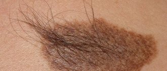
Causes of nevus formation on the eye
A nevus on the eye is an accumulation on a very small area of the tissue of the eyeball of a special pigment - melanin, which is responsible for the color of hair, skin and eyes. The reasons for this can be a variety of factors, but most often it is caused by changes in hormonal levels.
Often, a nevus on the eye occurs during pregnancy and lactation (breastfeeding), against the background of severe stress, infectious diseases, taking oral contraceptives, inflammatory skin diseases, natural aging of the body and as a result of ultraviolet and/or ionizing radiation.
It has been proven that there is also a genetic predisposition to the formation of a nevus on the eyeball. It is the hereditary factor that usually explains the occurrence of this pathology in children.
Clinical picture
Patient complaints depend on the size and location of choroidal melanoma, as well as on the presence of accompanying complications, which include: secondary retinal detachment, the appearance of degenerative processes in the retina, and clouding of the lens.
At the initial appointment with an ophthalmologist, a decrease in visual acuity, the appearance of blind spots (scotomas) in front of the eye, and hemianopsia (loss of half the visual field) are usually determined. In case of late treatment, patients complain of pain in the eye (secondary glaucoma), dilation of the vascular network. Also, a pigment spot (extraocular growth of a neoplasm) can be detected on the sclera.
Book a consultation 24 hours a day
+7+7+78
Types and types of nevi
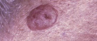
Choroidal nevi can be stationary or progressive. In addition, experts divide them into typical, atypical and suspicious. A stationary typical choroidal nevus is a flat or slightly protruding neoplasm in the fundus. Its shape is round or oval with clear or slightly blurry edges, the color is gray or greenish, the diameter, for the most part, does not exceed 6 mm. A feature of such nevi is the uniform color, as well as the lack of growth. In some cases, drusen are detected on the surface of the formation, which are an accumulation of products of intracellular metabolism. Such nevi do not cause changes in retinal tissue or visual impairment.
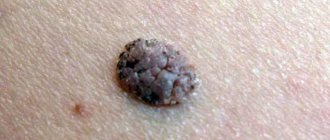
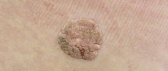
Progressive nevus of the choroid is characterized by growth and increase in volume. Changes gradually occur in its shape and boundaries, and the uniformity of color may also be disrupted. Progressive nevi often cause compression of the choroidal vessels and dystrophic changes in the adjacent retina. Sometimes the dystrophy processes are complicated by serous retinal detachment. The formations seriously impair the quality of vision: its acuity may decrease, visible images may be distorted, and blind spots may appear in front of the eyes. Specialists at the Vision Correction Center classify progressive nevi as a group with an increased risk of degeneration into a malignant neoplasm. A choroidal nevus with the signs described above, detected for the first time, is considered “suspicious.” Progressive nevi are detected during dynamic observation.
Choroidal nevi devoid of pigment, as well as “halo-nevi” located within the zone of its atrophy with a paler color, are recognized as atypical. Such formations include cells with signs of degeneration, which refers to symptoms of malignant tumor growth.
Stages of development of choroidal melanoma
According to the international classification, there are 4 stages of development of this tumor. Criteria for the prevalence of the tumor process:
- T1 - melanoma size 10 mm or less, thickness - 2.5 mm or less.
- T2 - the size of the neoplasm is 10–16 mm, the greatest thickness is 2.5–10 mm.
- T3 - measuring 16 mm and/or thickness more than 10 mm without spreading beyond the eyeball.
- T4 - the largest tumor size is 16 mm and/or thickness more than 10 mm with extension beyond the eyeball.
There are also 4 clinical stages of choroidal melanoma. Each of them is characterized by certain symptoms of the disease:
- The first, so-called “quiet eye” stage, is characterized by the absence of significant clinical manifestations and complaints. Retinal clouding may be present, and visual field defects may also be detected.
- The second stage is characterized by the appearance of pain in the eyes, inflammation, redness of the eyeball, and swelling of the eyelids.
- At the third stage, choroidal melanoma extends beyond the boundaries of the eyeball, exophthalmos is formed, and the sclera loses its integrity.
- The fourth stage is accompanied by generalization of the process. The patient's general condition is deteriorating. Patients complain of severe pain, body weight decreases, and intoxication increases. Metastases of melanoma appear in internal organs: liver, lungs, bones. Damage to one or another organ provokes the appearance of corresponding symptoms. A further decrease in visual acuity, a feeling of veil or fog before the eyes may be detected. These manifestations are caused by bleeding into the vitreous body and clouding of the lens.
Symptoms of the second and third stages of choroidal melanoma are pronounced when the tumor is located in the central or paracentral part of the fundus. Peripheral localization of the tumor is characterized by a long absence of subjective sensations. In this case, melanoma is detected either by chance or at the stage of tumor disintegration and its secondary manifestations.
Treatment of nevi
For a typical stationary choroidal nevus, special treatment or long-term observation is not required, since the risk of malignant transformation of its cells is extremely low. However, these entities are subject to mandatory documentary registration.
For suspicious and atypical nevi, mandatory photographic recording of the fundus picture, repeated ultrasound examinations and frequent re-examinations (several times annually) are required. Establishing a diagnosis of a progressive nevus makes it necessary to choose immediate treatment, since a progressive nevus is a potentially malignant tumor. For the treatment of such nevi, photo and laser coagulation are recommended. With timely treatment, the prognosis is favorable.
At what stage the malignancy of the nevus begins is impossible to predict. Therefore, the key to the patient’s health is to carefully follow the doctor’s recommendations, regular follow-up visits to a specialist, etc. treatment started as early as possible, if indicated.
In the ophthalmology department, you can undergo a full examination using the latest equipment if you suspect a nevus of the choroid and further treatment (if necessary) from leading retina specialists in Moscow.
To find out the cost of a particular procedure or make an appointment at our clinic, you can call in Moscow 8(499)322-36-36 or number 8(800)777-38-81 (toll-free from mobile phones and for regions of the Russian Federation) daily from 9:00 to 21:00. You can also use the online registration form.
Types of eye melanoma
A classification of choroidal melanoma based on morphological characteristics has been developed. Depending on the cellular structure, the following types of this tumor are distinguished:
- Spindle cell.
- Epithelioid.
- Mixed (mixed melanoma).
- Fascicular.
- Necrotic.
This classification has certain disadvantages, since necrotizing choroidal melanoma is determined clinically, but it is impossible to determine its cellular identity due to extensive necrosis. Fusiform and fascicular types have a similar prognosis. In this regard, it is currently customary to distinguish only 2 types morphologically: spindle cell and epithelioid. The mixed form occupies an intermediate position. Its prognosis depends on the predominance of certain cells. Epithelioid cell melanoma of the choroid is considered to have the least favorable prognosis.
Symptoms of retinitis pigmentosa
Manifestations of retinitis pigmentosa can occur in a carrier of the disease at any age. In early childhood, as a rule, symptoms of recessive and X-factor-linked forms of pathology develop more often, and autosomal dominant varieties often manifest themselves much later - in people of mature and even old age.
The first “bell” of the onset of the disease is usually a deterioration in dark adaptation and day blindness in excess light. Such symptoms may remain the only signs of the disease for several weeks in a fast-acting form or years in a slow-onset retinitis pigmentosa.
Further progression of the disease is accompanied by night blindness (nyctalopia), although daytime vision remains almost normal. This occurs because in the retina, it is mainly the rod photoreceptors involved in the process of light perception in low light conditions that undergo degenerative changes.
The next phase in the course of the disease is a gradual narrowing of the visual fields and the appearance of a peripheral scotoma - a blind spot with loss of areas of peripheral vision. This is explained by further pathological changes in the rods, mainly located at the edges (periphery) of the retina. Severe forms of the disease lead to “tunnel” vision with very low acuity, and patients with retinitis pigmentosa are at risk of disability.
Dystrophic changes begin to gradually affect the vessels of the eye, due to which the cone photoreceptors are also destroyed, the lens of the eye and the vitreous body become cloudy, and the sclera becomes thinner. The listed processes together lead to blindness. True, this outcome is not inherent in every form of the disease. Many autosomal dominant types of retinitis pigmentosa are characterized by long-term day blindness (hemeralopia) and narrowing of the visual fields of an unexpressed nature.
Diagnostics
Considering the clinical features of choroidal melanoma, its diagnosis, especially in the initial stages, presents certain difficulties. In addition to the analysis of patient complaints and clinical and anamnestic data, the results of the following instrumental studies are taken into account:
- Biomicroscopy.
- Ophthalmoscopy.
- Ultrasound examination of the eye.
- Diaphanoscopy, etc.
Choroidal melanoma is a neoplasm with a high risk of metastases. Therefore, when examining a patient, it is also necessary to use methods for diagnosing metastatic foci: ultrasound of the abdominal organs and lymph nodes, lung radiography, CT, MRI.
Material and methods
A total of 80 patients with NH (84 eyes) are under observation; the average age is 65.33±3.26 years. Men - 23 (23 eyes), average age 63.05±5.75 years, women - 57 (61 eyes), average age 66.71±3.96 years. The average follow-up period was 1.74±0.2 years (12-48 months). In 26 cases, a combined lesion was detected: NC and age-related macular degeneration (AMD).
In all cases, NH was discovered accidentally during a clinical examination or during ophthalmoscopy due to patient complaints of decreased vision.
All patients underwent a standard ophthalmological examination and, if necessary, specialized methods were used: ultrasound examination (ultrasound) with targeted scanning in the area of localization of the intestinal tract and digital photographic recording of the fundus. The results were compared with data from archival fundus photographs. If there were additional inclusions on its surface, autofluorescence (AF), fundus fluorescein angiography (FAG) and retinal optical coherence tomography (OCT) were performed. The following parameters of the nevus were assessed: shape, maximum diameter, prominence and its localization (including distance from the macular zone), degree of color intensity, presence or absence of drusen on the surface of the nevus and other inclusions.
Statistical processing was performed in Microsoft Excel.
Treatment options for choroidal melanoma
There are organ-preserving methods of treating this tumor and a surgical method without preserving the eye. In cases where it is not possible to save the eye, enucleation is performed - isolated removal of the eyeball or exenteration - excision of the entire contents of the orbital cavity along with the eyeball.
Indications for enucleation:
- The tumor is of significant size.
- Spread of melanoma to the optic disc.
- Complete absence of visual function.
- Extrabulbar tumor growth.
- Secondary glaucoma.
After removal of the eyeball, an internal prosthesis is implanted and subsequent external prosthetics is performed. These measures not only allow you to achieve a good cosmetic result, but also prevent facial deformation.
Organ-preserving methods of treating choroidal melanoma include:
- Radiation therapy. Depending on the method of delivering radiation, radiation therapy for this disease is carried out by contact or remote methods. Contact radiation, or brachytherapy, is the implantation of radioactive elements near the site of melanoma.
Indications for brachytherapy:
- No signs of decay.
The diameter of the neoplasm is up to 15 mm.
- The distance from the optic disc is at least 2 times the diameter of the disc itself.
- Laser coagulation in combination with thermotherapy. Transpupillary thermotherapy is a type of laser treatment for melanoma using deep local hyperthermia. The method is based on the possibility of deep penetration of infrared radiation through chorioretinal tissue. Transpapillary thermotherapy is an effective independent treatment for small choroidal melanomas (up to 4 mm in diameter).
- Cryodestruction. This method is based on extreme cooling of small melanoma lesions to −78 °C.
Brachytherapy is the most effective method of organ-preserving treatment for choroidal melanoma. Its use can reduce the likelihood of tumor metastases.
As part of systemic treatment, immune therapy is important. Also, when providing care to patients with choroidal melanoma in the later stages, the features of its metastasis are taken into account. This tumor is characterized by isolated liver damage by metastases. In such cases, chemoembolization of this organ is successfully used.
Such a common method of treating skin melanoma as targeted therapy is not used for choroidal melanoma, since this type of tumor does not have specific BRAF mutations.
Prevention
There are no specific rules for preventing the development of such neoplasms. Until now, the etiology of such moles is not known. To avoid the development of negative consequences, you should consult a doctor in a timely manner.
Timely diagnosis will help determine the presence of such neoplasms and help get rid of them, especially if the nevi are malignant. Therefore, every person should visit an ophthalmologist 1-2 times a year.
In addition, do not forget to monitor eye and eyelid hygiene. If suspicious symptoms develop, you should consult a doctor. Progressive nevi can affect visual acuity. If the patient observes clouding or distortion of the image, then it is necessary to contact an ophthalmologist.
After a full diagnosis, the doctor will be able to determine the classification of formations and the method of eliminating them. As an additional prevention, you should adhere to a healthy lifestyle. Sleep should be complete, nutrition balanced and rich in vitamins. It is necessary to give up bad habits.
Prognosis for life with choroidal melanoma
Life expectancy for this type of cancer depends on the location and size of the tumor, the patient’s age, the morphology of the tumor, the treatment performed and other features. The five-year survival rate in the initial stages of choroidal melanoma after the use of organ-preserving radical methods is 93%, and the ten-year survival rate is 89%. In later stages, when liver metastases are detected, the median survival is only 4-6 months. For patients with metastatic disease to other organs, the one-year survival rate is 76%.
Book a consultation 24 hours a day
+7+7+78